Signalment:
Gross Description:
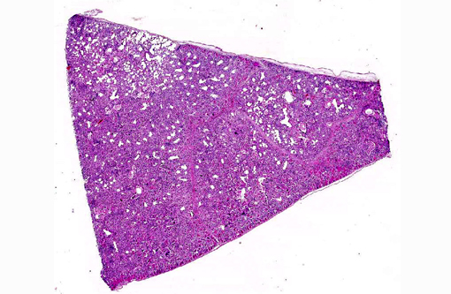
The liver had pale severely fibrotic bile ducts, diffusely. Bilateral mild hyperemia of carpal joints was noted as well as mild meningeal hyperemia.
Histopathologic Description:
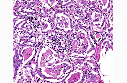
There is severe type II pneumocyte hyperplasia and hypertrophy, characterized by typical cuboidal cells outlining the septal walls.Â
Bronchioli are often difficult to separate from alveoli, since the bronchiolar epithelium is hyperplastic and sometimes necrotic. Bronchiolar lumina are also filled with an eosinophilic exudate and nuclear debris, with degenerating eosinophils. The bronchiolar walls show moderate hypertrophy of the smooth muscles.
The interstitial fibrous tissue septa exhibit mild to moderate diffuse infiltration of eosinophils, dilated blood vessels and moderate edema.Â
There is diffuse mild subpleural edema.
Occasionally within alveoli there are transverse and longitudinal sections of nematode larvae admixed with eosinophilic debris. In some sections rare adult lung worms are present in alveolar lumina. The distinctive pointed tail that is characteristic of a Protostrongylidae was not identified; however, only a single transversely cut adult was seen.
In addition, there is eosinophilic and granulomatous lymphadenitis of the mediastinal and tracheobronchial lymph nodes, as well as lymphohistiocytic (subependymal) encephalitis and meningitis.
Morphologic Diagnosis:
Lung: Pneumonia, interstitial, eosinophilic, moderate, multifocal with few intralesional larvae and adults of small lung worm, (family Protostrongylidae).
Etiology: CAEV (caprine arthritis-encephalitis virus).
Lab Results:
Condition:
Contributor Comment:
The disease is characterized by dyspnea, and the respiratory form is closely related to a small ruminant lentivirus infection in sheep called maedi, meaning dyspnea in Icelandic. CAEV and maedi-visna virus have also been found as co-infections in naturally infected goats.(4)
The SRLVs cause chronic multi-systemic inflammatory disease and mammary gland epithelium is often infected. Transmission to young animals is through milk (colostrum) via the intestine, or, less commonly, horizontally via the respiratory tract. Transmission from infected domestic goats through milk to Rocky Mountain goats has recently been described,(3) and in these cases a direct transmission between animals also was reported.Â
The natural infection of the SRLVs is with virus or virus-infected cells through the mucous membranes such as respiratory or intestinal tracts. The tropism of SRLVs is primarily the monocytes/macrophages and dendritic cells. Infected dendritic cells migrate to lymph nodes and transmit the virus to macrophages. The macrophages are then responsible for systemic infection. Persistent infection can be established in bone marrow cells, and infected monocytes continue the transmission of infection to mainly lung, mammary gland, CNS and joints. The immune activation to the viral antigen causes an inflammatory response with mononuclear cells such as lymphocytes, macrophages and plasma cells, which gives the virus access to more macrophages, continuing the inflammation, hence the multi-systemic disease of SRLVs is immunopathogenic (for review see Blacklaws).(1)
The most common tissues involved in SRLV infection are lung, CNS, mammary gland and joint. Other organs such as kidney, liver and heart can also be involved.Â
The time between infection and clinical disease is dependent on the strain and amount of the virus as well as the genetic background of the host.
CAEV is characterized by chronic interstitial pneumonia with infiltration of lymphocytes into alveolar septa which are lined by a layer of hyperplastic cuboidal type II pneumocytes, and accumulation of eosinophilic fluid within alveoli.Â
The exudative form of the acute phase is followed by regenerative lesions characterized mainly by hyperplasia as well as hypertrophy of the type II pneumocytes.
The differential diagnosis for interstitial pneumonia is extensive;(2) however, the typical type II reaction together with accumulation of eosinophilic fluid within alveoli and diffuse infiltration of lymphocytes is typical for CAEV infection. This pneumonia, together with multisystemic involvement including arthritis and encephalitis, further strengthens the diagnosis, which was confirmed by immunological testing.
Muellerius capillaris is a common small (12 -24 mm adults) lungworm of goats and sheep. Infection with this nematode is often characterized by nodular inflammation with eosinophils and giant cells; however, the inflammation depends on the amount of larvae/adults present in the alveoli. Eggs are rapidly hatched and first-stage larvae are found in alveolar spaces. These are coughed up, swallowed and eventually found in the feces. Intermediate hosts are snails and slugs and the life cycle of the nematode is completed when the goat digests the intermediate host. The larvae will migrate via lymphatics to the lungs and adults can be seen in the alveolar spaces.
Subpleural nodules are a characteristic finding in heavy worm infections, but this was not seen in the present case.Â
Another member of the family Protostrongylidae is Protostrongylus rufescens, which measure 16-35 mm. These small lung worms have a similar life cycle to Muelleris capillaris and also cause inflammation in the lungs with pulmonary nodules.Â
The few larvae/adults found in the present case could not be classified on histological slides as Muellerius or Protostrongylus.
The clinical symptoms and severity of the inflammatory reaction depend on the number of invading worms, the stage of the larvae and the immune status of the animal. The severity of the pneumonia in this case does not correlate with the animals low parasite burden; thus CAEV infection is a much more likely diagnosis.
JPC Diagnosis:
1. Lung: Pneumonia, interstitial, lymphohistiocytic, chronic, diffuse, severe, with marked type II pneumocyte hyperplasia and alveolar proteinosis.
2. Lung, alveoli: Trichostrongyle larvae, multiple.
Conference Comment:
References:
2. Panciera JR, Confer AW. Pathogenesis and pathology of bovine pneumonia. Vet Clin Food Anim. 2010;26:191-214.
3. Patton MK, Bildfell RJ, Anderson Ml, et al. Fatal caprine arthritis encephalitis virus-like infection in 4 Rocky Mountain goats (Oreamnos americanus). J Vet Diagnostic. 2012;24(2):392-396.
4. Pisoni G, Bertonei G, Puricelli M, et al. Demonstration of coinfection with recombination by caprine arthritis-encephalitis virus and maedi-visna virus in naturally infected goats. J Virol. 2007;81(10):4948-4955.