CASE I:
Signalment:
Late-term aborted, female lamb from lowland sheep (Ovis aries).
History:
Eight aborted lambs from six abortions were submitted for necropsy. Twenty-two late-term abortions had occurred in a group of 230 lowland ewe lambs (1-year-old) over an 8-day period. The ewe lambs were due to start lambing approximately 1 week later. Twin pregnancies had been particularly affected and occasionally one twin was born alive and survived. The ewe lambs had been kept outdoors on grass only, with no access to silage or concentrates, appeared in good body condition and had been vaccinated against toxoplasmosis and ovinecampylobacteriosis, but not against ovine enzootic abortion (syn. enzootic abortion of ewes (EAE)).
Gross Pathology:
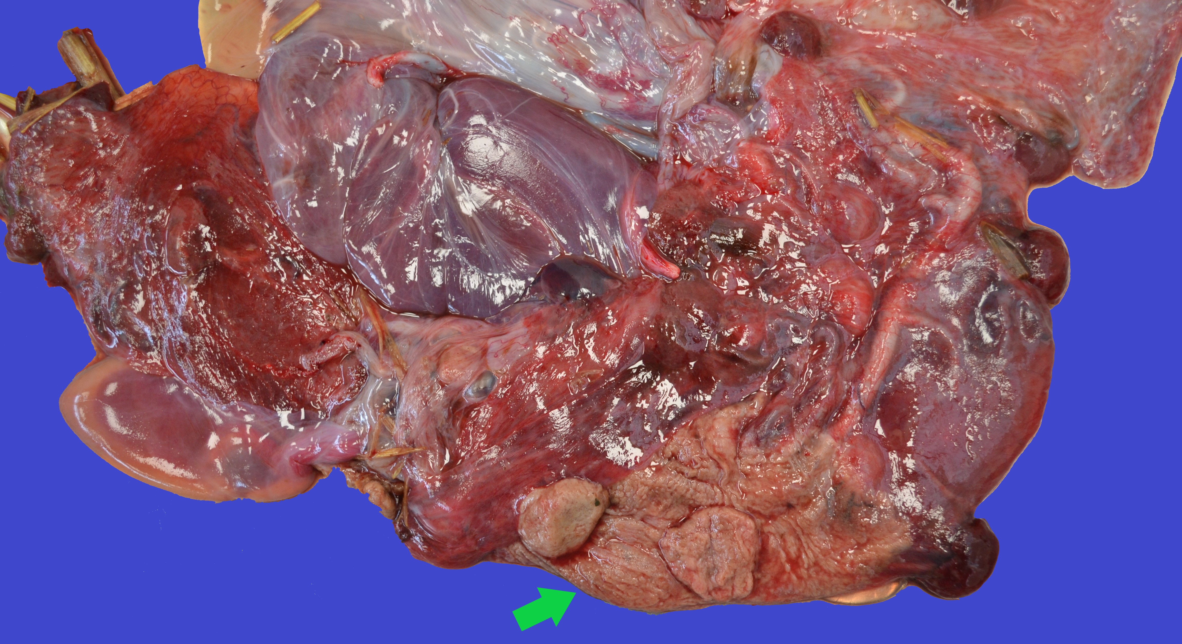
Six out of the eight submitted aborted lambs were received with all or part of their placentas. Three placentas were grossly unremarkable. In the other three placentas, there was a focally extensive area of moderately thickened, non-translucent, slightly dry and brown intercotyledonary tissue and similarly dry and brown cotyledons. Large adjacent areas were moderately edematous with injected blood vessels. The aborted lambs were well-preserved with no evidence of maceration or mummification. Some aborted lambs had small amounts of serosanguinous fluid in the abdominal cavity and occasional subcutaneous or perirenal bruising.
Laboratory Results:
Laboratory testing was carried out on four of the six abortions. Culture of fetal stomach contents from three cases revealed a very heavy pure growth of hemolytic Gram-positive rods that were identified as Listeria ivanovii. Enriched culture was negative for Campylobacter spp. Placental smears stained with modified Ziehl-Neelsen were negative for Brucella spp., Chlamydia spp. and Coxiella spp. PCR for Toxoplasma gondii and border disease virus were both negative.
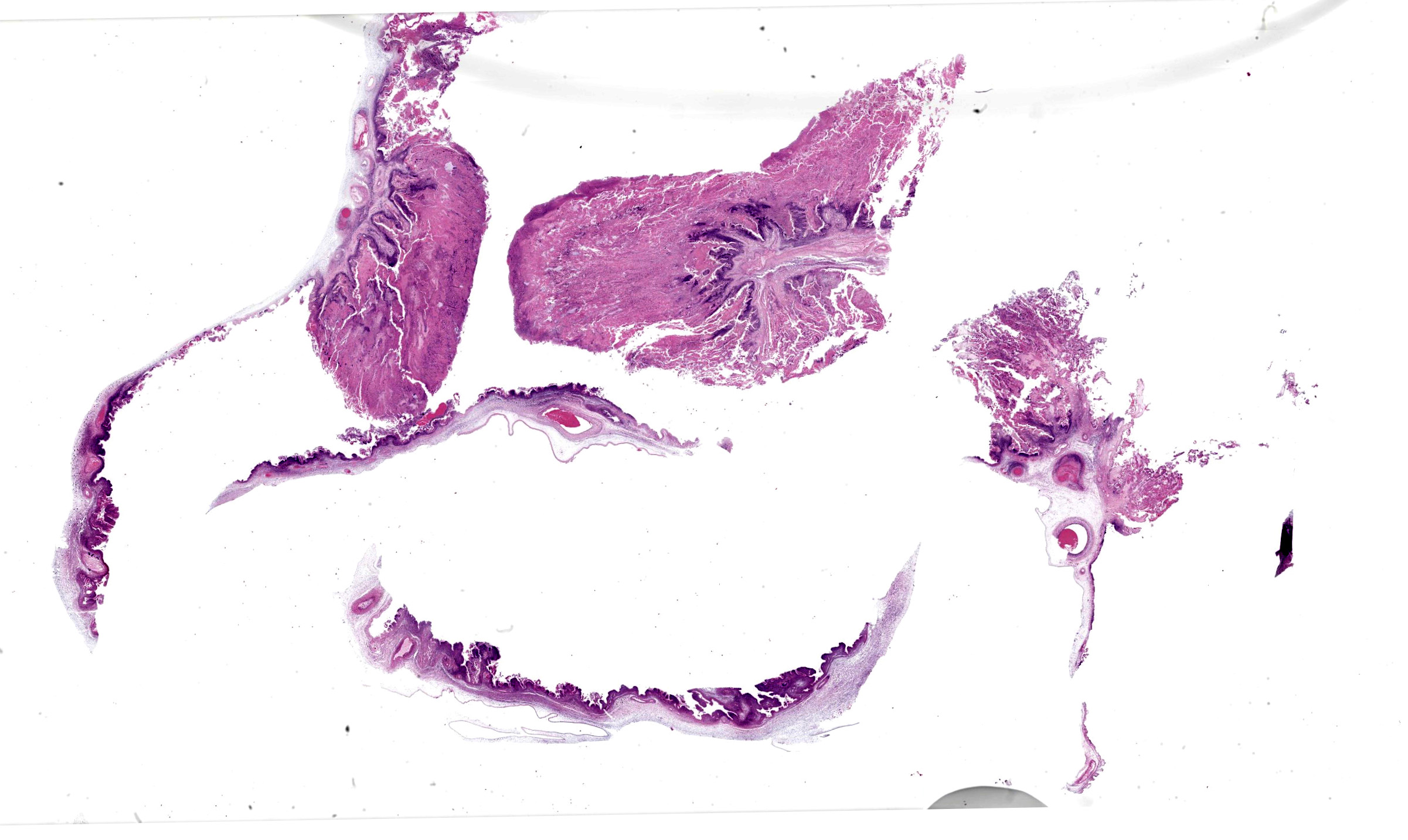
Microscopic Description:
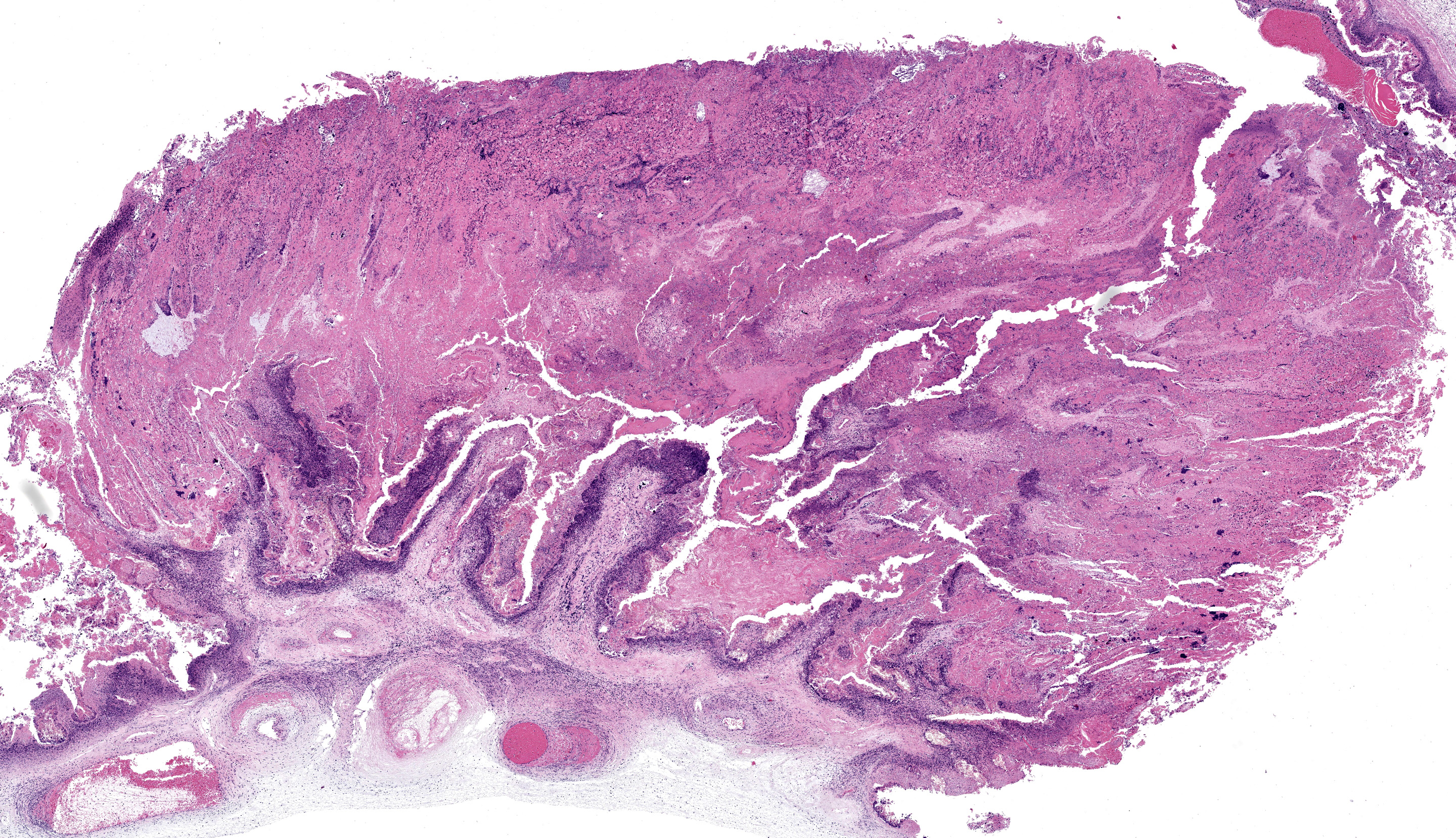
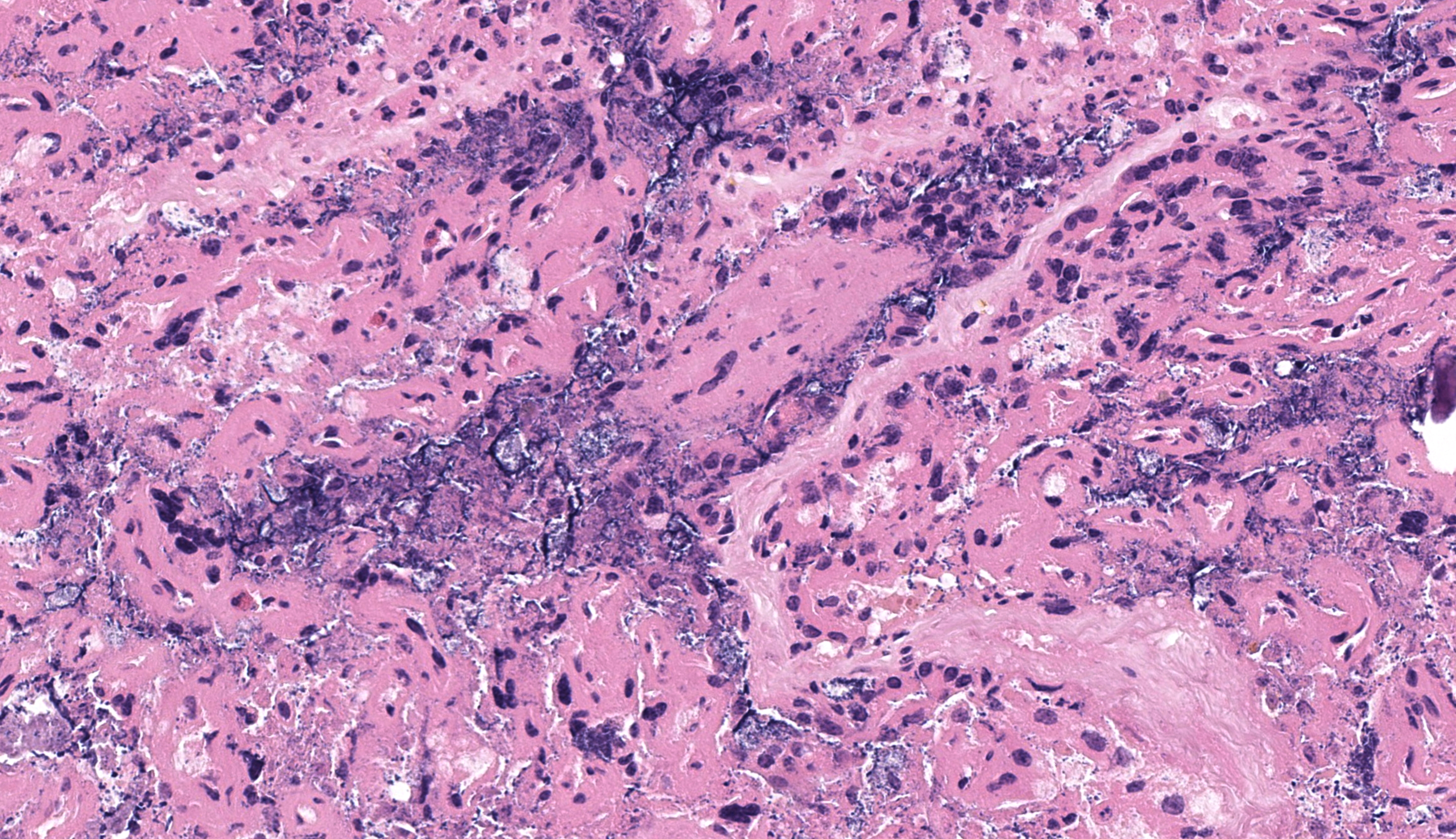
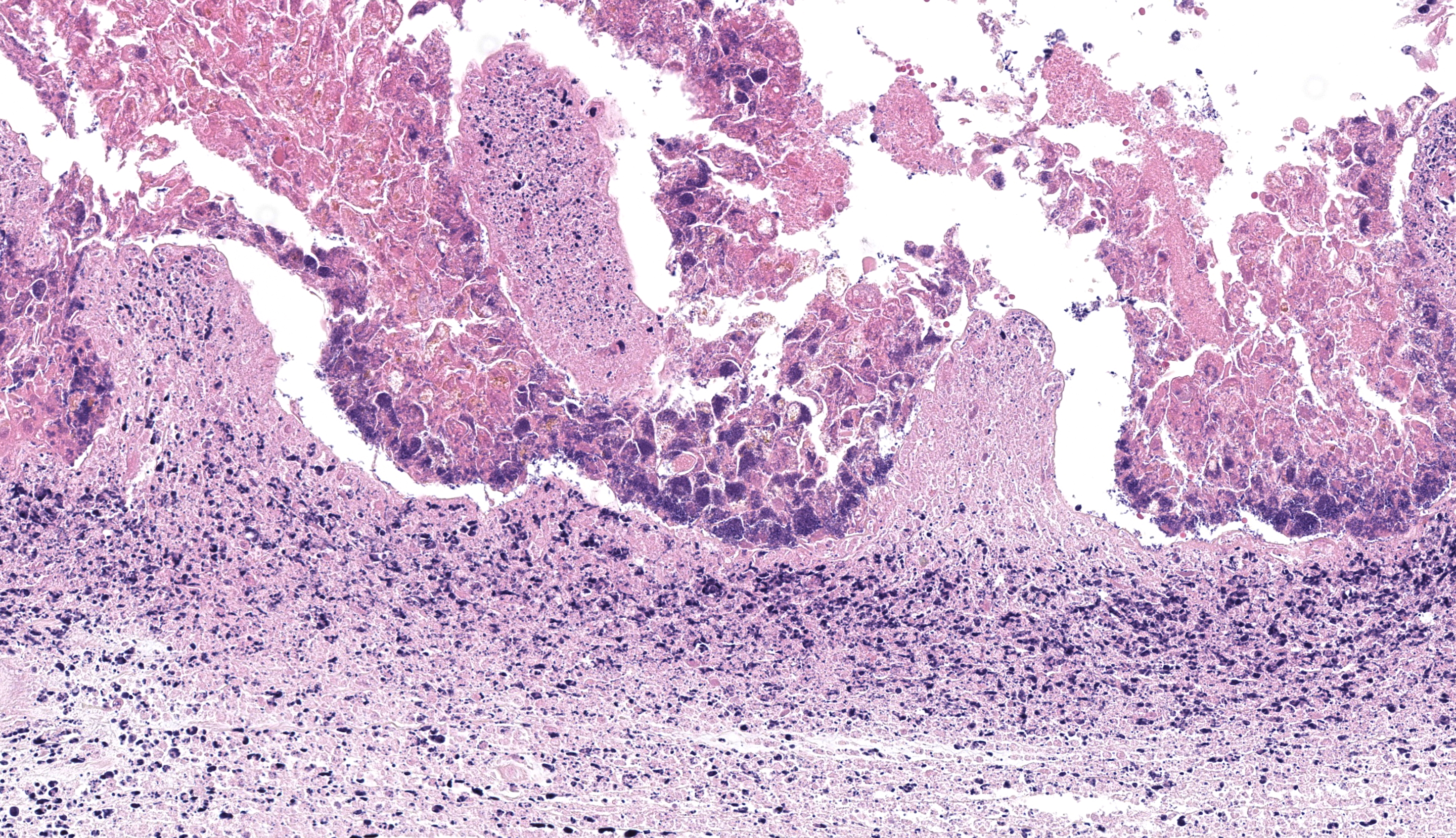
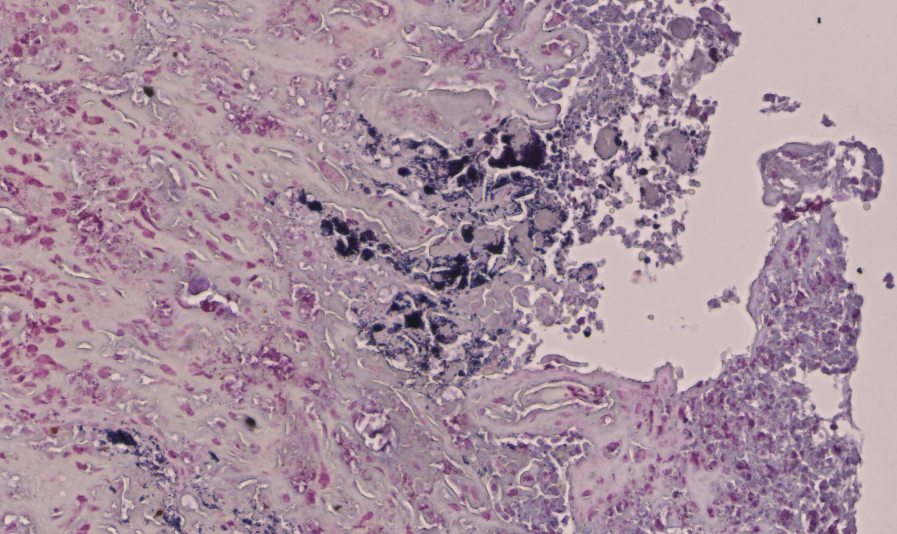
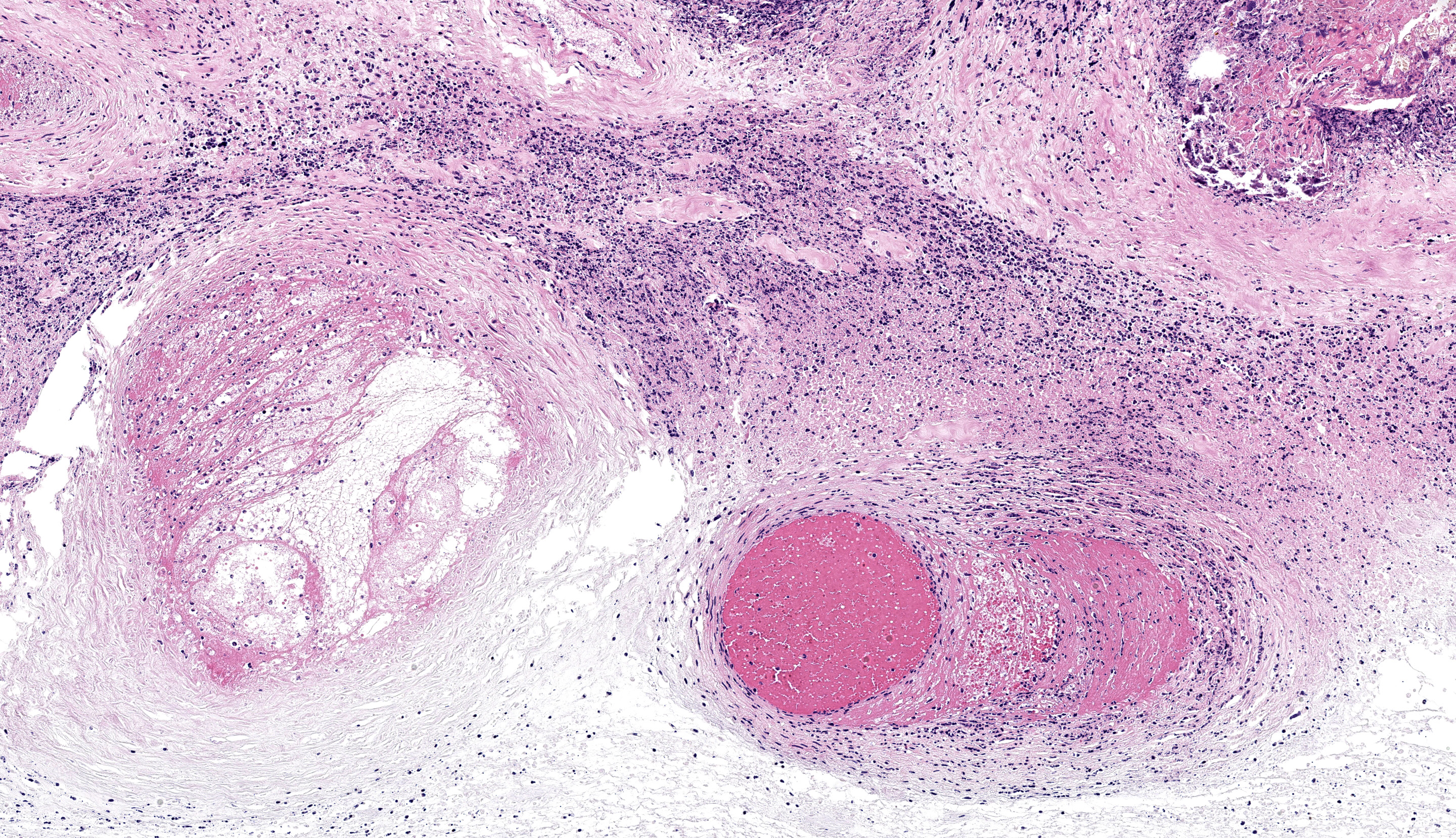
Chorioallantois, cotyledons and intercotyledonary tissue: The cotyledons and intercotyledonary tissue are extensively necrotic, expanded by edema and multifocal fibrin deposits and infiltrated by high numbers of viable and degenerate neutrophils, fewer macrophages and several prominent colonies of short, Gram-positive, rod-shaped bacteria (bacilli), measuring 0.5–2 μm in length (Figure 2). Multifocally, there are degenerate and necrotic trophoblasts, some of which contain intracytoplasmic Gram-positive bacilli. Several blood vessel walls, mainly arteries, contain intraluminal fibrin thrombi and the blood vessel walls are moderately to markedly infiltrated and expanded by high numbers of viable and degenerate neutrophils and fibrin.
Contributor’s Morphologic Diagnosis:
Chorioallantois, cotyledons and intercotyledonary tissue; acute, focally extensive, marked, necro-suppurative placentitis with large numbers of intralesional Gram-positive bacilli and multifocal, marked, necrotizing and neutrophilic vasculitis with acute intraluminal fibrin thrombi
Contributor’s Comment:
The microscopic findings in the chorioallantois and the pure growth of Listeria ivanovii from fetal stomach contents support this abortion outbreak to be the result of listerial infection. L. ivanovii, formerly known as L. monocytogenes, serotype 5 8 is a recognized cause of ovine,1,6,9 caprine5 and bovine3 abortions, but rarely causes other conditions. It appears to be especially pathogenic for sheep but is less commonly isolated than L. monocytogenes.7
The most common lesions in abortions caused by L. monocytogenes are necro-suppurative cotyledonary and intercotyledonary placentitis, autolysis of aborted fetuses, miliary foci of necrosis in the liver and spleen, and marked necrotizing enteritis,4,7 whereas less is known about typical lesions in abortions caused by L. ivanovii.
The present case had necro-suppurative placentitis and fibrino-suppurative vasculitis, which has not been previously described in cases of ovine abortions due to L. ivanovii,1,6,9 but has been described in a case of caprine abortion. 5 Interestingly, only rare, microscopically evident, necrotic foci in the liver were identified in this case, even though grossly apparent necrotic hepatitis is commonly described in ovine and bovine abortions due to L. monocytogenes 7 and also in previous cases of ovine abortions due to L. ivanovii.1,6,9 Also, the lungs had moderate neutrophilic bronchopneumonia which was likely the result of inhaled amniotic fluid with admixed listerial bacteria. Similarly, bronchopneumonia has been described in an ovine,6 caprine5 and bovine3 abortion case caused by L. ivanovii.
In abortion outbreaks caused by L. monocytogenes, infection most often spreads by ingestion of food or water contaminated by feces, urine, placenta or vaginal discharge from affected ewes. Also, consumption of contaminated poor-quality silage is a well-known and important source of infection with L. monocytogenes.7 Previously reported ovine abortion outbreaks caused by L. ivanovii often coincided with periods of cold and wet weather1,6,9 and feeding of spoiled hay9 or moldy hay and barley.6 In the herein described
abortion outbreak, the ewe lambs had only been on grass, suggesting that L. ivanovii infection may have been directly acquired from the pasture environment. This route of exposure has been proposed in a case series of weaned lambs in the United Kingdom with visceral L. ivanovii infections.2 Interestingly, in this submission, the farmer reported that some abortions consisted of one dead lamb and one live unremarkable lamb including one of those submitted from which L. ivanovii was isolated. This is in contrast to previous reports.1,6,9
The present case highlights the importance of testing for common as well as less common abortion pathogens and sampling of several tissues for histology to confirm isolated pathogens microscopically.
Contributing Institution:
Department of Pathobiology and Population Sciences, Royal Veterinary College, Hawkshead Lane, North Mymms, Hertfordshire, United Kingdom, AL9 7TA. The website address is www.rvc.ac.uk: https://vet.purdue.edu/cpb/
JPC Diagnosis:
Placenta: Placentitis, necrosuppurative, subacute, diffuse, severe, with vasculitis, thrombosis, and numerous extracellular and intratrophoblastic bacilli.
JPC Comment:
This week’s moderator was Dr. Maggie Highland from the University of Wisconsin Veterinary Diagnostic Laboratory. Dr. Highland emphasized the diagnostic workup of small ruminant abortion in her pre-conference lecture and revisited these concepts in this first case which we excerpt again here.
From subgross magnification, tissue identification of the placenta in this case is facilitated by recognition of ruminant cotyledonary structure. Although there is marked coagulative (and subsequently lytic necrosis) present in this tissue, the state of tissue preservation is representative of typical abortion cases. The major diagnostic features present include vasculitis and innumerable intratrophoblastic cytoplasmic bacilli which provide a clear mechanism for abortion (interruption of blood flow and placental insufficiency). Conference participants considered several possible etiological agents including Chlamydia (which would also have vasculitis and intracytoplasmic bacteria) as well as Brucella (vasculitis) and Coxiella (lacks vasculitis, but centered on trophoblasts).7 Other potential rule outs for small ruminant abortion include Border disease virus (and BVDV) as well as toxoplasmosis, though the latter is associated with cotyledonary necrosis without vasculitis.7
Our Gram stain was helpful for resolving Listeria as the probable agent given the size and slight curvilinear shape – we noted large numbers of gram-positive bacteria both free and within trophoblasts. Conference participants wondered if the large number of organisms present on this slide represented a particular tropism/niche (and corresponding fitness) of L. ivanovii for placenta. Likewise, we speculate that Listeria hitting twins harder than singleton lambs reflects a decreased placental reserve for twins over singletons. Although not performed at the JPC, Giemsa and/or Gimenez stains to enhance visualization of trophoblastic organisms is a helpful part of the diagnostic workup.7 Culture of fetal abomasal contents may prove vital in instances where the placenta lacks diagnostic information. Dr. Highland emphasized reporting PCR/culture results as “not detected” or cultures as “no growth: rather than ‘negative’ in both instances– this distinction carries some weight in animals previously treated with antibiotics and precludes potential client misinterpretation of “negative” results.
References:
- Chand P, Sadana JR. Outbreak of Listeria ivanovii abortion in sheep in India. Vet Rec. 1999;145(3):83-84.
- Dunnett E, Florea L, Thurston L, Floyd T, Collins R, Otter A. Deaths of weaned lambs with visceral Listeria ivanovii infections. Vet Rec Case Rep. 2020;8: e001254.
- Gill PA, Boulton JG, Fraser GC, Stevenson AE, Reddacliff LA. Bovine abortion caused by Listeria ivanovii. Aust Vet J. 1997;75(3):214.
- Low JC, Donachie W. A review of Listeria monocytogenes and listeriosis. Vet J. 1997;153(1):9-29.
- Moeller RB. Causes of caprine abortion: diagnostic assessment of 211 cases (1991-1998). J Vet Diagn Invest. 2001;13:265-270.
- Sahin M, Beytut E. Abortions in sheep due to Listeria ivanovii in the Kars Region. Turk J Vet Anim Sci. 2006;30:503-506.
- Schlafer DH, Foster RA. Listeriosis. Female Genital System. In: Maxie MG, ed. Jubb, Kennedy, and Palmer’s Pathology of Domestic Animals. Vol 3. 6th ed. St. Louis, MO: Elsevier; 2016:398-402, 402-406, 408-409,414-416, 420-421.
8. Seeliger HPR, Rocourt J, Schrettenbrunner A, Grimont PAD, Jones D. Listeria ivanovii sp. nov. Int. J. Syst. Bacteriol. 1984;34(3):336-337. - Sergeant ESG, Love SCJ, McInnes A. Abortions in sheep due to Listeria ivanovii. Aust Vet J. 1991;68(1):39.