Signalment:
Gross Description:
Histopathologic Description:
Morphologic Diagnosis:
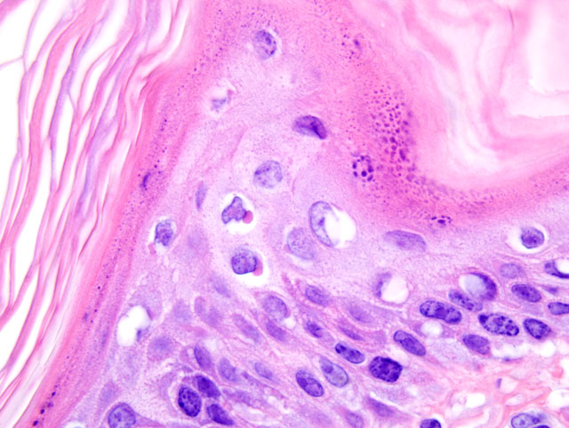
1. Haired skin: Hyperkeratosis, orthokeratotic, marked, with acanthosis and intracytoplasmic and intranuclear inclusions consistent with canine distemper virus infection.
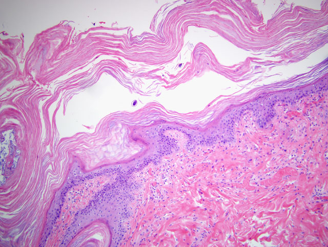
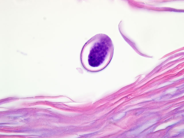
2. Haired skin: Multifocal serocellular crusting of the skin surface with intralesional nematode embryonated eggs consistent with Dracunculus insignis (only in some slides).
Lab Results:
Condition:
Contributor Comment:
Histologic lesions of CDV include lymphoid atrophy and necrosis, bronchointerstitial pneumonia, non-suppurative encephalomyelitis, demyelination of white matter tracts, enamel hypoplasia, conjunctivitis, keratitis, retinitis, optic neuritis, hyperkeratosis, persistence of primary spongiosa of bones, and myocardial necrosis. Viral inclusions are most prominent in the brain and epithelial tissues. Intracytoplasmic inclusions tend to be more common in epithelial cells, while intranuclear inclusions are more common in the brain.(2) Skin lesions of CDV are most commonly found on the foot pads, nose, eyelids, lips, and anus,(8) although the haired skin can also be affected.(2,5) Histologic findings in the epidermis of haired or non-haired skin include hyperkeratosis and/or parakeratosis, acanthosis, rete ridge formation, epidermal syncytial cells, cytoplasmic and nuclear inclusions, pustular dermatitis secondary to hyperkeratosis, and periadnexal and perivascular mononuclear inflammation.(2) Immunohistochemistry and viral inclusions visible on H&E-stained slides demonstrate that viral particles are primarily located within the stratum spinosum and stratum granulosum.(5) The pathogenesis of hyperkeratosis in CDV is suggested to be due to defective differentiation of infected keratinocytes.(5)
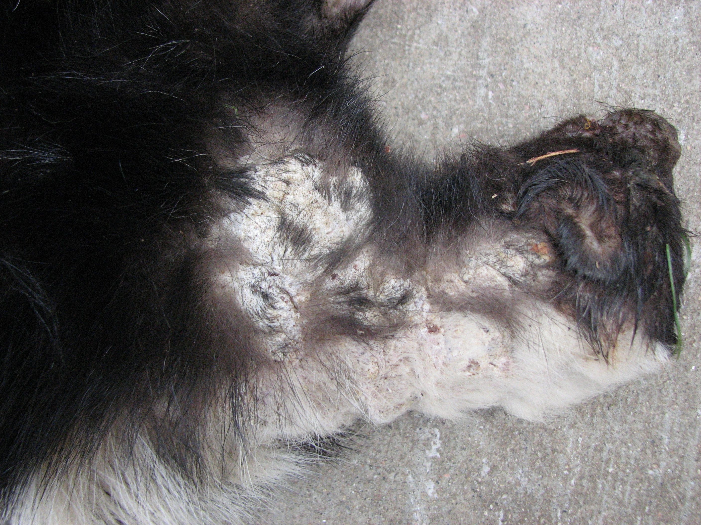
In the case presented here, the infected skunk demonstrated multiple gross and histologic features of systemic infection by CDV, including oculonasal discharge, interstitial pneumonia, evidence of diarrhea, focal myocardial necrosis, and the submitted lesion of hyperkeratosis of the haired skin overlying the shoulders, neck, and head. Interestingly, the epidermis of the foot pads was not noticeably thickened in this case. Fluorescent antibody testing of the brain confirmed infection with CDV.
The additional finding in some slides of multifocal serocellular crusting of the skin surface with intralesional cross sections of nematode eggs is consistent with the gross finding of subcutaneous nematodes suggestive of Dracunculus insignis infection. Definitive identification of the parasite was pending at the time of case submission. Dracunculus insignis is a member of the spirurid group of nematodes and is commonly found in wild mammals (predominantly raccoons), but can also be transmitted to domestic mammals including dogs and cats.(1) For this parasite to reproduce, the female uterus is prolapsed through an ulcer in the skin to release larvae. The larvae are then ingested by Cyclops spp. (an aquatic copepod) where they develop to infective third stage larvae. The copepod is ingested either directly by the definitive host, or can be ingested first by a paratenic host such as a frog. Once ingested by the definitive host, the copepod dies, the infective stage is released, and the larvae penetrate the hosts stomach and intestinal wall to mature and reproduce. Once fertilized, the female parasites migrate to the subcutaneous tissues, typically of the lower limbs, to expel ova.(1)
JPC Diagnosis:
1. Haired skin and subcutis: Epidermal hyperplasia, diffuse, marked, with epidermal and follicular orthokeratotic hyperkeratosis, intracorneal pustules, and few embryonated filarid nematode eggs, etiology consistent with Filaria taxideae.
2. Haired skin and subcutis, epidermis, hair follicles, and apocrine glands: Epithelial degeneration, multifocal, mild, with rare intracellular edema and numerous intracellular eosinophilic viral inclusion bodies, etiology consistent with canine distemper virus.
Conference Comment:
Two surveys of badgers in Wyoming found F. taxideae in 81%(6) and 39%(4) of badgers examined, respectively, and an apparent seasonal occurrence of associated dermatitis, with active lesions peaking during the summer. In badgers, gross lesions are most pronounced in the inguinal area, proximal thigh, and ventral abdomen and are characterized by blood-tinged fluid-filled vesicles and/or areas of serocellular crusting and ulceration.(6) In the single published report of filarial dermatitis attributed to F. taxideae in a striped skunk, the lesions consisted of large confluent areas of alopecia and marked thickening of the skin overlying the head, thoracolumbar region, and tail base; the description of the lesions in the case report bear striking resemblance to those depicted in the image submitted by the contributor for this case.(7)
Adult filarids are white, often tightly coiled, nematodes ranging from 122-131 mm (males) to 285-420 mm (females) in length; they are readily visualized in the subcutis and fascia, where they elicit little inflammatory response. Lesions result when gravid females deposit eggs in the upper dermis or between the epidermis and cutaneous basement membrane. Early microscopic findings in badgers consist of subepidermal vesiculobullous dermatitis extending into follicular infundibulae, with numerous embryonated filarid eggs occupying vesicles; lesions progress to ulceration and dermatitis, with orthokeratotic hyperkeratosis and acanthosis in the adjacent epidermis.(6) In skunks, reported lesions include marked epidermal hyperplasia with orthokeratotic hyperkeratosis, and numerous dermoepidermal pustules containing larvated eggs.(7)
Conference participants attributed much of the acanthosis and hyperkeratosis observed in the submitted case to filarial dermatitis, rather than CDV infection, but acknowledged that the CDV inclusions in the epidermis, follicular epithelium, and apocrine glands are numerous and prominent. Infection with CDV may have predisposed this skunk to nematode infection; in addition to the microscopic lesions described by the contributor, several participants slides contained few pigmented fungal hyphae in the superficial layers of the epidermis, interpreted as opportunistic dematiaceous fungi.
The contributor provides a useful synopsis of CDV infection, which participants reviewed in conference. Attendees briefly reviewed other noteworthy morbilliviruses of importance in veterinary medicine, including measles virus, rinderpest virus, peste-des-petits-ruminants virus, phocine distemper virus, and cetacean morbillivirus.
References:
2. Caswell JL, Williams KJ: Respiratory system. In: Jubb, Kennedy, and Palmers Pathology of Domestic Animals, ed. Maxie MG, 5th ed., vol. 2, pp. 635-638. Elsevier Saunders, Philadelphia, PA, 2007
3. Gardiner CH, Loomis MR, Britt JO Jr., Montali RJ: Dermatitis caused by Filaria taxideae in a lesser panda. J Am Vet Med Assoc 183:1285-1287, 1983
4. Keppner EJ: The pathology of Filaria taxideae (Filarioidea: Filariidae) infection in the badger, J Wildl Dis 7:317-232, 1971
5. Koutinas AF, Baumg+�-�rtner W, Tontis D, Polizopoulou Z, Saridomichelakis MN, Lekkas S: Histopathology and immunohistochemistry of canine distemper virus-induced footpad hyperkeratosis (hard pad disease) in dogs with natural canine distemper. Vet Pathol 41:2-9, 2004
6. OToole D, Williams ES, Welch V, Nunamaker CE, Lynn C: Subepidermal vesiculobullous filarial dermatitis in free-ranging American badgers (Taxidea taxus). Vet Pathol 30:343-351, 1993
7. Saito EK, Little SE: Filarial dermatitis in a striped skunk. J Wildl Dis 33:873-876, 1997
8. Williams ES: Canine distemper. In: Infectious Diseases of Wild Mammals, eds. Williams ESW, Barker IK, 3rd ed., pp. 50-55. Iowa State University Press, Ames, IA, 2001