Signalment:  

2-year-old, female Giant marine toad (Bufo marinus)Found dead; no observable problems prior to death. Two cagemates have died of mycobacteriosis in the past.


Gross Description:  

Expanding the caudal portion of the base of the tongue and laryngeal/pharyngeal area are multiple, up to 5 mm cross sectional diameter, white to tan, caseous nodules that extend to the cranial portion of the left lung. There are multiple, up to 4 mm in diameter, white to tan, caseous nodules throughout the tissue of both lungs. The spleen is enlarged and measures 1.0 cm x 1.0 cm x 0.7 cm; there are multiple, up to 2 mm in diameter, caseous, white to tan nodules throughout the splenic parenchyma. The kidneys are uniformly dark red to purple and contain a few, small, up to 1 mm in diameter, white to gray foci, predominantly on the surface of the cranial portion of both kidneys.


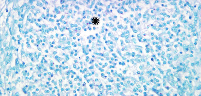
Histopathologic Description:

Lung: Multifocally expanding the intersitium are many granulomas that are up to 2 mm in diameter and are composed of a core of macrophages and few lymphocytes surrounded by a variably thick capsule of loose, fibrous connective tissue. Epithelium overlying these granulomas is often thinned and has lost the ciliated border. Occasionally, the epithelium is ulcerated. Between the faveolar walls are frequent infiltrates of macrophages, few lymphocytes and neutrophils and rare multinucleated giant cells. Rarely macrophages have peripheralized nuclei and peripheral cytoplasmic pallor.

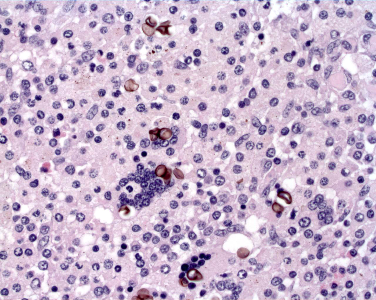
Acid fast: There are scattered, positive bacilli throughout granulomas.


Morphologic Diagnosis:  

Lung: Pneumonia, granulomatous, chronic, multifocal, moderate, with intralesional acid fast bacilli consistent with Mycobacterium sp., giant marine toad (Bufo marinus)


Lab Results:  

Lung cytology revealed acid fast bacilli
Culture of the lung grew Mycobacterium gordonae


Condition:  

Mycobacterium gordonae


Contributor Comment:  

Mycobacteriosis is common in animals and may be associated with a variety of pathogenic or environmental species. Historically, Mycobacterium tuberculosis and M. leprae most commonly affect humans, but M. tuberculosis has also been isolated from a number of non-human primates and elephants, and M. leprae may be enzootic in 9-banded armadillos5. Mycobacterium gordonae is commonly found in water and is not considered pathogenic5. However, compromise of the immune system can leave an individual open to opportunistic invasion by this organism. The toad in this case had systemic chromoblastomycosis which may have been associated with immunocompromise.

In amphibians, Mycobacterium induces granulomas similar to those precipitated by M. tuberculosis in humans. Key differences for amphibians include few multinucleated giant cells, a lack of mineralization, and a propensity for development in the skin4. Additionally, cytology and histology of amphibian lesions reveal numerous acid fast organisms, in contrast to the paucibacillary lesions of human TB. Cutaneous mycobacteriosis in the amphibian is most often associated with M. marinum, another water-borne mycobacterial species. The initial inflammatory response is disorganized and histiocytic and can be seen histologically by 2 weeks post-infection1. Mature granulomas composed of sheets of interdigitating epithelioid macrophages surrounded by a fine, fibrous capsule are seen by 8 weeks post-infection. Caseation of the core may occur in some granulomas3 effectively walling off the bacteria from the host immune system. In this way, latent infection may be maintained.


JPC Diagnosis:  

Lung: Pneumonia, granulomatous, multifocal, moderate, with acid fast bacilli consistent with Mycobacterium sp., giant marine toad (Bufo marinus), amphibian.


Conference Comment:  

Mycobacteria are non-motile, nonspore-forming, pleomorphic, acid-fast, weakly Gram-positive coccobacilli.3 Their cell walls contain a large hydrophobic layer of mycolic acids which allows environmental and antimicrobial resistance. Increased amounts of trehalose dimycolate (cord factor) within the cell wall is also associated with increased virulence.3 The Mycobacterium genus contains numerous strict pathogens (i.e. M. tuberculosis and M. leprae), as well as opportunistic pathogens (i.e. M. avium and M. marinum).5 Tuberculosis is a term reserved for diseases caused by M. tuberculosis or M. bovis with all other conditions referred to as mycobacteriosis.3

The initiation of the cell-mediated immune response consists of activated macrophages secreting interleukin-12, which in turn skews the immune system to secrete interferon-γ and interleukin-2 by CD4+ T-helper-1 lymphocytes.3 Tumor necrosis factor-_ and interferon-γ act together to promote the formation of the tuberculoid granuloma.3

Granulomas in the amphibians induced by Mycobacterium marinum share many features with human tuburculosis granulomas.1 They contain mature macrophages, epithelioid cells, and extracellular matrix components. Yet caseation does not appear to be a feature in frog granulomas even though it is seen in M. marinum infections of humans, goldfish and toads.1

Hyphae due to the systemic chromoblastomycosis were not present in all lung sections, so the entity was not discussed by the contributor. Chromoblastomycosis can be caused by a wide variety of brown pigmented, dematiaceous fungi, and infections have been seen in mammals and amphibians.2 In amphibians the infection is often systemic with lesions in the skin, liver, lungs, and kidneys of stressed animals.2


References:

1. Bouly, DM, N Ghori, KL Mercer, S Falkow, L Ramakrishnan: Dynamic nature of host-pathogen interactions in Mycobacterium marinum granulomas. Infection and Immunity 69:12 p 7820-7831, 2001
2. Bube A, Burkhardt E, Weiss R: Spontaneous chromomycosis in the marine toad (Bufo marinus). J Comp Path 106:73-77, 1992
3. Caswell JL, Williams KJ: Respiratory system. In: Jubb, Kennedy, and Palmers Pathology of Domestic Animals, ed. Maxie MG, 5th ed., vol. 2, pp. 606-610. Elsevier Limited, St. Louis, MO, 2007
4. Green, DE: Infectious etiologies Mycobacteria Mycobacteriosis. In: Amphibian Medicine and Captive Husbandry, eds. Wright KM, Whitaker BR, pp. 427-429. Kreiger Publishing Co., Malabar, FL, 2001
5. Rastogi N, Legrand E, Sola C: The mycobacteria: an introduction to nomenclature and pathogenesis. Rev Sci Tech 20:21-54, 2001
6. Shively JN, Songer JG, Prchal S, Keasey MS 3rd, Thoen CO: Mycobacterium marinum infection in Bufonidae. J Wildl Dis 17:3-8, 1981
7. Taylor SK, Williams ES, Thorne ET, Mills KW, Withers DI, Pier AC: Causes of mortality of the Wyoming toad. J Wildl Dis 35:49-57, 1999

A virtual slide is not available for this case.


Fig. 4-1 Lung


Fig. 4-2 Lung


Back | VP Home | Contact Us |