Joint Pathology Center
Veterinary Pathology Services
Wednesday Slide Conference
2019-2020
Conference 4
18 September 2019
CASE III: UMN VDL(JPC 4116936)
Signalment: 3-year-old ewe (Ovis aries) breed unknown.
History: Pesistent nasal discharge and labored breathing. This animal was euthanized.
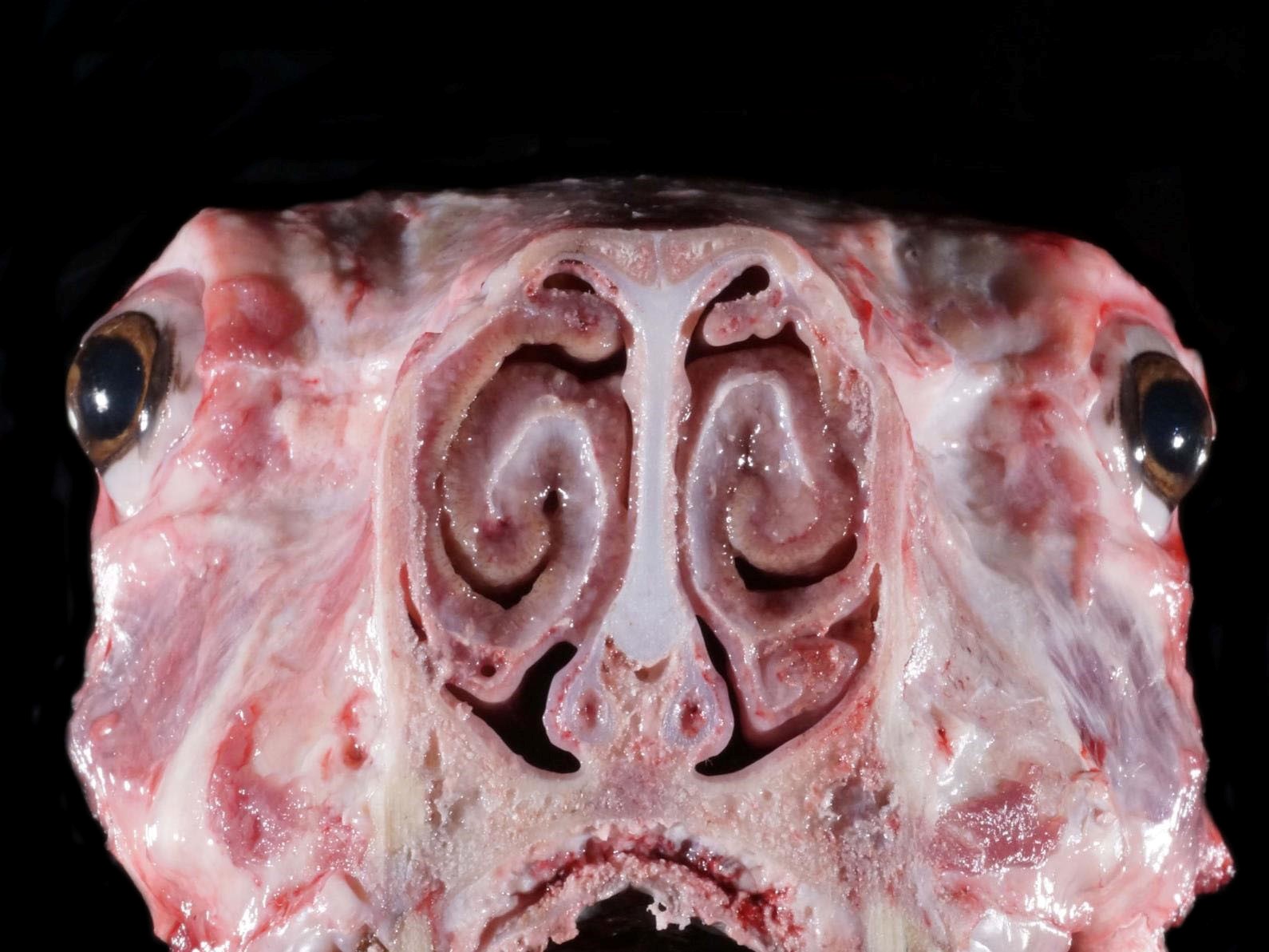
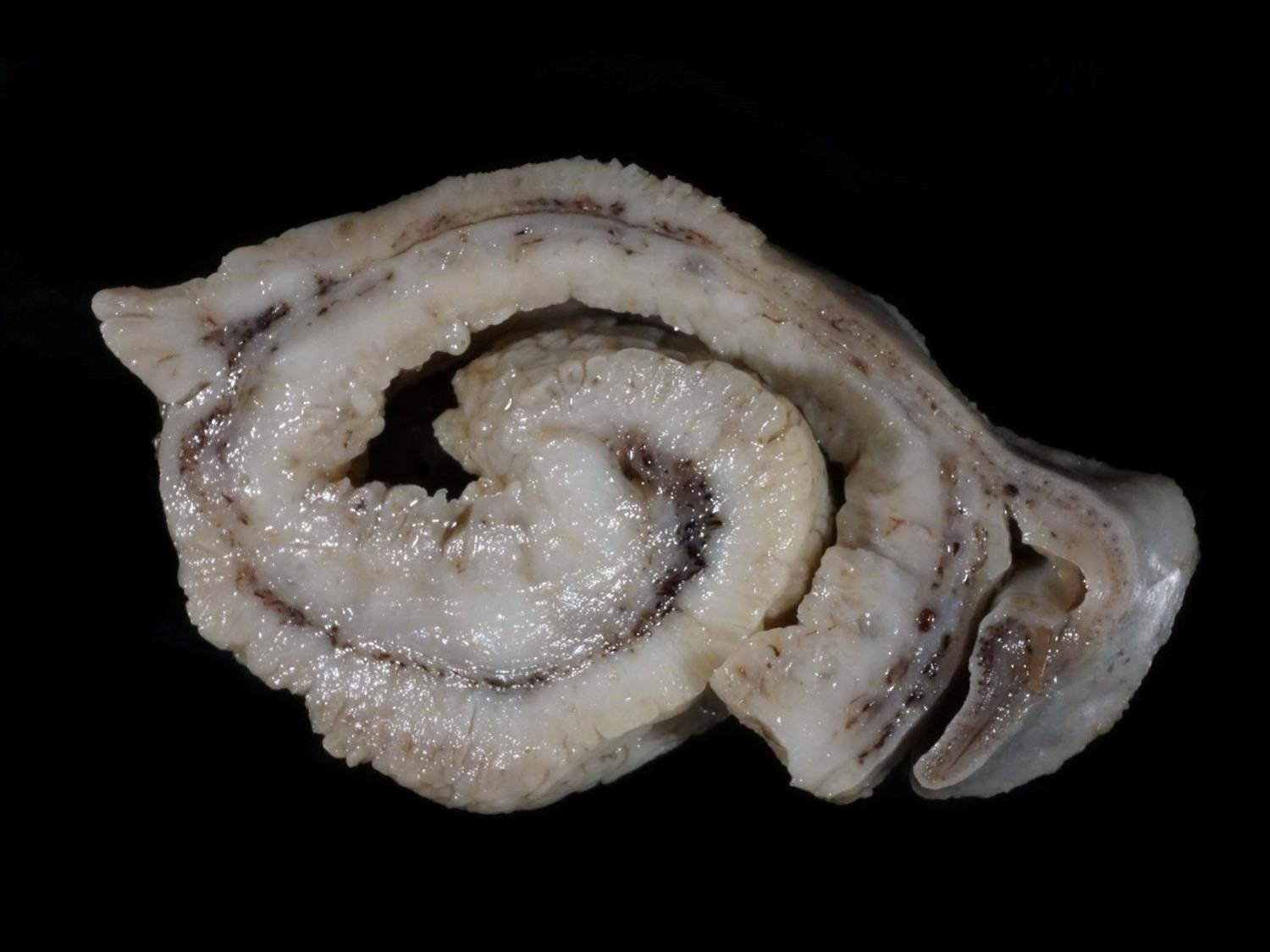
Gross Pathology: There was a moderate amount of mucoid discharge in the nares. On coronal section, the nasal turbinates (conchae) were diffusely and markedly thickened, with a roughened appearance and the meatuses were narrowed.
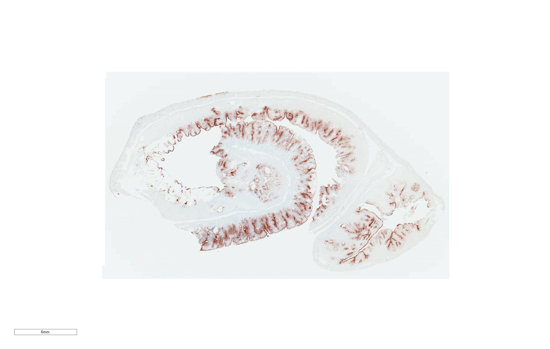
Laboratory results: Aerobic culture of a nasal swab yielded a 4+ predominant Salmonella sp. serotyping of this isolate performed by NVSL identified it as a serotype III 61:k:1,5,7 (S. enterica subspecies arizonae). Immunohistochemistry for Salmonella sp. showed strong positive immunoreactivity distributed throughout the nasal mucosa and submucosa, with epithelial cells, goblet cells, macrophages, and extracellularly.
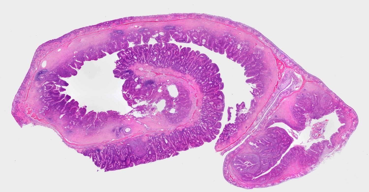
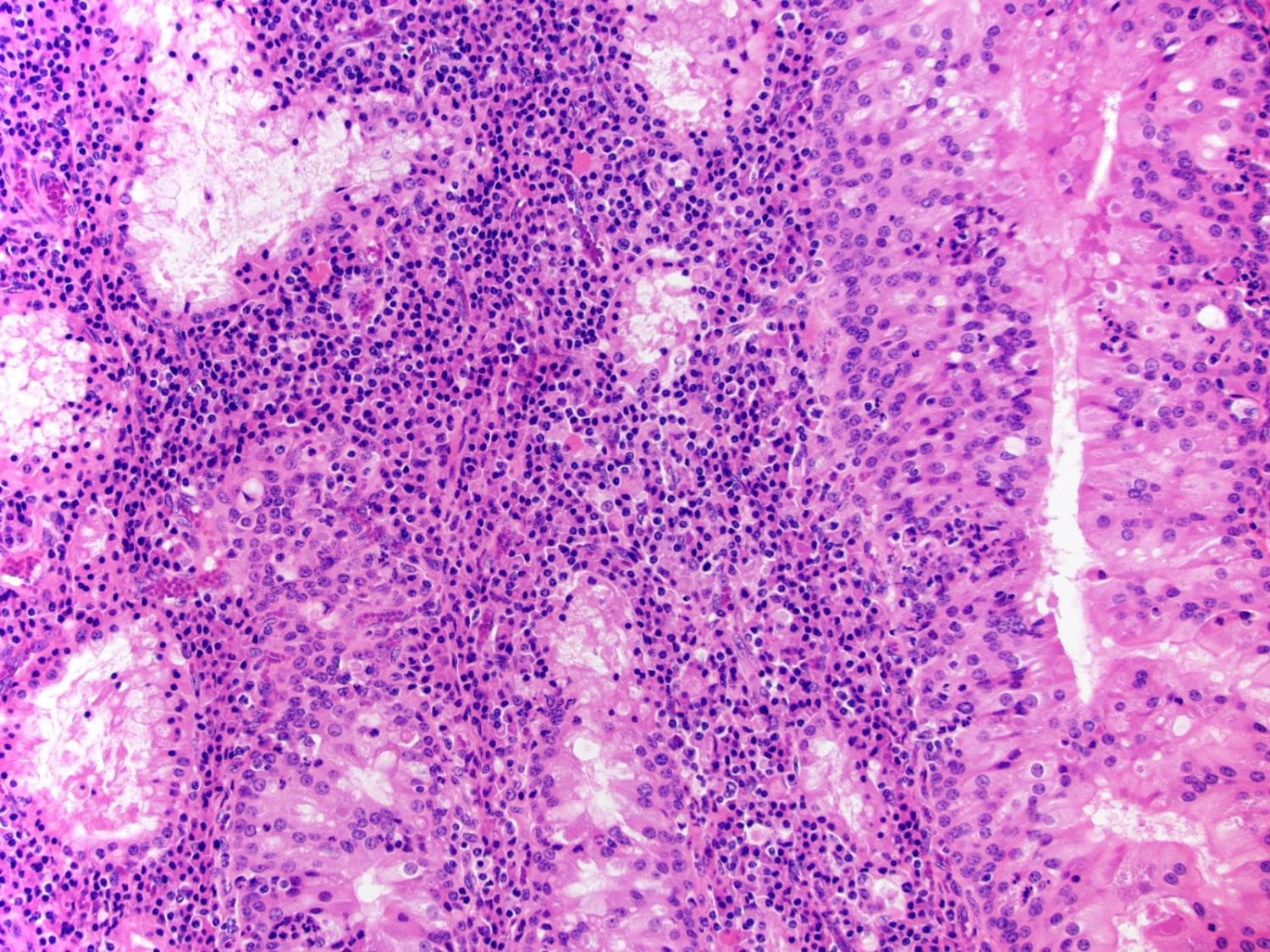
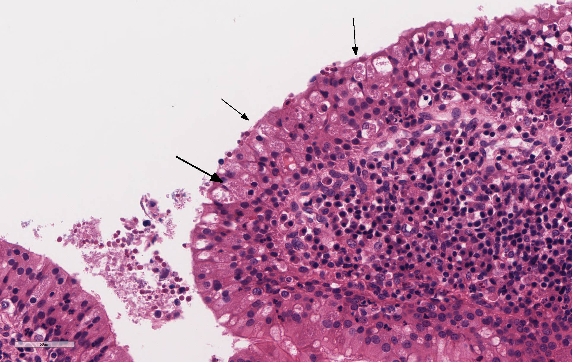
Microscopic Description: The mucosa of the nasal turbinate is markedly thickened by polypoid to finger-like projections of hyperplastic epithelial cells and dense infiltrates of plasma cells and lymphocytes admixed with few neutrophils, macrophages, and siderophages within the lamina propria and submucosa. The infiltrates within the lamina propria and submucosa separate and surround hyperplastic seromucous glands, which are often ectatic and contain intraluminal mucin and cellular debris. Glandular and surface epithelial cells often contain intracytoplasmic aggregates of indistinct, 1-2um diameter eosinophilic coccobacilli. There are moderate numbers of intraepithelial neutrophils, lymphocytes, and macrophages. There is multifocal follicular lymphocid hyperplasia within the submucosal lymphoid tissue and there are few coalescing pyogranulomas within the lamina propria and submucosa, which replace and compress adjacent glandular tissues. There is dense fibrous tissue expanding the submucosa (fibrosis).
Contributor Morphologic Diagnosis: Nasal cavity – Rhinitis, proliferative, lymphoplasmacytic and pyogranulomatous, diffuse, marked, chronic with bacteria.
Contributor Comment: Chronic proliferative rhinitis has been reported in sheep
from the United States, Spain, and most recently Switzerland.1,2,5 Affected
breeds in published reports include Columbian, Dorset, Aragonasa, and Texel
sheep. Clinical signs and associated lesions manifest n animals older
than 1 year of age, and most published cases are between 4 and 7 years of
age. The earliest published report was associated with S. enterica
spp. Arizonae (serotype III).1 Other reports have identified Salmonella
diarizonae (serotype IIIb: 61:k:1, 5,(7))
as the associated organism.2,5 This latter serotype is
commonly isolated from sheep in several countries and is considered
host-adapted.3
In a recent survey of US sheep flocks by the USDA, 66.4% of flocks
were culture-positive for Salmonella spp. and 94.6% of the
isolates were identified as serotype IIIb61:k:1, 5,(7)4 The
intestine and tonsils are commonly colonized without any clinical signs or
pathologic findings. There are few sporadic reports of associated
diarrhea in lambs, abortions, and a report of epididymo-orchitis in a ram.3
The bacterium can be detected in fecal swabs of healthy animals and is
likely maintained in sheep largely due to fecal-oral transmission and
colonization.3 The bacterium can also be detected in nasal swabs
from animals with chronic proliferative rhinitis making this another possible
source of infection for other animals in a given flock.5
In all case
reports which included clinical findings, nasal discharge, respiratory
distress, and dyspnea of increasing severity over weeks to years were reported.1,2,5
Depending on severity and chronicity, “striking mouth-breathing”
and loss of body condition may also occur.3 This may be fatal unto
itself or lead to euthanasia.
The gross and microscopic appearance of this case is consistent with all
published cases. The most prominent light microscopic features are the polypoid
projections formed by hyperplastic epithelium and the infiltrate within the
lamina propria and submucosa composed of large numbers of plasma cells, lymphocytes,
neutrophils, and macrophages. Particularly prominent in this case were
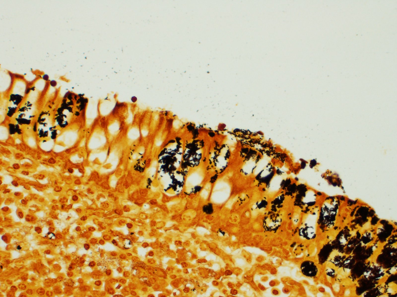
abundant, poorly visualized intracytoplasmic bacilli/coccobacilli within epithelial
cells. Additional extracellular bacteria are evident with the aid of
IHC. The factors which lead to this striking lesion in association with
this common bacterium remain unknown. One recent study attempted to
preproduce the disease in a controlled study with limited success.3 Intranasal
inoculation of bacteria led to elevated IFN-gamma over several months and
bacteria were maintained in the respiratory tract for at least one year;
however, the proliferative lesion was not reproduced. Upper respiratory
tract diseases in sheep are most often seen related to infection by larvae of
the bot fly Oestrus ovis (nasal myiasis; osteosis), leading to mild
eosinophilic, catarrhal rhinitis. Enzootic nasal tumor, caused by
enzootic nasal tumor virus -1 and -2 (oncogenic betaretrovirus) is another
clinical differential, but is likely to cause a focal mass lesion and have a
strikingly different histology appearance. Other sporadically occurring
nasal tumors or space occupying lesion (e.g. lymphoma, non-viral adenocarcinoma
and fungal granuloma) may also have similar clinical signs or gross
lesions. Lastly viral pneumonias (e.g. respiratory syncytial virus, ovine
adenovirus, parainfluenza 3) may present clinically as primarily upper
respiratory signs.)
Contributing Institution: The University of Minnesota, College of Veterinary Medicine, Veterinary Diagnostic Laboratory. https://www.vdl.umn.edu
JPC Diagnosis: Nasal mucosa: Rhinitis, proliferative, and lymphoplasmacytic, chronic, severe, with multifocal mucosal erosion and numerous intracytoplasmic bacilli.
JPC
Comment: The contributor does an excellent and thorough writeup
on the current knowledge of this uncommon but very characteristic lesion
associated with Salmonella in sheep. This entity has made one previous
appearance in the Wednesday Slide Conference (and must have made quite the
impression during the AFIP residency of the moderator, who chose this
counter-intuitive and unique lesion for a conference 29 years later.)
According to one report, there is concern over zoonotic potential for this
bacterium within this particular presentation, although there is no evidence
demonstrating actual occurrence. This particular serovar is most commonly seen
following transmission from infected snakes with vertebral osteomyelitis and
oophoritis.3
References:
1. Meehan IT, Brogden KA, Courtney C et al. Chronic proliferative rhinitis associated with Salmonella arizonae in sheep. Vet Pathol 1992; 29:556-559.
2. Lacasta D, Ferrer LM, Ramos JJ et al. Chronic proliferative rhinitis associated with Salmonella enterica subspecies diarizonae serovar 61:k:1, 5,(7) in sheep in Spain. J Comp Path 2012; 147: 406-409.
3. Lacasta D, Figueras L, Bueso JP et al.
Experimental infection with Salmonella enterica subsp. diarizonae serotype
61:k:1, 5,(7)in sheep: Study of cell-mediated immune response. Small
Rum Res 2017; 149:28-33.
4. Salmonella on US Sheep Operations, 2011. USDA-APHIS, Veterinary
Services Info Sheet. June 2013.
5. Stokar-Regenschiet N, Overesch G, Giezendanner R et al. Salmonella enterica subsp. diarizonae serotype 61:k:1, 5,(7) associated with chronic proliferative rhinitis and high nasal colonization rates in a Texel sheep in Switzerland. Prev Vet Med 2017; 145:78-82.