Signalment:
14-week-old male Toggenburg goat (
Capra hircus).This goat kid presented to the veterinarian with hind limb paresis of 6 weeks duration with progression to
ataxia, recumbency, facial tremors, and strabismus. The goat kid was euthanized.
Gross Description:
The goat was in good postmortem and nutritional condition.Â
Musculoskeletal: There was
redness and swelling of the tendons and muscles of the caudal aspect of the right rear leg extending from the hoof to
the distal femur. There were two areas of hemorrhage and dark friable muscle along the left lateral thorax and
focally in the left cervical muscles.Â
Respiratory: There was a mild amount of green purulent fluid in the frontal
sinuses. There was redness, wetness and consolidation of 50% of the left cranial, left middle and accessory lung
lobes.Â
Nervous: The lateral ventricles were slightly dilated. The meninges were multifocally congested and wet.
Integumentary, cardiovascular, alimentary, hemolymphatic, endocrine, urinary and reproductive systems had no
lesions.
Morphologic Diagnosis:
Severe, focally extensive leukomyelitis, perivascular, lymphoplasmacytic
and histiocytic, caprine arthritis-encephalitis virus.
Lab Results:
Bacteriology: There was no significant growth from aerobic cultures of the brain, joint, lung,
liver, meninges, skeletal muscle or sinuses. There was no
Salmonella spp. isolated.Â
Molecular Diagnostics: A PCR
test for pestivirus (border disease virus) on the tissue homogenate was negative.Â
Toxicology: A liver mineral
analysis was performed. The liver copper concentration of 8.4 ppm was interpreted as deficient. The liver selenium
concentration of 1.43 was interpreted as adequate.Â
Immunohistochemistry: The CAEV IHC on the lung and spinal
cord was positive.
Condition:
Small rumnant lentivirus; CAEV
Contributor Comment:
Caprine arthritis encephalitis virus (CAEV) is a retrovirus in the subfamily
Lentivirinae.(2) Phylogenetic analyses demonstrate similarity to maedi/visna virus (MVV) of sheep, one of the first
lentiviruses to be discovered.(2) Both MVV and CAEV are characterized clinically by slow progressive and
debilitating illnesses of sheep and goats.(3) Subclinical infections with CAEV occur commonly in goats and
subclinically infected animals can be sources of virus transmission to others in the herd.(5) Infection with CAEV
results in persistent infection of macrophage-monocyte cells, and virus is expressed and then shed upon transition of
the precursor monocyte to its mature macrophage type.(4)
Several clinical entities of CAEV infection are recognized: progressive arthritis in adults, progressive weight loss in
adults, indurative mastitis or hard bag in adult does, interstitial pneumonia and a leukoencephalomyelitis in kids 2 to
6 months of age.(3,4,6) The pathological feature of each is lymphocytic/lymphoproliferative inflammation due to a
host immune response with the expressed viral proteins.(4)
The goat kid in this case, in addition to having leukomyelitis, had focal area of lymphoplasmacytic and histiocytic
inflammation of the cerebral white matter; moderate, multifocal, interstitial pneumonia with lymphoplasmacytic and
histiocytic thickening of the alveolar septa, perivasculitis and peribronchitis; and mild multifocal lymphoplasmacytic
and histiocytic synovitis. Postmortem diagnosis can be confirmed through detection of virus, with
immunohistochemistry and polymerase chain reaction tests being more rewarding than virus isolation.(4)
Antemortem diagnosis is commonly achieved by detecting antibodies using agar gel immunodiffusion (AGID) or
ELISA (4,6) and much of the disease control efforts are focused on isolation or culling of seropositive animals.(4,5)
JPC Diagnosis:
Spinal cord, white matter: Meningomyelitis, necrotizing and lymphohistiocytic, focally
extensive, severe, with spheroids and gliosis.
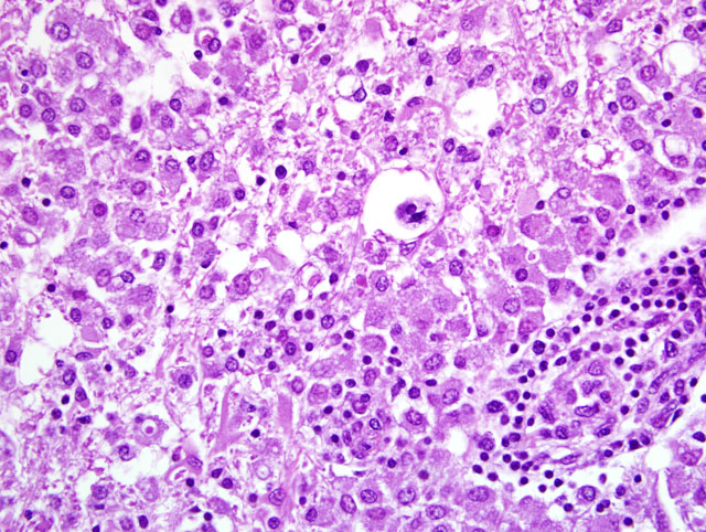
Conference Comment:
There is significant slide variation, with the distribution ranging from unilateral and
confined primarily to the white matter to bilateral and nearly diffuse. Predominantly within the white matter, but
also extending into the adjacent gray matter and overlying leptomeninges, a perivascular infiltrate composed of
numerous lymphocytes and fewer histiocytes and plasma cells expands Virchow-Robin space and extends variably
into the adjacent neuropil. Affected blood vessels are lined by hypertrophied reactive endothelium. Affected white
matter is often lost and replaced by myriad macrophages with abundant foamy cytoplasm (gitter cells), astrocytes
that often contain phagocytic debris, and fewer lymphocytes and reactive microglia (
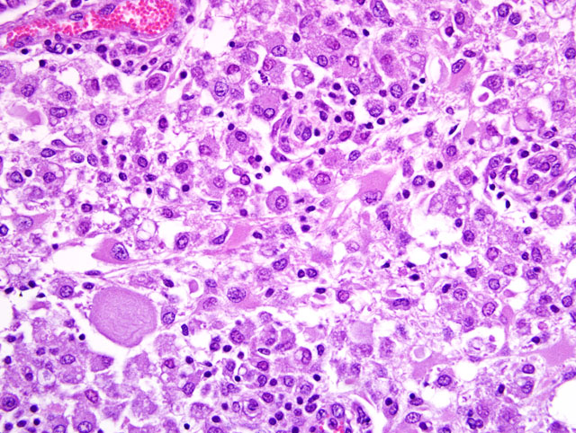
fig. 3-1). Remaining myelin sheaths are
frequently dilated, occasionally mineralized, and either empty or expanded by swollen axons (spheroids) (
fig. 3-2). There is
mild gliosis and rare neuronal necrosis within the gray matter. Numerous lymphocytes and fewer histiocytes and
plasma cells multifocally expand the leptomeninges.
Conference participants discussed the differential diagnosis for neurologic disease in goats (1, 4):
- Copper deficiency (enzootic ataxia, swayback)
- Neuronal vacuolar degeneration of Angora goats
- Cerebrospinal nematodiasis
- Polioencephalomalacia
- Spinal abscess
- Listeriosis
- Rabies
- CAEV
The contributor provided a concise overview of the pathogenesis and clinical manifestations of CAEV. Small
ruminant lentiviruses are enveloped, single-stranded RNA viruses that are more homologous to the feline
immunodeficiency virus (FIV) than to other retroviruses, but differ from feline and primate lentiviruses in that they
are not immunosuppressive.(1) Like other retroviruses, CAEV expresses the following viral genes (1):
- gag: encodes nucleocapsid and matrix glycoproteins that are detected by antibody-based diagnostic tests
- pol: encodes a variety of viral enzymes, including reverse transcriptase
- env: encodes surface glycoprotein that mediates binding to cell receptors and entry into cells
References:
1. Caswell JL, Williams KJ: Respiratory system.Â
In: Jubb, Kennedy, and Palmers Pathology of Domestic Animals,
ed. Maxie MG, 5th ed., vol. 2, pp. 618-620. Elsevier Saunders, Philadelphia, PA, 2007
2. Desrosiers, RC: Nonhuman lentiviruses.Â
In: Fields Virology, eds. Knipe DM, Howley, PM, 4th ed., vol. 2, pp.
2095-2122. Lippincott, Williams & Wilkins, Philadelphia, PA, 2001
3. Maxie MG, Youssef S: Nervous system.Â
In: Jubb, Kennedy, and Palmers Pathology of Domestic Animals, ed.
Maxie MG, 5th ed., vol. 1, pp. 426-428. Elsevier Saunders, Philadelphia, PA, 2007
4. Radostits OM, Gay CC, Hinchcliff KW, Constable PD: Veterinary Medicine, A Textbook of the Diseases of
Cattle, Horses, Sheep, and Goats, 10th ed., pp. 1410-1413. Saunders Elsevier, Philadelphia, PA, 2007
5. Rowe, JD, East, NE: Risk factors for transmission and methods for control of caprine arthritis-encephalitis virus
Infection. Vet Clin North Amer: Food An Prac
13(1):35-53, 1997
6. Wolf, CB: The difficult diseases of CAEV, Johnes disease, and caseous lymphadenitis. Proc. 141st AVMA Ann
Mtg, 2003