Signalment:
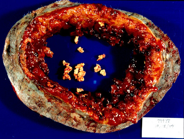
Gross Description:
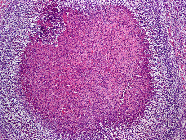
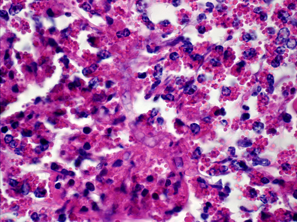
Histopathologic Description:
Morphologic Diagnosis:
Lab Results:
Condition:
Contributor Comment:
Cutaneous disease in horses is the most common infection seen in animals, but infection is reported in many other animal species and in other locations, such as the intestinal and respiratory tracts (1). Cutaneous lesions usually occur on the distal extremities or ventrum, areas most likely to be in contact with stagnant water.Â
The zoospores produce an ulcerative lesion that rapidly expands to a large mass of granulation tissue containing draining sinus tracts. Distinct yellow to tan masses, 2-10 mm in diameter, form within the sinus tracts and are known as kunkers. Kunkers represent areas of tissue necrosis containing hyphae and necrotic eosinophilic inflammation.
JPC Diagnosis:
Conference Comment:
Pythium insidiosum and Lagenidium sp. are aquatic dimorphic water molds of the kingdom Protista. Lagenidium sp. have only been described in dogs.(4) The infective stage of pythium is a biflagellate zoospore that is attracted chemotactically to injured tissue. Upon contact with host tissue the zoospores lose their flagellae and form germ tubes to allow penetration and invasion of tissue.(7)
Histologic features of pythiosis are granulomatous inflammation with foci of liquefactive necrosis and extensive fibrosis with eosinophilic coagula. Circulating monocytes are recruited from the circulation by several chemotactic stimuli such as C5a, TGF-α, and platelet derived growth factor. The tissue macrophages are then activated primarily by IFN-γ, or by endotoxin.(5)
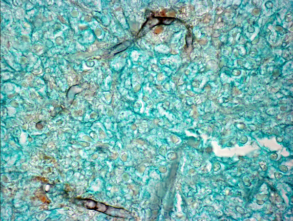
Pythium sp. stain well with GMS but do not stain well with PAS.(7) Fungal hyphae or hyphal-like structures are distinctive between various entities and are often described according to the following structural features.(6)
| Species | Hyphael type | Septae | Width | Branching | Walls | Misc |
| Aspergillus sp | Hyphae, +/- fruiting bodies | Regularly septate | 2.5-4.5 um | Acute angle, dichotomous | Parallel walls | Conidia not seen in tissue |
| Candida sp. | Hyphae, pseudohyphae & budding yeast | septate | 3-6 um | Irregular branching | ||
| Pseudallescheria boydii | Hyphae with conidiophoresseptate | 5 um | Less acute, highly branching, intertwined | 15-20um intercalary chlamydoconidia (swollen cells) | ||
| Pythium insidiosum | Hyphal like structures | Rarely septate | 2-9 um | Irregularly branching | Non-parallel | Kunkers or leeches |
| Zygomycetes | May occur as 5-30um chlamydoconidia | Few septae | 3-25 um | Non-dichotomous irregular branching | Non-parallel | Mucor sp., Basidiobolus sp., Rhizopus sp. |
Eosinophils are a prominent feature of equine pythiosis. Residents discussed the four primary differentials for eosinophilic and granulomatous dermatitis in a horse:
1. Pythiosis Hyphal-like structures, negative images on H&E that are highlighted with GMS stain, necrotizing vasculitis
2. Habronema Nematode larvae within eosinophilic granulomas
3. Mast cell tumors monomorphic population of mast cells surrounding eosinophilic granulomas
4. Eosinophilic collegenolytic granulomas Usually centered on collagen bundles
References:
2. Ginn PE, Mansell JEKL, Rakich PM: Skin and appendages. In: Jubb, Kennedy and Palmers Pathology of Domestic Animals, ed. Maxie MG, 5th ed., pp 704-707. Elsevier Ltd, Philadelphia, PA 2007
3. Gross TL, Ihrke PJ, Walder EJ, Affolter VK: Skin diseases of the dog and cat, 2nd ed., pp. 303-309. Blackwell Science, Oxford, UK, 2005
4. Hargis AM, Ginn PE: The integument. In: Pathologic Basis of Veterinary Disease, eds. McGavin MD, Zachary JF, 4th ed., pp. 1196. Elsevier, St. Louis, MO, 2007
5. Kumar V, Abbas AK, Fausto N: Acute and chronic inflammation. In: Robbins and Cotran Pathologic Basis of Disease, 7th ed., pp. 877-937. Elsevier Saunders, Philadelphia, PA, 1999
6. Rippon JW: Medical Mycology: The Pathogenic Fungi and the Pathogenic Actinomycetes, pp. 569, 638, 653, 687, 739. W.B. Saunders Company, Philadelphia, PA, 1988
7. Scott DW, Miller WH: Equine Dermatolgy, pp. 287-293. Elsevier Science, St. Louis, MO, 2003