Signalment:
Gross Description:
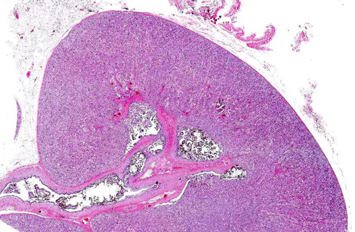
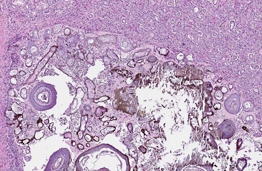
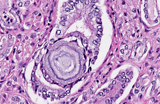
Histopathologic Description:
Morphologic Diagnosis:
Lab Results:
| Serum Chemistry | Value | Reference Range | Units |
| Albumin | 3.7 | 2.8-3.8 | g/dL |
| Total Protein | 7.7 | 6.7-8.8 | g/dL |
| Globulin | 4.0 | 3.3-6.3 | g/dL |
| BUN | 148 | 10-23 | mg/dL |
| Creatinine | 10.3 | 0.7-1.2 | mg/dL |
| Cholesterol | 310 | 157-393 | mg/dL |
| Glucose | 246 | 41-74 | mg/dL |
| Calcium | 6.7 | 9.1-11.3 | mg/dL |
| Phosphorus | 14.8 | 5.1-8.7 | mg/dL |
| Chloride | 132 | 93-100 | mEq/L |
| Potassium | 5.6 | 4.1-5.5 | mEq/L |
| Sodium | 157 | 136-148 | mEq/L |
| Alk Phos | 225 | 23-96 | U/L |
| ALT | 11 | 15-43 | U/L |
| Total Bilirubin | 0.4 | 0.0-0.2 | mg/dL |
| CBC | Value | Reference Range | Units |
| HCT | 31 | 24-38 | % |
| WBC | 19.4 | 5.3-14.9 | K/uL |
| Neutrophils | 16498 | 2200-8073 | per ul |
| Lymphocytes | 2328 | 1431-8694 | per ul |
| Monocytes | 582 | 0-774 | per ul |
| Eosinophils | 0 | per ul | |
| Basophils | 0 | per ul | |
| Platelets | 1692 | K/ul |
Kidney stone analysis (Minnesota Urolith Center): 100% xanthine
Condition:
Contributor Comment:
Xanthine is a metabolite of purines normally converted to uric acid by xanthine oxidase. Xanthinuria and xanthine urolithiasis result from loss of function of this enzyme, which can be the result of a primary enzyme defect inherited as an autosomal recessive condition or from secondary inhibition of enzyme function.(3,7) Cases of the secondary form are most often caused by treatment with allopurinol, an inhibitor of xanthine oxidase, and are thus most commonly seen in Dalmatian dogs treated for urate urolithiasis.(2,3) Dietary deficiency of molybdenum, a component of an essential cofactor of xanthine oxidase, was thought to account for xanthine urolithiasis in sheep.(3) The Cape buffalo calf in this report had not been treated with allopurinol and had normal levels of molybdenum on postmortem analysis of liver. It is therefore thought to have had a primary (hereditary) form of the disease, similar to cases in domestic cattle.(1,4,8) A half-sibling of this calf (same sire, different dam) had previously died at 2 months of age with nearly identical lesions, although stone analysis was not performed in that case. Four prior calves from the same dam and sire were unaffected. Characterization of the hypothesized genetic defect in this group of Cape buffalo has not yet been attempted.
JPC Diagnosis:
Conference Comment:
The important types of urinary calculi in various species are listed in table 1; it is also worthwhile to remember that many uroliths are of mixed composition (although one mineral may predominate). Silica calculi are a significant cause of urinary tract obstruction in pastured ruminants and are occasionally seen in male dogs, especially German shepherds and old English sheepdogs or dogs receiving a ration high in plant-derived ingredients (i.e. corn gluten or rice/soybean hulls). They are hard, white to dark brown, radiopaque and spherical/ovoid (in the urinary bladder) or angular/irregular (when in renal calyces) with a friable core.(3)
Struvite calculi are composed of magnesium ammonium phosphate hexahydrate; historically, they were inappropriately identified as triple phosphate calculi. Grossly, these radiopaque stones appear white to gray, chalky, smooth and easily fractured. They typically contain additional compounds such as calcium phosphate (which often forms a shell around the struvite), ammonium urate, oxalate or carbonate.(3) Struvites are most important in dogs, cats and ruminants where they are commonly associated with infection. Ureases produced by bacteria such as Staphylococcus spp. or Proteus spp. increase the urine pH, which decreases struvite solubility, thus favoring the formation of calculi. Miniature schnauzers appear predisposed, probably due to a familial susceptibility to urinary tract infections. Sterile struvite urolithiasis has been reported in a line of English cocker spaniels and beagles. The formation of struvite calculi within the urinary bladder of cats is one manifestation of an idiopathic condition known as feline lower urinary tract disease (FLUTD); Russian blues, Himalayans, Persians and castrated/spayed males and females are predisposed. Of note, struvite crystalluria is often seen in cats even without calculi; the reasons for this phenomenon are poorly understood. It should also be noted that amorphous urethral plugs containing Tamm-Horsfall mucoprotein, albumin, globulins, cellular debris and struvite crystals are a much more important cause of urethral obstruction in male cats, typically associated with concurrent urinary tract inflammation. Struvite calculi also occur in feedlot steers and sheep on a high grain ration. Much like cats, obstruction in ruminants is typically due to formation of a gritty sludge, rather than discrete calculi, while a high phosphate diet predisposes sheep to struvite urolithiasis.(3,5)
Oxalate calculi tend to develop as large, solitary, white to yellow bladder stones covered with sharp spines. They are composed of calcium oxalate monohydrate or calcium oxalate dihydrate, and although the pathogenesis is poorly understood, it likely involves some combination of hypercalciuria and hyperoxaluria. The major causes of hypercalcemia/hypercalciuria are briefly discussed in WSC 2013-2014, conference 13, case 3. Oxalic acid may be ingested in some foods or synthesized from glyoxylic and ascorbic acid. The formation of calcium oxalate uroliths is inhibited by dietary magnesium, which forms soluble complexes with oxalate, and citrate, which forms a similar complex with calcium. Oxalate uroliths are the second most common type of calculi encountered in dogs and have been associated with hyperparathyroidism, hypercalcemia, hyperadrenocorticism and exogenous steroid administration. Male miniature schnauzers, bichon frises, lhasa apsos, Yorkshire terriers, shih tzus and miniature poodles are predisposed. The prevalence of oxalate urolithiasis is increasing in cats and is likely related to dietary factors, although the specifics are not clear. Since oxalate is metabolized in the rumen, oxalate calculi are not generally considered important in ruminants; however, they are reported in sheep grazing grain stubble, although the source of the oxalates in these cases remains unknown.(3,5)
Uric acid and urates are products of purine metabolism; calculi usually contain ammonium urate in combination with uric acid and phosphate. Uric acid/urate calculi are typically multiple, hard, laminated green-brown, radiodense spherical stones found within the urinary bladder. Urate stones are rarely reported in swine and cats but are most common in Dalmation dogs. In neonatal pigs in a negative energy balance, there is increased production of purine catabolites, which also predispose the formation of urate calculi. Urate urolithiasis in Dalmations results from an autosomal recessive defect in the hepatic uptake of uric acid, leading to its incomplete metabolism and accumulation. This defective transport system also inhibits tubular reabsorption of uric acid from the glomerular filtrate, resulting in urine that is supersaturated with urates.(3,5)
Xanthine is another purine metabolite, which, as noted by the contributor, is normally degraded via xanthine oxidase to uric acid. Xanthine stones are irregular, yellow to brown-red, laminated, friable and radiolucent.(3,5) This interesting case provides an excellent example of an uncommon condition (xanthine urolithiasis) in an unusual species. The contributor provides a thorough description of the gross pathology, histology, clinical-pathology and pathogenesis of xanthine urolithiasis in veterinary medicine.
Table 1. Composition of urinary calculi in select domestic animal species.(3)
| Species | Common Types | Uncommon Types |
| Ox | Silica Struvite Carbonate | Xanthine |
| Sheep | Silica Struvite Oxalate Clover stones Carbonate | Xanthine |
| Dog | Struvite Oxalate Purines (urate, uric acid, xanthine) | Silica Cystine Calcium phosphate |
| Cat | Struvite Oxalate | Urate Cystine |
| Horse* | Carbonate | |
| Pig* | Urate (neonates) |
References:
1. Hayashi M, Ide Y, Shoya S. Observation of xanthinuria and xanthine calculosis in beef calves. Jap J Vet Sci. 1979;41:505-510.
2. Ling GV, Ruby AL, Harrold DR, Johnson DL. Xanthine-containing urinary calculi in dogs given allopurinol. J Am Vet Assoc. 1991;198:1935-1940.
3. Maxie MG, Newman SJ. Urinary system. In: Maxie MG, ed. Jubb, Kennedy and Palmers Pathology of Domestic Animals. Vol 2. 5th ed. Edinburgh, UK: Elsevier Limited; 2007:508-514.
4. Miranda M, Rigueira L, Suarez ML, et al. Xanthine nephrolithiasis in a Galician blond beef calf. J Vet Med Sci.2010;72(7);921-923.
5. Newman SJ. The urinary system. In: Zachary JF, McGavin MD, eds. Pathologic Basis of Veterinary Disease. 5th ed. St. Louis: Elsevier; 2012:643-645.
6. Tsuchida S, Kagi A, Koyama H, Tagawa M. Xanthine urolithiasis in a cat: a case report and evaluation of a candidate gene for xanthine dehydrogenase. J Feline Med Surg. 2007;9:503-508.
7. Van Zuilen CD, Nickel RF, van Dijk TH, Reijngoud D-J. Xanthinuria in a family of Cavalier King Charles spaniels. Vet Quart. 1997;19:172-174.
8. Watanabe T, Ihara N, Itoh T, Fujita T, Sugimoto Y. Deletion mutation in Drosophila ma-l homologous, putative molybdopterin cofactor sulfurase gene is associated with bovine xanthinuria type II. J Biol Chem. 2000;275(29);21789-21792.