CASE 1: 7950-19 (4137581-00)
Signalment:
Two-year-old, male, Doberman pinscher mix dog (Canus lupus familiaris)
History:
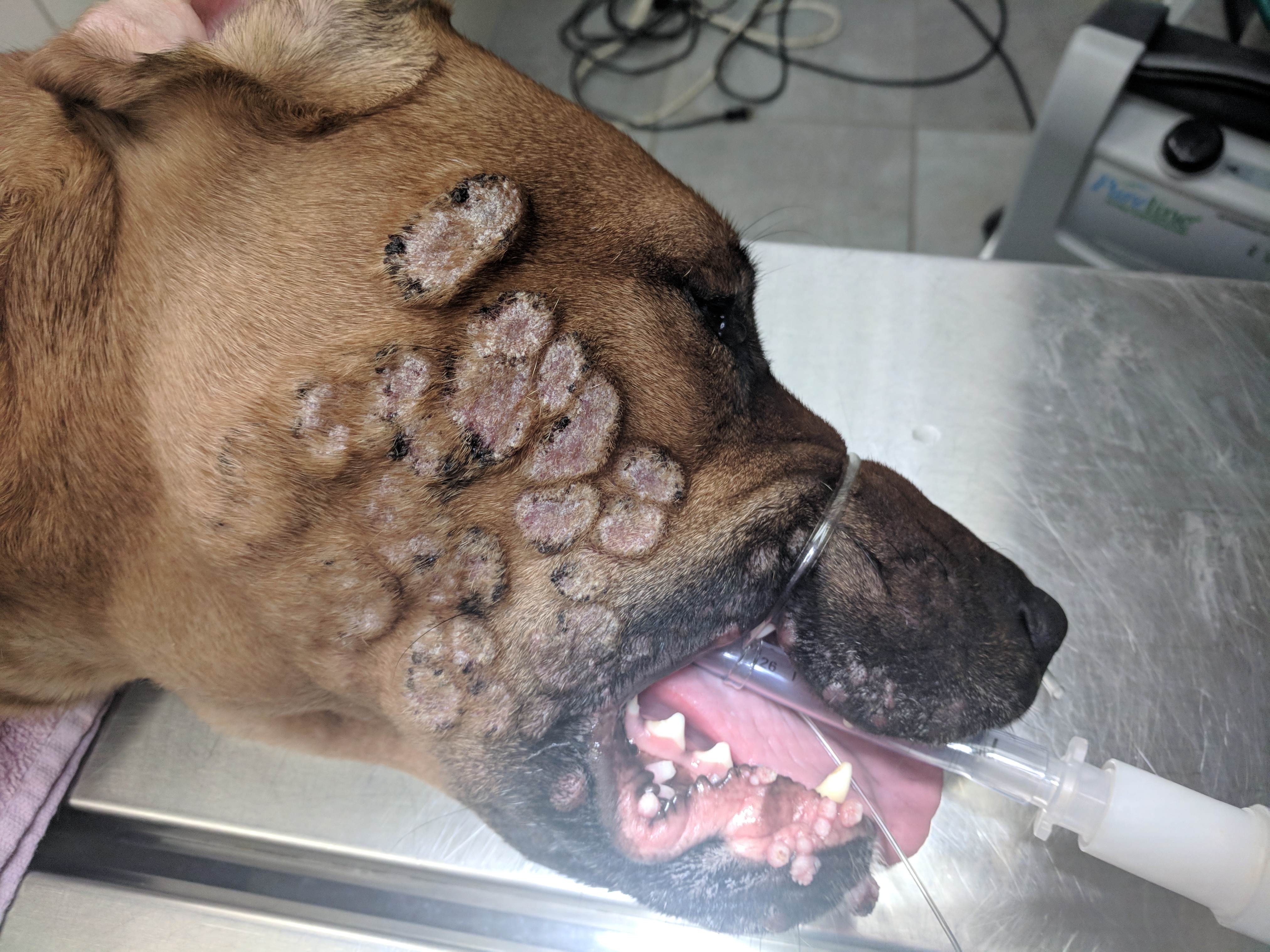
The animal initially presented to the referring veterinarian 90 days prior and over the course of two months was treated with cefdopoxime and clindamycin for suspected folliculitis/ furunculosis. During the two-month treatment period, the animal displayed no clinical signs of systemic illness. Approximately 75 days after initial presentation, the animal presented with acute lethargy, inappetence, and lymphadenomegaly. Numerous skin biopsies of exophytic facial lesions were received for examination as well as lymph node aspirates from the enlarged prescapular and popliteal lymph nodes.
Gross Pathology:
N/A
Laboratory results:
N/A
Microscopic description:
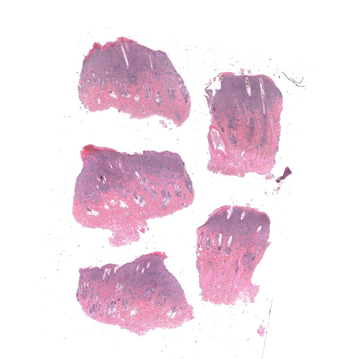
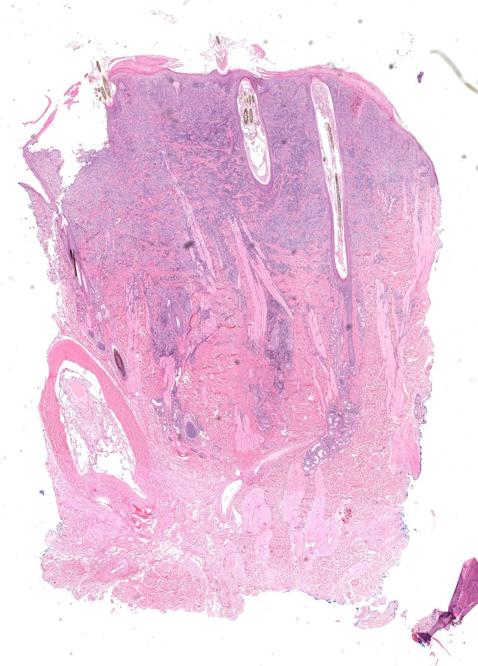
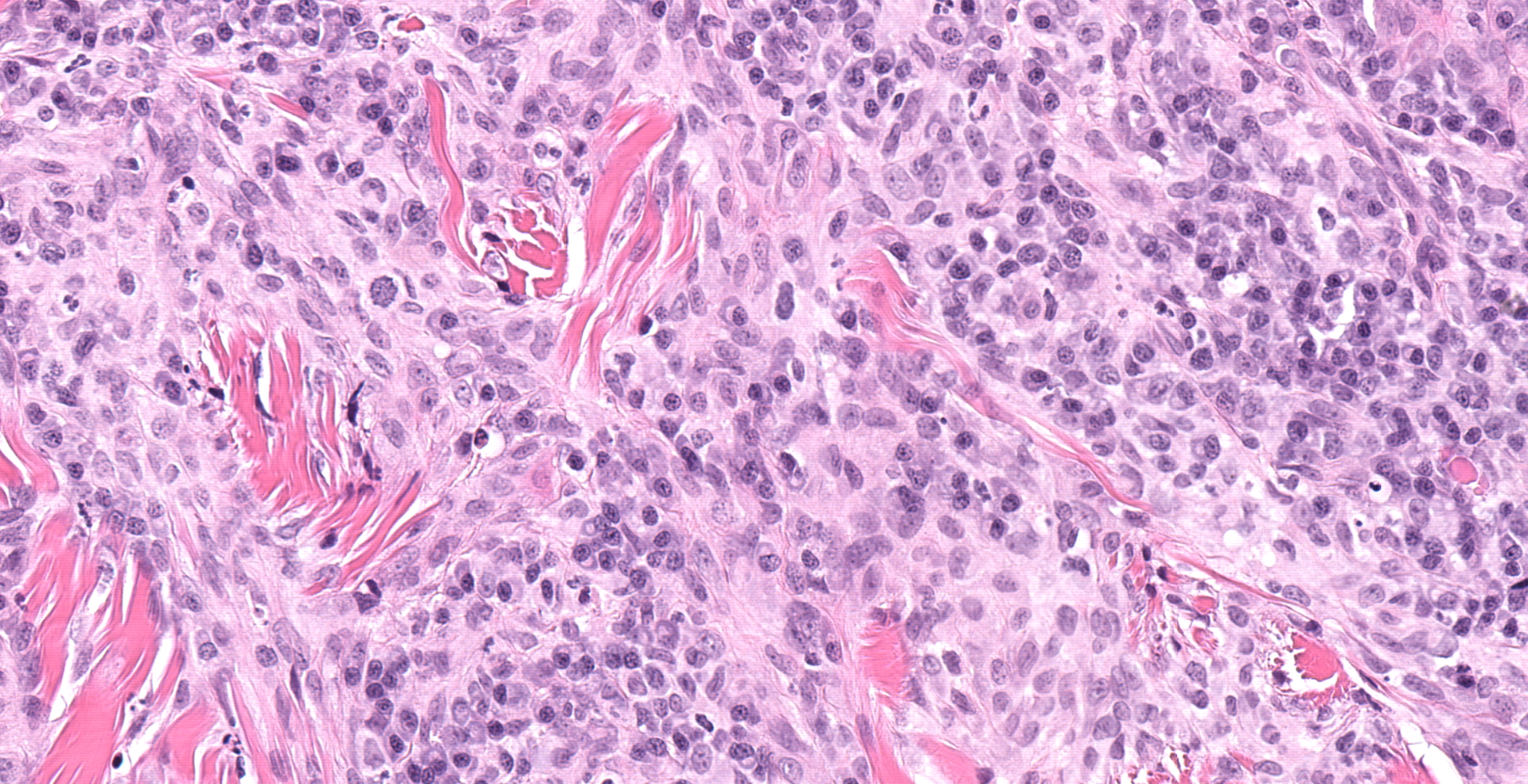
Multiple tissue biopsies are received and evaluated in sixteen sections. Within the sections of haired skin are multiple masses that are predominantly within the superficial to middle dermis, lack a Grenz zone with the epidermis, and in some sections extend deep along pilosebaceous units. The masses consist round cells arranged in sheets which dissect through the dermal collagen. The round cells are large, with distinct cellular margins and moderate to large amounts of finely granular lightly eosinophilic cytoplasm. The nuclei are large and round to oval to reniform with stippled chromatin and 1-3 small nucleoli. Mitotic figures are common and range from 1-5 per high powered field, including a few atypical mitotic figures. There is moderate anisocytosis and anisokaryosis. Many lymphocytes are present as are moderate numbers of plasma cells, although they are more prevalent along the periphery and they are more prevalently associated with the adjacent pilosebaceous glands. There is a small amount of scarring in the superficial dermis. The overlying epidermis is hyperplastic and contains a small amount of pseudoepitheliomatous hyperplasia.
Contributor's morphologic diagnosis:
Langerhans cell histiocytosis
Contributor's comment:
Cutaneous histiocytoma is one of the most frequently diagnosed skin tumors in the dog that commonly occurs in younger dogs with breeds like the boxer, English cocker spaniel, Doberman pinscher, and Scottish terriers (among others) overrepresented in diagnosed cases.3 Traditionally, histiocytomas are considered a benign lesion that will often spontaneously regress with or without surgical intervention.2 Langerhans cell histiocytosis (LCH) however, represents a rare malignant form of histiocytic disease in the dog that can mimic histiocytoma grossly, but that often occur as multiple cutaneous lesions and can lead to metastasis of neoplastic cells and a rapid course of disease.4 The prognosis for LCH with lymph node involvement is poor as the majority of dogs are euthanized due to rapid clinical deterioration associated with metastatic disease.2
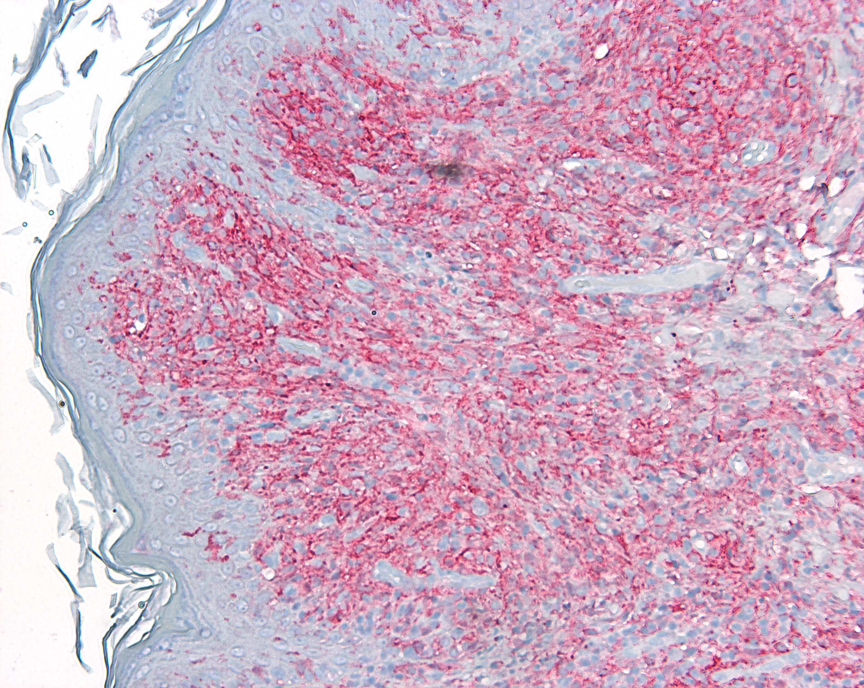
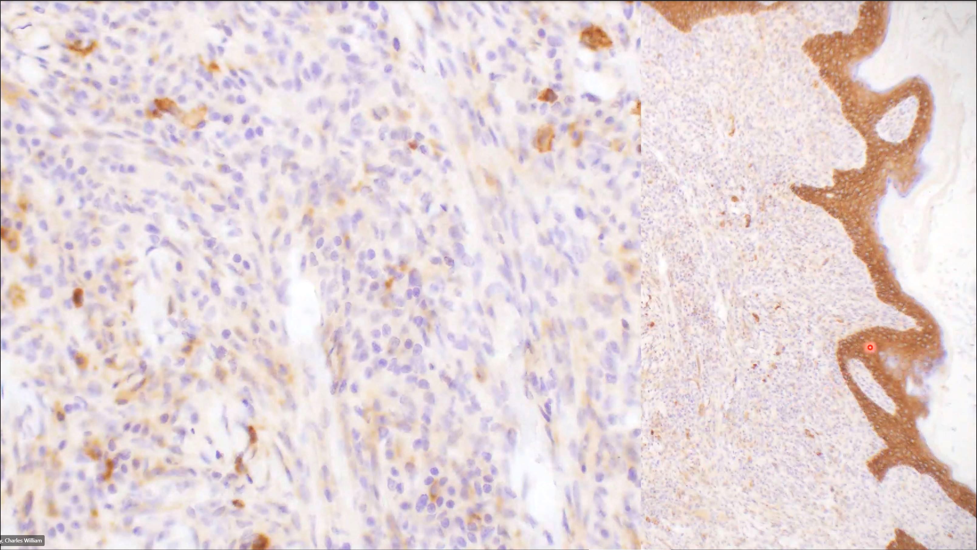
Histologically, the histiocytoma and LCH can display roughly the same characteristics with LCH tending to exhibit more pronounced anisocytosis and anisokaryosis. Often, as in this case, the clinical history can be very helpful in leading to an accurate diagnosis. It appears likely that if given a single piece of the biopsies submitted without any clinical history pertaining to numerous lesions, even a well-trained diagnostician could mistake these lesions for a routine histiocytoma. The reliable expression of ionized calcium-binding adapter molecule 1 (IBA-1) is well characterized in proliferative histiocytic diseases of dogs and cats.5 As such, an IBA-1 IHC was performed to confirm the neoplastic cells were of monocyte/macrophage origin and the cells exhibited widespread, intense staining among the neoplastic cells.
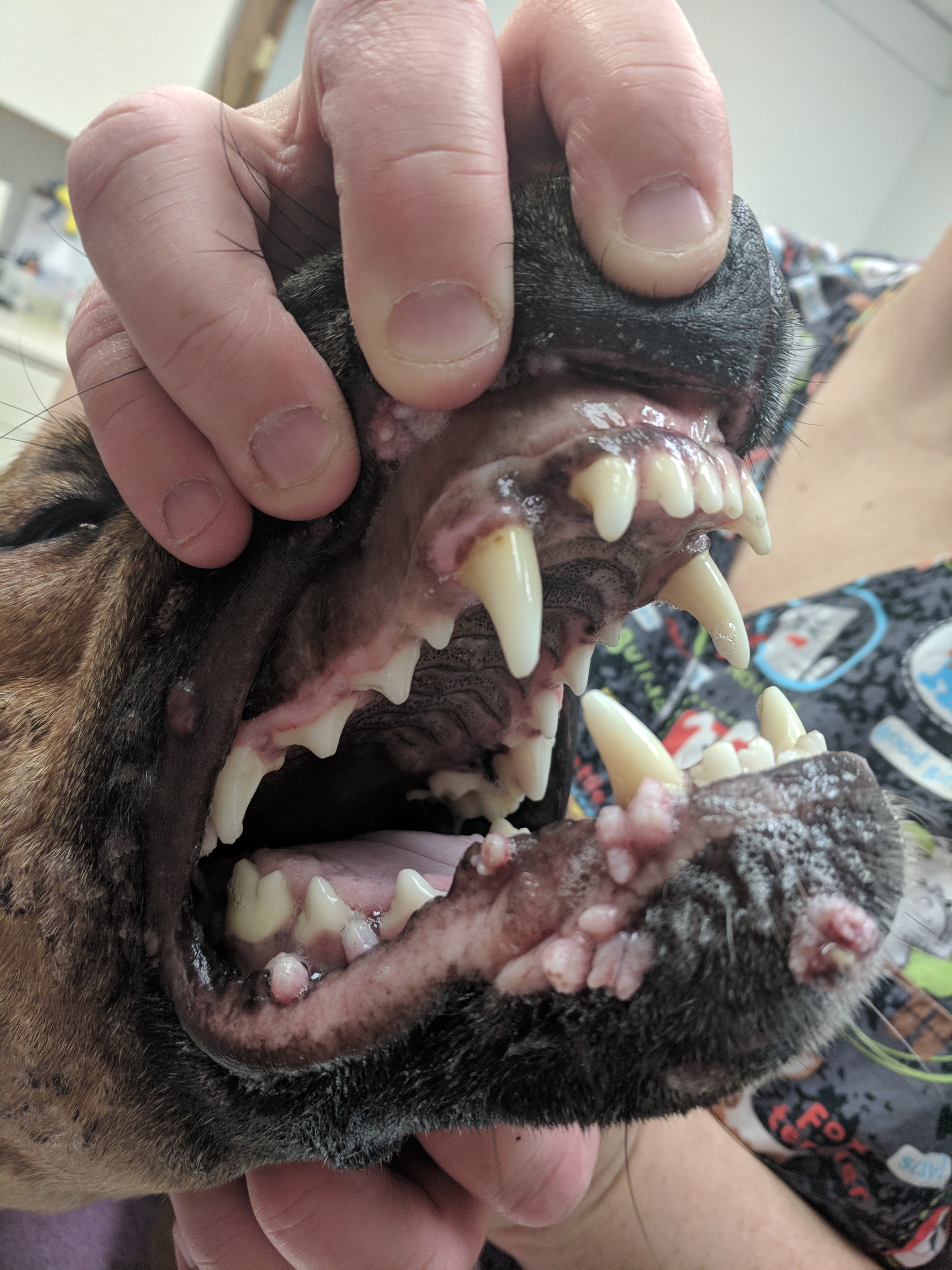
A review of histiocytic diseases in dogs and cats describes LCH lesions often observed at mucocutaneous junctions and within the oral cavity.4 Such lesions were observed in this case. The animal in this case continued to worsen clinically and died on his own within two weeks of the biopsy resection or approximately 105 days after the initial presentation to the referring veterinarian. The referring veterinarian described an easily palpable spleen postmortem, but an autopsy was not performed.
Contributing Institution:
University of Nebraska ? Lincoln
Nebraska Veterinary Diagnostic Center
Lincoln, NE
https://vbms.unl.edu/nvdls
JPC diagnosis:
Haired skin, dermis: Langerhans cell histiocytosis, focally extensive, marked.
JPC comment:
In human pathology literature, there is still debate as how to best classify Langerhans cell histiocytosis: inflammatory, or neoplastic? The histologic appearance of the Langerhans cells is benign, the typical accompanying inflammatory infiltrate, and the local and systemic cytokine storm are suggestive of an inflammatory disease. Yet there is clonality of the cell population, identified mutations in the MAPK pathway, and some identified mutations shared with hematopoietic precursors provide support for a neoplastic classification. Approximately 57% of human cases of LCH express the BRAF V600E mutation, a key kinase in the RAS-RAF-MEK signaling pathway. The BRAF V600E mutation forces the MAPK pathway to be constitutively active, leading ultimately to increased expression of downstream transcription targets.1
Based on the presence of somatic MAPK mutations in myeloid precursor cells provides the current basis of classification of Langerhans cell histiocytosis as a myeloid neoplastic disorder. While there are differences between human and veterinary species, this classification appears valid until additional research in animals can be pursued.1
This case also provides the context to discuss immunohistochemistry to differentiate histiocytic diseases. As the contributor stated, IBA-1 is reliably expressed on histiocytes of all lineages. In addition, Langerhans cells in the dog and cat express CD1a, CD18, and E-cadherin. Crucially, E-cadherin is differentially expressed on Langerhans cells and not on interstitial dendritic cells, the origin of systemic histiocytosis, cutaneous histiocytosis, histiocytic sarcoma, feline progressive histiocytosis, and dendritic cell leukemia. Additionally, if available, CD207 (Langerin), would be a specific marker for Langerhans cells, consistent with histiocytoma, cutaneous Langerhans cell histiocytosis, or feline pulmonary Langerhans cell histiocytosis.4
References:
- Allen CE, Merad M, McClain KL. Langerhans-Cell Histiocytosis. The New England Journal of Medicine. 2018;379:856-68.
- Gross, T.L., Ihrke, P.J., Walder, E.J., Affolter, V.K. Skin Diseases of the Dog and Cat, 2nd ed. 2005; Ames, IA; Blackwell Publishing.
- Hendrick, M.J. Tumors in Domestic Animals, 5th ed. 2017; Ames IA; Iowa State Press.
- Moore, P.F. (2014). A review of histiocytic diseases of dogs and cats. Vet Pathol. 51(1):167-184.
- Pierezan, F., Mansell, J., Ambrus, A., Rodrigues, H.A. Immunohistochemical expression of ionized calcium binding adapter molecule 1 in cutaneous histiocytic proliferative, neoplastic and inflammatory disorders of dogs and cats. J Comp Pathol. 151(4):347-351.