Joint Pathology Center
Veterinary Pathology Services
Wednesday Slide Conference
2019-2020
Conference 10
20 November 2019
Dr. Elise Ladouceur
Chief, Extramural Projects
Veterinary Pathology Services
Joint Pathology Center
CASE II: N18-0170
(JPC 4132732).
Signalment: Adult (>4 years) female Oscar, cichlid (Astronotus ocellatus)
History: On August 9, 2018 the patient presented for rapid gilling. Upon physical examination the gill filaments were thickened, erythematous and mottled red to light pink. A black spot on the gill filaments of the first right gill arch was seen, and a trematode was observed on a gill clip. The patient was treated with 10 ppt NaCl baths, ceftazidime and praziquantel injections but no improvement was seen. Due to quality of life concerns, euthanasia with MS-222 was elected on August 19, 2018.
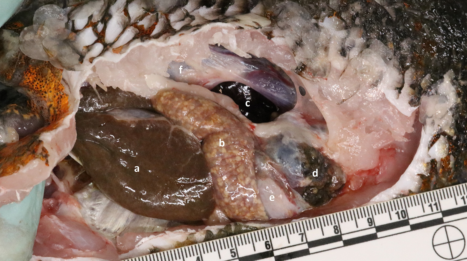
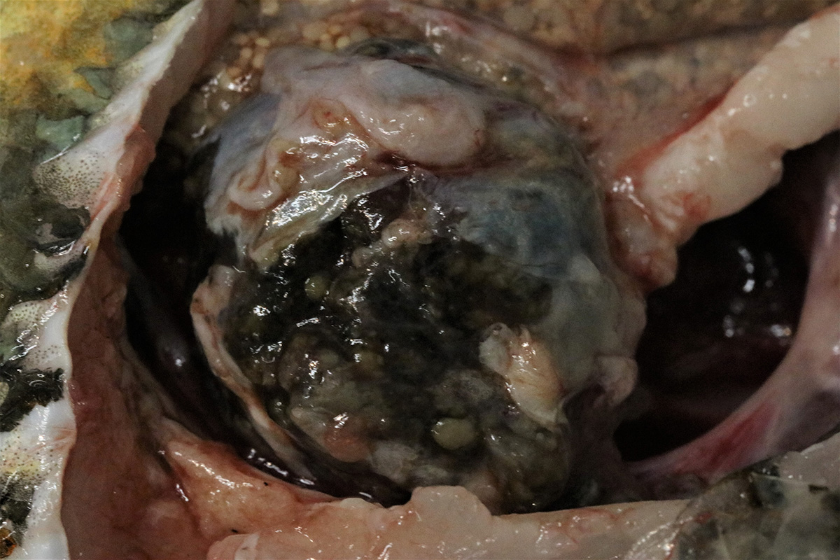
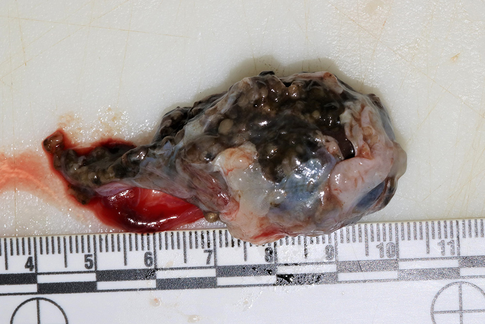
Gross Pathology: On gross examination there is abundant mucus coating the entire fish. The caudal pole of the kidney is expanded by a dark, mottled, red-brown to tan, 3.5 x 2.5 x 2.5 cm multinodular mass. The mass is partially encapsulated, soft, wet, friable, and located ventral to the swim bladder. The kidney is similarly mottled and attached by apparent renal tissue to the mass. No other abnormalities are seen.
Laboratory results:
Renal mass cytology - Rafts of epithelial cells. Cells occasionally surround acellular material (possible mineral) in a tubule-like formation.
Microscopic Description:
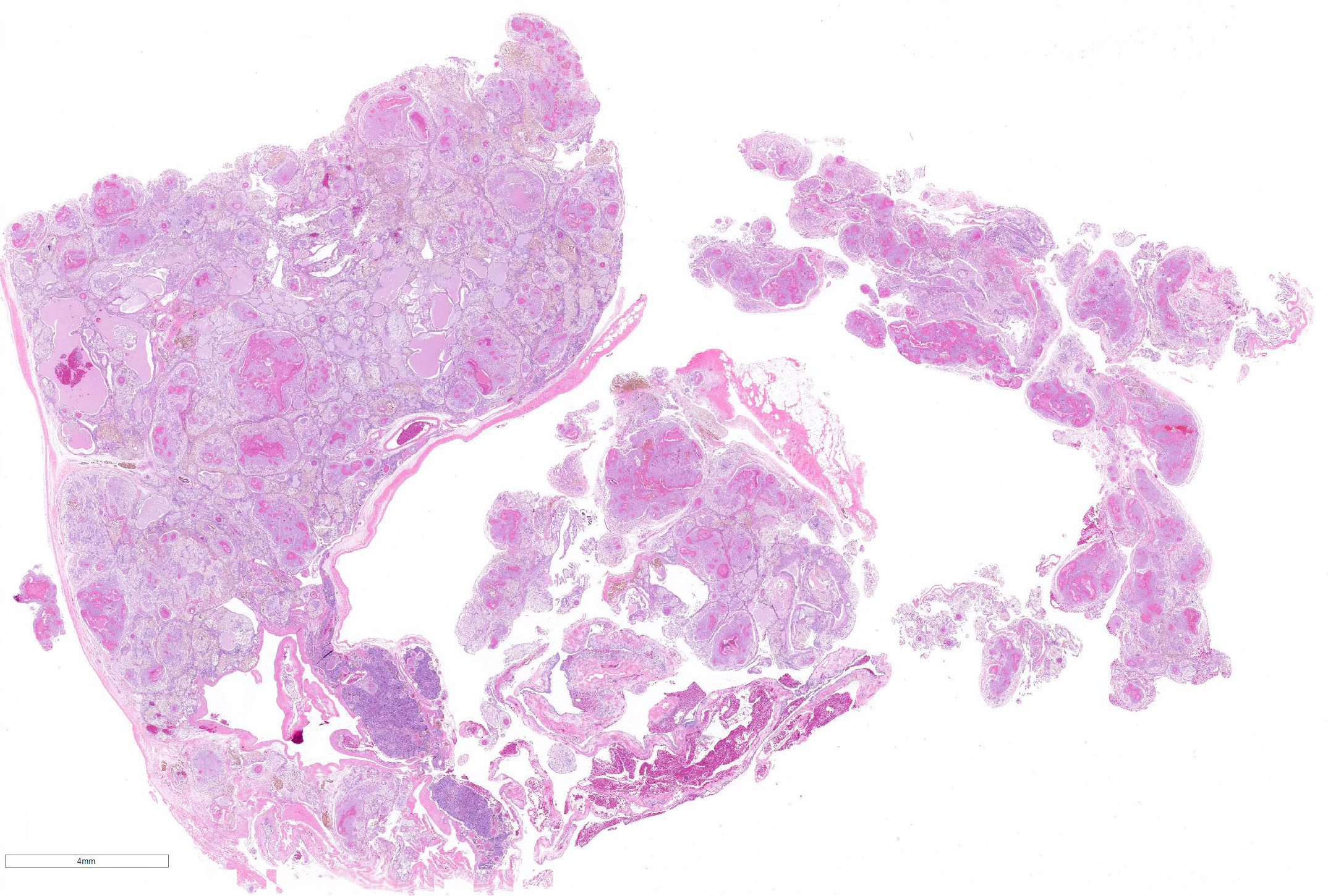
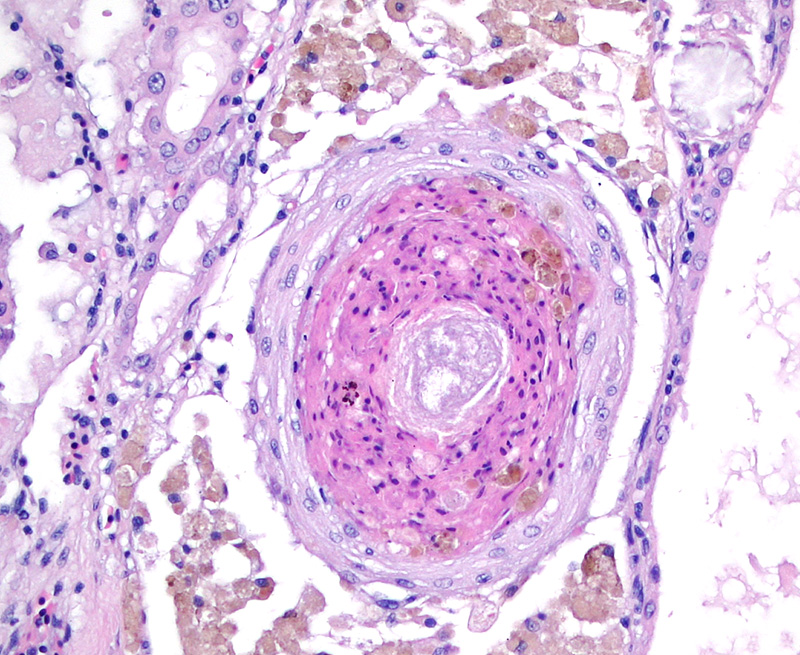
Preexisting renal tissue is identified by renal tubules and glomeruli. Greater than 90% of the normal renal tissue is expanded and obliterated within the original renal capsule by a neoplastic mass with lobules separated by a fibrovascular stroma. Neovascularization is noted throughout the mass within the interstitium. The mass is primarily cystic forming distinguishable lumina up to 1.8 mm in diameter containing viable and degenerate erythrocytes, indistinct cellular debris, and eosinophilic fluid. The cells lining these spaces are well-differentiated, cuboidal to occasionally columnar epithelium exhibiting mild anisocytosis and anisokaryosis. Each cell contains a moderate amount of eosinophilic occasionally vacuolated cytoplasm, and a single round, centrally to apically located nucleus. Nuclei have finely stippled to vesicular chromatin and 1-2 basophilic nucleoli. No mitotic figures are seen. The interstitium of the mass is moderately expanded by macrophages containing golden brown pigment (hemosiderin), lower numbers of lymphocytes and plasma cells and regions of neovascularization. Numerous coalescing organized granulomas are present within cystic lumina and the interstitium. They range from 0.1 mm solitary granulomas to 3 mm coalescing granulomas. The granulomas consist of a necrotic core of brightly colored eosinophilic cellular and karyorrhectic debris contained by an inner layer of concentric compressed epithelioid macrophages surrounded by a thicker layer of foamy macrophages. Hemosiderophages are prominent throughout the interstitium and occasionally within outer foamy layers of granulomas. Scattered mineralization is present within granulomas. There are areas within the mass where disorganized and coiled membranes of cuboidal epithelium are seen (likely handling artifact).
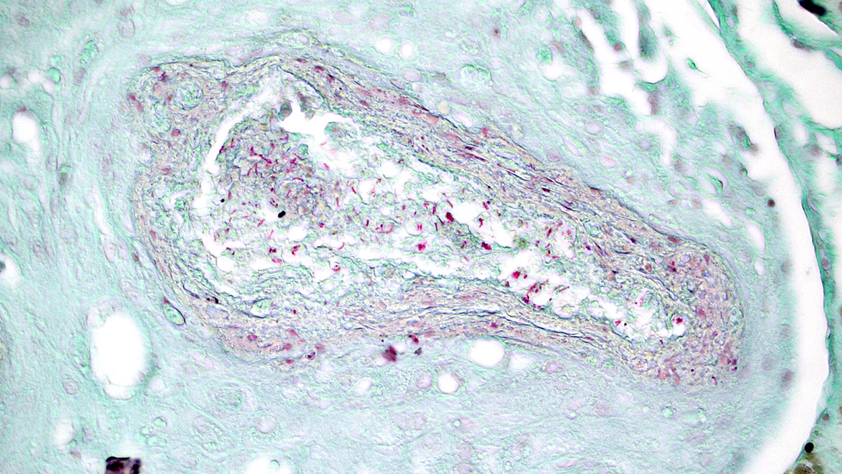
Slide N18-0170-2 Acid Fast âThere are abundant intracellular and extracellular robust acid-fast rods (1 x 3 um) within granulomas.
Contributor Morphologic Diagnosis:
Kidney, posterior: Cystadenoma with concomitant multifocal, subacute to chronic, granulomatous nephritis with acid-fast bacilli
Contributor Comment: Renal tumors in teleosts (bony fish) are generally quite rare with the most common reports being nephroblastic tumors both in rainbow trout (associated with toxin exposure) and in Japanese eels (Anguilla japonica).4,5 Most reports are of single cases, including a cystic adenocarcinoma in a European eel (Anguilla Anguilla), an adenoma in a brown bullhead catfish (Ameiurus nebulosus), and a papilliferous cystadenoma of the mesonephric duct in a Chinook salmon.4,5 However, the tumor featured in this case is a well described tumor of this species and has been seen in multiple A. ocellatus specimens including three from the National Cancer Instituteâs Registry of Tumors in Lower Animals, one from North Carolina State University College of Veterinary Medicine10 and one from the University of Veterinary Medicine Veterinarplatz, Vienna Austria.2 The clinical presentation was similar in all cases where recorded (abdominal distention and deteriorating condition despite medical therapy). While there were minor differences between reported tumor specimens (+/- granulomas, mineral crystals), all appeared cystic and originated from the posterior kidney and, with the exception of one case where there was invasion of surrounding tissue, all cases appeared benign.7 In addition to the cases listed above, another oscar in the same aquarium this one died on August 1, 2018. That fish was also an adult (>4 years) female that presented with a smooth, firm, white tissue was protruding from the vent which was not able to be reduced. On necropsy a mass that was similar grossly to the mass in this case fish was found. Histologic examination identified it to be a renal cystadenoma but there were no granulomas in that case. Based on the number of specimens of the same species with similar tumors and lack of literature to indicate otherwise, it can be inferred that oscar cichlids are predisposed to renal cystadenomas. It is unknown whether this tumor occurs in wild individuals or if its development is due in part to low genetic diversity of the captive-bred population. Other potential etiologies of this tumor cannot be ruled out; it could be a unique species response to an unidentified environmental factor or an as-yet-unidentified microbe such as in proliferative kidney disease in goldfish caused by the myxozoan Hoferellus carassii.7
Clinical signs attributable to the tumor are nonspecific, though it can be presumed that the neoplasm (and in this case infection with mycobacteria) may have affected renal function and that large tumor size may have had an impact on buoyancy. This tumor&146;s abnormal tissue likely provided a permissive environment predisposed to secondary infection. Smaller granulomas were noted in heart, liver, spleen, and ovary, and other animals in this tank had also been previously diagnosed with mycobacteriosis. In the case presented, opportunistic mycobacteriosis is an unsurprising, though florid, complication of both systemic and tissue-specific disease associated with the neoplasm.
Mycobacteria are slow growing, acid-fast staining bacilli that are natural occupants of water systems and opportunistic pathogens. They form biofilms within pipes and on surfaces of aquariums which makes them resistant to many forms of filtration and disinfectants. This makes eradication of mycobacteria from a system difficult. Opportunistic infections of fish within a contaminated system can occur when those fish are inflicted with external injuries or stress. Pathogenicity varies depending on the species of mycobacteria ranging from mild, chronic infections of a few fish to acute system wide infections with mortalities of 30-100%. Infection often results in granuloma formation, most notably within the liver and kidneys but diffuse disease can be seen, especially with the more pathogenic strains.11 This case provides an example of environmental, host, tissue, and pathogen interactions resulting in dramatic pathology.
Contributing Institution:
https://nationalzoo.si.edu/animals/veterinary-care
JPC Diagnosis: 1. Kidney: Renal cystadenoma.
2. Kidney: Granulomas, numerous
JPC
Comment: The contributor
has done an excellent job of describing renal adenoma/cystadenoma in fishes. Benign
renal neoplasms are rare tumors in animals, as they rarely cause clinical signs
and are primarily diagnosed at autopsy. Many reports, as in fish, are single
case reports, with large studies of renal neoplasms in dogs, where they
amounted to 3% of cases overall, and in slaughtered cattle with an incidence of
one in 13,500. Another survey of 6706 cattle at slaughter yielded no benign
tumors. Due to the rarity of benign tumors in domestic species, definitive
criteria of benignancy have not been well established.8 As
well-differentiated malignancies and benign tumors may overlap in histologic
appearance, arbitrary criteria of 2cm diameter is often used, but tumors of
this size may be problematic. Mitoses over 10 per 2.37mm suggest malignancy in
the absence of more definitive criteria, including invasion and metastasis.8
A similar problem has been encountered in human medicine, in which metastasis
has been documented repeatedly in tumors less than 3cm in diameter. Current
human protocols define adenomas as tumors less than 0.5cm with densely packed
tubules or papillary projections and bland nuclei with the low mitotic rate.
Benign renal neoplasms are also encountered in higher incidence in cases of
pyelonephritis and renal vascular disease and are reported in high frequency
along with renal cysts in von-Hippel-Landau syndrome in children.9
Mycobacteriosis
is a common, serious, and often lethal disease affecting fish and other
poikilotherms in aquatic environment. As mentioned by the contributor, the
ability for this species (and related species such as Nocardia) to survive
in biofilms in recirculating systems, results in recurrent infections which are
the bane of aquarists.1
This condition, which has appeared numerous times in poikilotherms in the WSC
over the years, has played primary and secondary roles (as in this case.) The
kidney appears to be one of the most common (if not the most common) organ
affected along with the spleen, and granulomas are often seen as well in the
liver, gills, mesentery, gonads, and choroid.1 In the kidney, both
discrete granulomas and diffuse granulomatous inflammation (as demonstrated in
this slide) are seen.1 The process of development of inflammatory
lesions in affected fish has been studied in detail, characterized, and divided
largely into temporal phases.6 Subacute infections are characterized
by diffuse infiltration of macrophages in infected organs with caseonecrotic
centers. The chronic proliferative form is characterized by the formation of "hard"
and "soft" granulomas, differentiated largely by the presence of fibrous
connective tissue in the "hard" granulomas.6 Calcification (as seen
in this slide) occurs in more chronic lesions.6
A range of studies have demonstrated several paths of transmission in fish, including cutaneous wounds (common in bettas raised for export), transovarial transmission, and ingestion (demonstrated by feeding infected fish carcasses).3 Commonly isolated species include M. marinum, M. fortuitum, M. chelonae, M. peregrinum and M. abscessus.1
The moderator discussed additional differentials for the neoplasm in this case, to include polycystic kidney disease and obstructive cystic disease. The lack of epithelial attenuation within the large cysts tends to rule out obstruction as a cause, but polycystic kidney disease, remained a concern based solely on the histology. The moderator reviewed pertinent renal anatomy and histology in the fish, as well as general pathology of mycobacteriois, and a quick review of gross lesions in fish disease.
References:
1. Frasca S, Wolf JC, Kinsel MJ, Camus AC, Lombardini ED. Osteichthyes. In Terio KA, McAloose D, St. Leger J. Pathology of Wildlife and Zoo Animals. London, UK. Associated Press, pp. 964-966.
2. Hochwartner O, Loupal G, Wildgoose WH, Schmidt-Posthaus H. Occurrence of spontaneous tumours of the renal proximal tubules in oscars Astronotus ocellatus. Dis Aquat Organ. 2010. 89:185â189.
3. Jacobs JM, Stine CB, Baya Am, Kent ML. A review of mycobacteriosis in marine fish. J Fish Dis 2009; 32(2): 119-130.
4. Lee BC, Hendricks JD, Bailey GS. Rare renal neoplasms in Salmo gairdneri exposed to MNNG (N-methyl-Nâ-nitro-N-nitrosoguanidine). Diseases of Aquatic Organisms. 1989. 6:105-111.
5. Lombardini ED, Hard GC, Harshbarger JC. Neoplasms of the Urinary Tract in Fish. Veterinary Pathology. 2014. 51:1000â1012.
6. Lewis, S, Chinabut S. Mycobacteriosis and nocardiosis. In: Woo PTK, Bruno DW, eds. Fish Diseases and Disorders vol 3, ed. 2. Cambridge MA; CAB International 2011; pp. 397-423.
7. Noga EJ. Fish Disease: Diagnosis and Treatment (2nd edition). 2013. Wiley-Blackwell. 239-240.
8. Meuten DJ, Meuten TLK. In Meuten DJ, ed. Tumors in Domestic Animals, 5th ed., Ames Iowa, John Wiley and Sons, Inc., 2017; pp 634-638.
9. Murphy WM, Grignon DJ, Perlman EJ. In: AFIP Atlas of Tumor Pathology: Tumors of the kidney bladder and related urinary structures. Washington DC, ARP Press, Inc., 2004, pp.160-162.
10. Petervary N, Gillette DM, Lewbart GA, Harshbarger JC. A spontaneous neoplasm of the renal collecting ducts in an Oscar, Astronotus ocellatus (Cuvier), with comments on similar cases in this species. Journal of Fish Diseases. 1996. 19:279-281.
11. Racz A, Dwyer T, Killen SS. Overview of a disease outbreak and introduction of a step by step protocol for the eradication of Mycobacterium haemophilum in a Zebrafish system. Zebrafish. 2018. DOI: 10.1089/zeb.2018.1628.