Signalment:
6-year-old, spayed female, mixed breed dog (
Canis familiaris)6-week history of progressive neurologic deficits that began as posterior paresis and progressed to cerebellar ataxia. Neurologic exam revealed cranial nerve V involvement and central vestibular signs.
Gross Description:
No gross lesions in brain
Histopathologic Description:
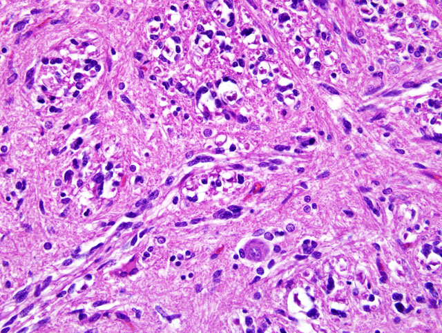
The pons and medulla have a diffuse infiltration of neoplastic elongate cells within gray and white matter. The cells have indistinct cytoplasm, elongate nuclei with dense, hyperchromatic chromatin, mild anisokaryosis and no mitotic figures. They resemble fibrillary astrocytes. The neoplasm does not form a distinct mass and has no borders, but infiltrates the neuropil diffusely without damaging the normal tissue. The cells tend to stream along the axis of white matter fiber tracts and often wrap around neurons, but no neuronal lesions are seen (
Fig. 1-1). Tumor cells form a thin subpial layer in the cerebellum and extend into the cerebellar folia with regional loss of Pukinje cells. An occasional perivascular cuff of lymphocytes is observed.
Morphologic Diagnosis:
Gliomatosis cerebri
Lab Results:
MRI and CSF analysis are both normal. Immunohistochemistry for GFAP was negative.
Condition:
Gliomatosis cerebri
Contributor Comment:
Gliomatosis cerebri is a neoplasm of unknown cell origin but is classified as a glial cell tumor in the WHO classification of tumors of the nervous system. The tumor cells resemble fibrillary astrocytes but GFAP immunostaining is variable; some tumors stain negative and others have few positive staining cells. Gliomatosis in man is divided into two subtypes. Type I is the most common and presents as a diffuse infiltration of the brain with no mass lesion. Type II gliomatosis presents as a mass lesion.Â
Gliomatosis cerebri was established in dogs in a report of 6 cases (5) and based upon lesions that are very similar to those in man. Four of the cases in dogs were type I, and two cases were type II. The case presented here represents type I gliomatosis. The neoplasm in dogs has many similarities to the disease in man, but GFAP staining has been consistently negative in dogs, as it was in this case.Â
JPC Diagnosis:
Brainstem: Gliomatosis cerebri
Conference Comment:
The differential diagnosis including gliosis, diffuse astrocytoma, lymphoma, primitive neuroectodermal tumors (PNETs) and microgliomatosis was discussed at the conference. Widespread infiltration of the central nervous system, atypia and mitotic activity are typical for gliomatosis cerebri and help to differentiate it from gliosis. Gliomatosis cerebri is also more widespread than diffuse astrocytoma and, in dogs, is usually negative for GFAP.(5,6) In lymphoma, neoplastic cells are round with round to irregularly round nuclei and scant to small amounts of cytoplasm, and lymphomas tend to efface tissue. CD3 or CD79 positivity helps to identify lymphoma and prevent an incorrect diagnosis of gliomatosis cerebri. PNETs also tend to efface tissue and are usually seen in young animals. Microgliomatosis is a neoplasm of microglial origin, the resident macrophage of the central nervous system, and should be positive for CD18.
References:
1. Burger PC, Scheithauer BW: Tumors of neuroglia and choroid plexus.Â
In: AFIP Atlas of Tumor Pathology, Tumors of the Central Nervous System, ed. Silverberg SG, Sobin LH, Series 4, pp. 83-86. ARP Press, Washington, DC, 2008
2. Koestner A, Bilzer T, Fatzer R, Schulman FY, Summers BA, Van Winkle TJ: Histological classification of tumors of the nervous system of domestic animals.Â
In: World Health Organization Histological Classification of Tumors of Domestic Animals, ed. Schulman FY, Second Series, vol. 5, pp. 21. Armed Forces Institute of Pathology, Washington, DC, 1999
3. Koestner A, Bilzer T, Fatzer R, Schulman FY, Summers BA, Van Winkle TJ: Lymphomas and hematopoietic tumors.Â
In: World Health Organization Histological Classification of Tumors of the Nervous Sytstem of Domestic Animals, ed. Schulman FY, Second Series, vol. 5, pp. 32. Armed Forces Institute of Pathology, Washington, DC, 1999
4. Maxie MG, Youssef S: Nervous system.Â
In: Jubb, Kennedy, and Palmer's Pathology of Domestic Animals, ed. Maxie MG, 5th ed., vol. 1, pp. 294-295, 448. Saunders Elsevier, Philadelphia, PA, 2007
5. Porter B, de Lahunta A, Summers B: Gliomatosis cerebri in six dogs. Vet Pathol
40:97-102, 2003
6. Vandevelde M, Fankhauser R, Luginbuhl H: Immunocytochemical studies in canine neuroectodermal brain tumors. Acta Neuropathol
66:111-116, 1985
7. Louis DN, Ohgaki H, Wiestler OD, Cavenee WK, Burger PC, Jouvet A, Scheithauer BW, Kleihues P: The 2007 WHO classification of tumours of the central nervous system. Acta Neuropath
114(2):164-172, 2007