Conference #25
Case I:
Signalment: 26 y 9 m, male, Tiliqua scincoides intermedia, northern blue-tongued skink
History: Physical exam performed due to continued lethargy, complete anorexia, weight loss noted by keeper staff. Physical exam showed a lethargic, weak, pale 6-10% dehydrated blue-tongue skink. No masses noted on coelomic palpation, but significant muscle catabolism noted. Bloodwork showed increasing serum phosphorus, and uric acid indicative of renal disease. Due to quality of life concerns euthanasia performed via IV injection in the tail vein of Euthasol (pentobarbital/phenytoin solution) and potassium chloride mixed 1:1.
Gross Pathology:
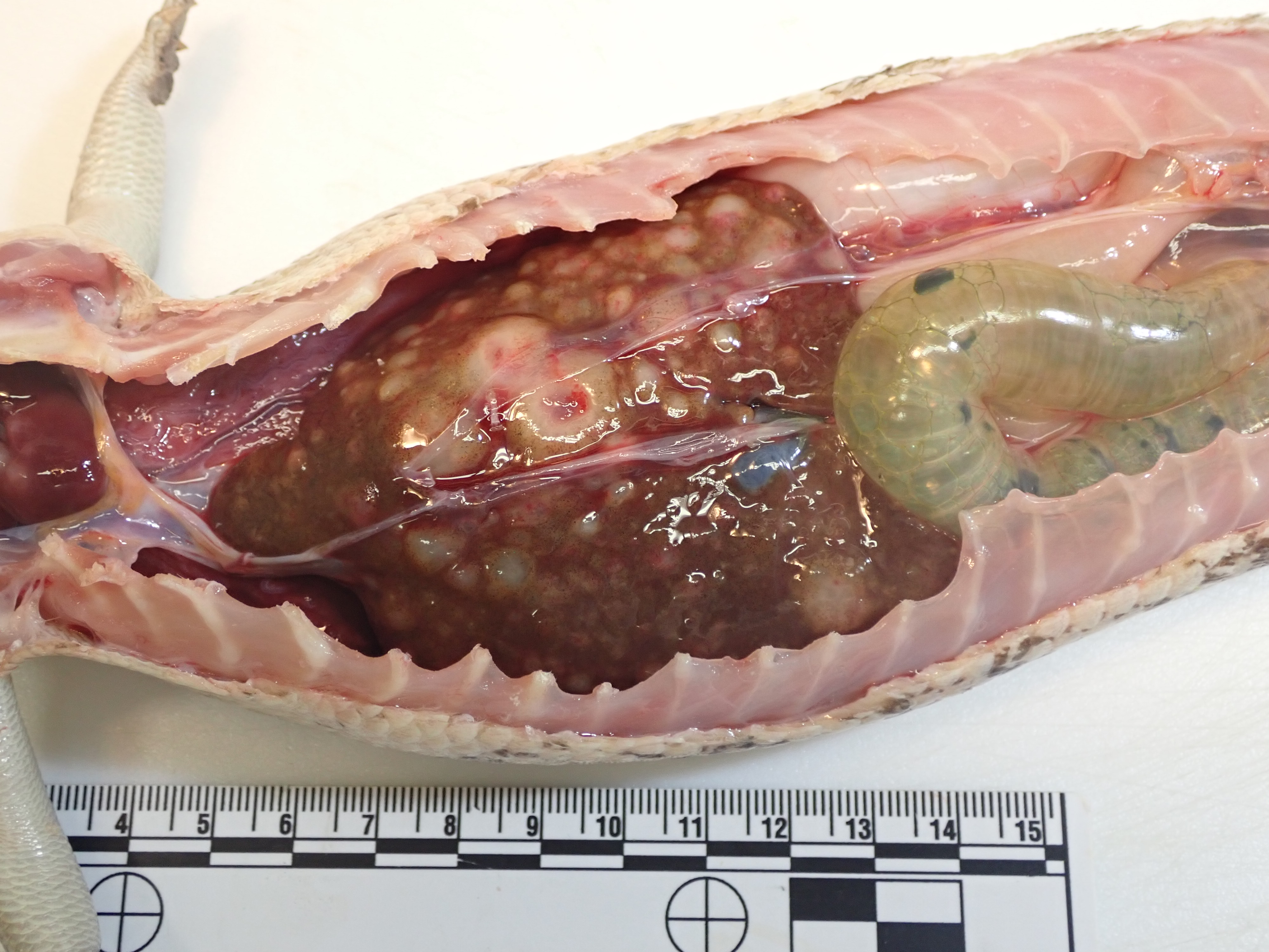
Body condition was reduced with minimal coelomic fat and only remnants of shrunken, orange fat bodies. There was a moderate amount of clear coelomic fluid. Approximately 70% of the liver was expanded and replaced by numerous white, well-demarcated, soft nodules up to 14 mm in diameter. The spleen was mottled with tan nodules up to 3 mm in diameter.
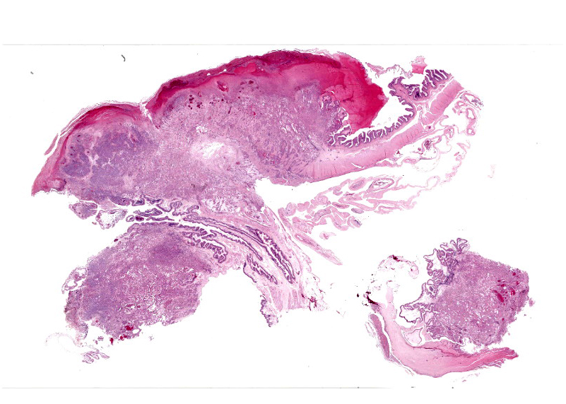
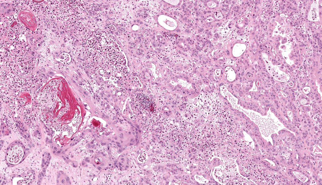
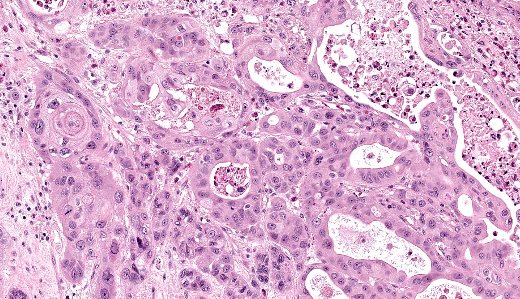
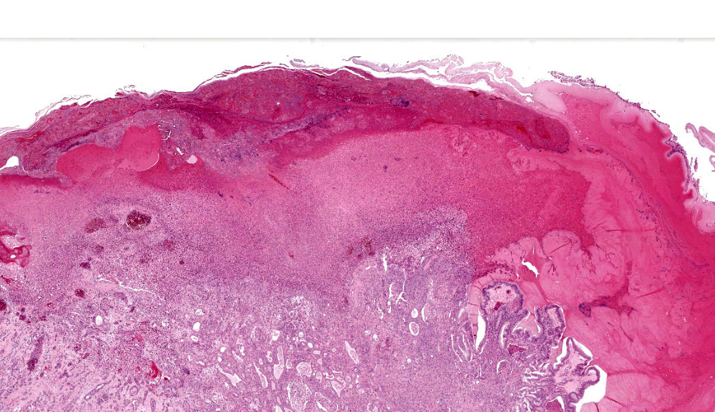
There were white, chalky areas within both kidneys, up to 1.5 cm in diameter expanding representation of both glandular and squamous differentiation.5,6,8 Adenosquamous is a descriptor that is most commonly used in respiratory, alimentary, and mammary tumors with mixed glandular and squamous features and is not specific to any one cell of origin. The malignancy in this case clearly demonstrates glandular formations common in colonic adenocarcinomas, but some areas show nesting, keratinization, and even keratin pearl formation characteristic of squamous cell carcinomas. This tumor was highly metastatic - most likely due to hematogenous spread - and established itself in the liver, spleen, lungs, heart, urinary bladder, pancreas, and fat bodies of the animal. Tumor cells at metastatic sites did not maintain squamous differentiation and only occasionally formed vaguely glandular arrangements, with the vast majority of metastases composed of sheets of malignant cells.
Colonic malignancies of any type are uncommon in reptiles. In a 2004 retrospective of 5353 reptile submissions by Garner et al., neoplasia was not uncommon, with a prevalence of 9.8% across all species submitted (527 tumors).1 Lizards (n= 1909) had a neoplastic prevalence of 8.5%. No cases of colonic adenocarcinoma were reported in any lizard, chelonian, or crocodilian. Only in snakes were cases reported, and in that taxon only 6/325 tumors were diagnosed as colonic adenocarcinomas, indicating these tumors are quite uncommon at ~1.1% of reptile tumors in the retrospective. There was one case of intestinal carcinoma in lizards (0.6% of lizard tumors), but it was small intestine.
In records from the Smithsonian's National Zoo and Conservation Biology Institute, seven cases of colonic adenocarcinoma were identified in lizards (contributor's unpublished research). Three species were represented: 1 blue-tongued skink (Tiliqua spp., this case), 4 Gila monsters (Heloderma spp), and 2 African plated lizards (Gerrhosaurus validus). While these are all heavy-bodied arid-environment lizards, they are not taxonomically closely related, and the overrepresentation should be interpreted in light of their long lifespans and popularity in zoo collections. Interestingly, the cases represent a prominent proportion of adult necropsy findings in these species (1/6 blue-tongued skinks, 2/5 plated lizards, and 4/12 Gila monsters). No cases of colonic adenosquamous carcinoma were described in the National Zoo's records for any species, though an adenosquamous carcinoma was described on the hemipenis of a Mexican lance-headed rattlesnake (Crotalus polystictus) and the esophagus of a wood duck (Aix sponsa).
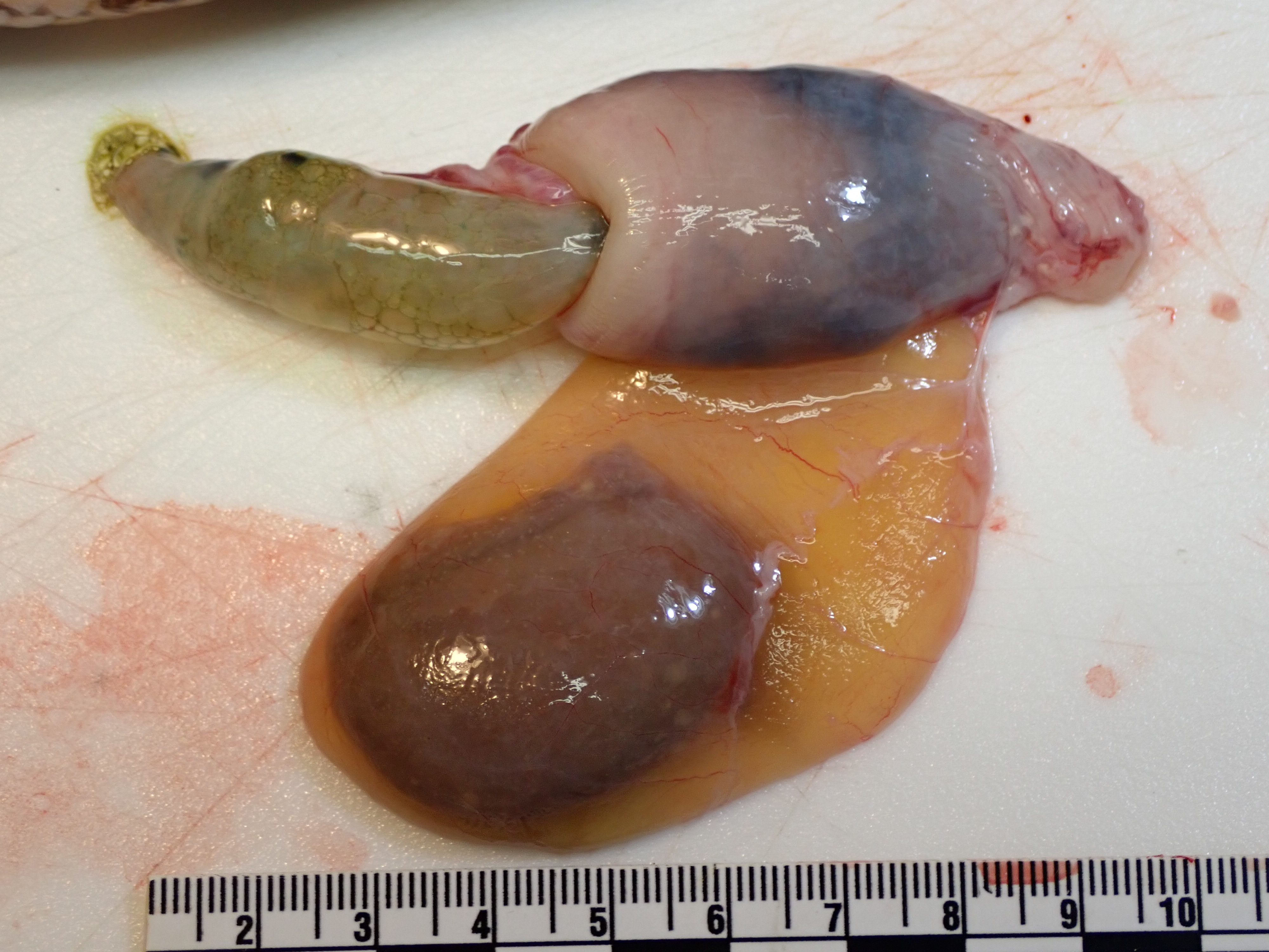
Grossly, the section of colon including the tumor had an identifiable intussusception. Intussusception is the telescoping of a section of bowel (the intussusceptum) within an adjacent segment (the intussuscipiens), generally due to abnormal motility of the bowel smooth muscle. Intussusception is most common in young animals, where it is frequently associated with parasitism or inflammation. In the less frequent adult cases, tumors are often contributory. In humans, a retrospective of adult intestinal intussusception found that over 77% of cases were associated with a tumor, and nearly 60% of the intussusceptions were associated with a malignancy (74% of the tumors).2 In the case presented here, the adenosquamous carcinoma is presumed to be the inciting cause of the grossly identified intussusception.
Contributing Institution:
Smithsonian's National Zoo and Conservation Biology Institute, Wildlife Health Sciences
https://nationalzoo.si.edu/animals/veterinary-careJPC Morphological Diagnosis:
Colon: Adenosquamous carcinoma
JPC Comment:
We crossed the finish line of this year's conferences with the amazing Dr. Kali Holder from the Smithsonian National Zoo! Dr. Holder brought her signature passion for pathology and exceptional comedic timing, setting the tone for a highly entertaining discussion on this first case. The neoplasm in this skink was diagnosed as an adenosquamous carcinoma, likely arising from the colon, with clear areas of both glandular and squamous differentiation. One participant noted that squamous differentiation in reptiles can also be due to hypovitaminosis A and chronic irritation,7 postulating whether this could represent an adenocarcinoma with secondary squamous metaplasia rather than a true adenosquamous carcinoma. The organization of the squamous and glandular components convinced most attendees to support the ultimate diagnosis, however. When considering the disseminated metastasis of the primary carcinoma seen in this skink, Dr. Holder boldly exclaimed, "Don't put butt cancer in your heart! That's no way to run a skink!" Indeed.
The gross photographs, which are not provided to participants prior to conference, were crucial to diagnosing the intussusception in this case. Although uncommon in reptiles, intussusception is a well-recognized sequela to intestinal masses in numerous species, as they disrupt the coordinated peristaltic rhythm required to keep the bowel moving in a single direction.4 Dr. Holder reviewed the pathophysiology of intussusception, which starts when a small fold of bowel telescopes into the adjacent segment, narrowing the lumen and causing partial or complete obstruction.4 This leads to proximal distension, vascular compromise, edema, congestion, and inflammation. As the process worsens, fibrin accumulates on the serosa of the intussusceptum, which may cause adhesions between the intussusceptum and intussuscipiens. Severe cases progress to segmental necrosis, or even sloughing of the affected intestine.4
The difference between "intussusceptum" and "intussuscipiens" is a common sticking point for residents. A good way to differentiate between the two is making a mental connection between the word "recipient" and "intussuscipiens", which is the "recipient" of the intussusceptum. "Recipient" is basically part of the word "intussuscipiens!"
In this skink, the neoplasm likely initiated a cascade of necrosis, inflammation, and mechanical disruption, culminating in the intussusception. This combination would likely have caused anorexia which, in reptiles, rapidly leads to dehydration.2 Dehydration, in turn, causes renal stress and the precipitation of urate crystal deposition (gout). 2 Increased uric acid and urolithiasis were reported in this animal. This combination of unpleasantness likely contributed to this animal's clinical decline.
Dr. Holder closed this case with a discussion on how intestinal carcinomas in reptiles may be more common than currently recognized as improvements in husbandry, veterinary care, and pathology understanding allow more reptiles to reach ages where neoplasia becomes more prevalent.1 Among reptiles, intestinal carcinomas are most common in snakes, less common in lizards, and rare in chelonians.1
References:
- Garner MM, Hernandez-Divers SM, Raymond JT. Reptile neoplasia: a retrospective study of case submissions to a specialty diagnostic service. Vet Clin North Am Exot Anim Pract. 2004 Sep 1;7:653-671.
- Halan M, Kottferova L, Racka K, Lam A. The Amount of Food Ingested and Its Impact on the Level of Uric Acid in the Blood Plasma of Snakes. Animals (Basel). 2022;12(21):2959.
- Honjo H, Mike M, Kusanagi H, Kano N. Adult Intussusception: A Retrospective Review. World J Surg. 2015 Jan 6 [cited 2021 Jul 29];39:134-138.
- Marsicovetere P, Ivatury SJ, White B, Holubar SD. Intestinal Intussusception: Etiology, Diagnosis, and Treatment. Clin Colon Rectal Surg. 2017;30(1):30-39.
- Munday JS, Löhr C V., Kiupel M. Tumors of the Alimentary Tract. In: Meuten DJ, ed. Tumors in Domestic Animals. Hoboken, NJ, USA: John Wiley & Sons, Inc.; 2016:499-601.
- Sunkara T, Caughey ME, Makkar P, John F, Gaduputi V. Adenosquamous Carcinoma of the Colon. Case Rep Gastroenterol. 2017 Sep 1 [cited 2021 Jul 19];11:791.
- Sykes JM 4th, Trupkiewicz JG. Reptile neoplasia at the Philadelphia Zoological Garden, 1901-2002. J Zoo Wildl Med. 2006;37(1):11-19.
- Toumi O, Hamida B, Njima M, et al. Adenosquamous carcinoma of the right colon: A case report and review of the literature. Int J Surg Case Rep. 2018 Jan 1;50:119-121.