WSC 23-24 CONFERENCE 10 CASE IV:
Signalment:
Hatch-year, female bald eagle, avian (Haliaeetus leucocephalus)
History:
The animal was emaciated. It was euthanized at admission to the wildlife clinic with suspected West Nile disease (neurologic signs and eye lesions consistent with chorioretinitis) and liver enlargement.
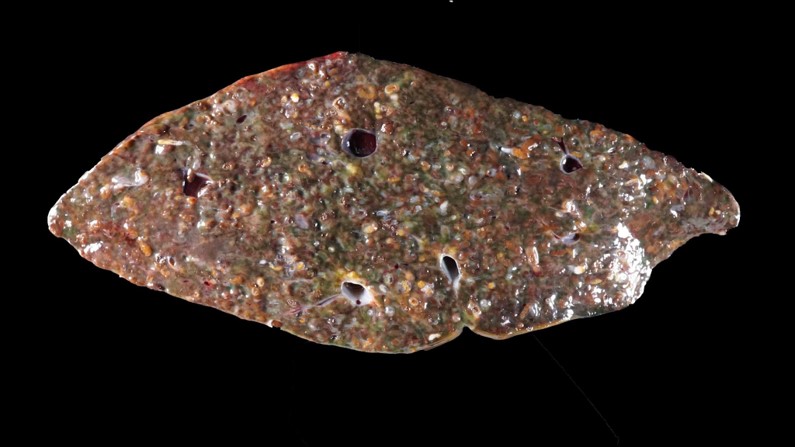
Gross Pathology:
The liver was markedly enlarged (weighing 149g) and discolored brown to green with numerous pinpoint, beige, poorly defined foci. Numerous fragile thin-walled parasites (flukes) less than 5.0 mm long were embedded in the liver.
Laboratory Results:
A liver sample tested positive for Erschoviorchis sp. by PCR and sequencing.
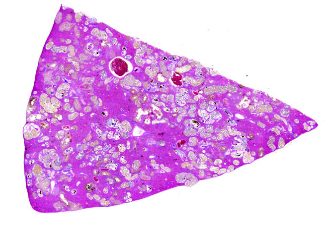
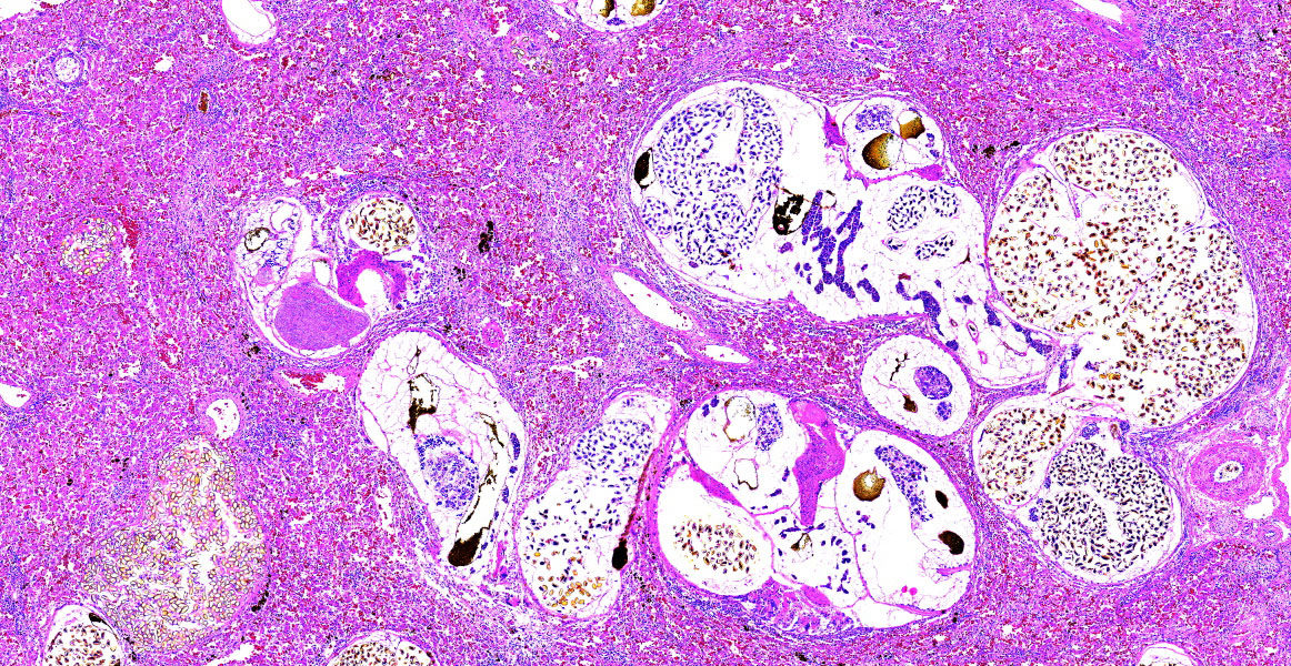
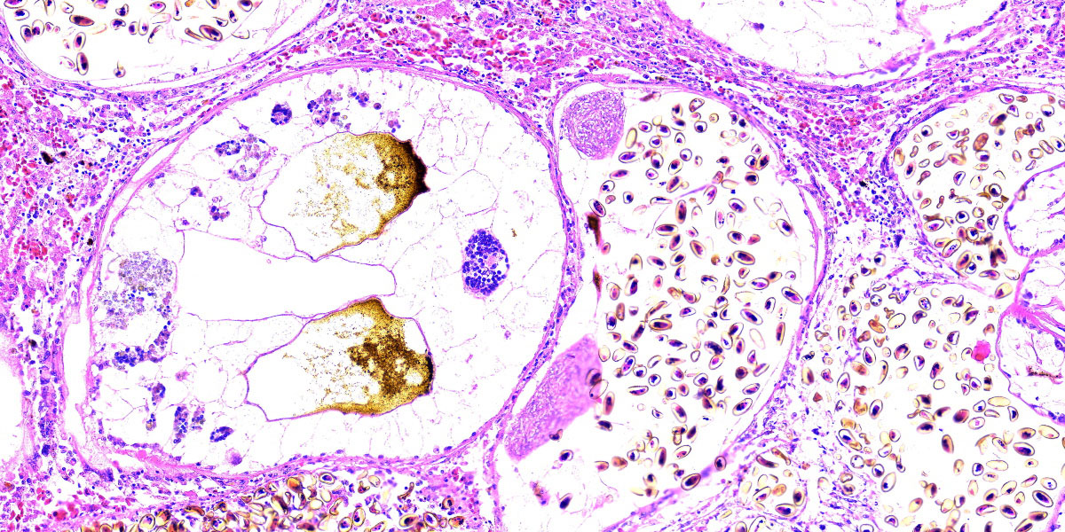
Microscopic Description:
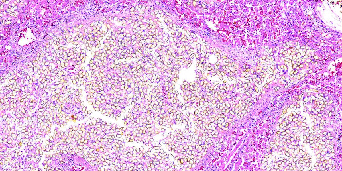
Approximately 75% of the liver section is infiltrated by myriad metazoan parasites that have a thin, smooth tegument, a variably discernible ventral sucker, and numerous intraparenchymal, golden, operculated eggs that are approximately 25.0 x 12.0 µm (morphologically consistent with adult trematodes). Hepatic cords are often dissociated, and sinusoids are frequently expanded by either brown to black fluke pigment, ova, fibrin, and/or numerous lymphocytes, heterophils, and macrophages, including multinucleated cells. Small numbers of lymphocytes, macrophages, and heterophils frequently surround trematodes and occasionally form small granulomas that are centered on areas of lytic necrosis.
Contributor’s Morphologic Diagnosis:
Liver: Granulomatous hepatitis, multifocal to coalescing, moderate, chronic with numerous intraparenchymal adult trematodes.
Contributor’s Comment:
Hepatic trematodosis is a sporadically reported disease in raptors, with a predominance for species in the Opisthorchiidae family. Opisthorchis sp., Metorchis bilis, and Amphimerus elongatus are all members of the Opisthorchiidae family and have been implicated in the previous cases of hepatic trematodosis of eagles. However, recently, a fluke of the genus Erschoviorchis has been reported to cause severe liver infections in bald eagles in the upper Midwest of the United States.3,5 Until recently, the only species within this genus was E. lintoni, which infects the pancreas of the common loon (Gavia immer) in North America.1E. anuiensis, the other species within this genus, was described from experimental infections of Muscovy ducks using metacercaria recovered from fish from the Amur River basin in the Russian Far East.4 Whether the Erschoviorchis sp. affecting the bald eagles was newly introduced to the United States or has long been established in North America and possibly confused with Amphimerus elongatus remains unknown.
Erschoviorchis flukes are notoriously difficult to tease out from the affected tissue due to their fragile nature. As a result, the parasitological features described in this case are based on fluke fragments. Recovering complete flukes would be necessary to assign a new species denomination.
The life cycle of opisthorchiid flukes includes two intermediate hosts, typically a snail and a fish as the first and second host, respectively. Adult trematodes release eggs that are passed in the feces. The first intermediate host, a gastropod snail, ingests the eggs, which then release miracidia. These miracidia undergo several stages of development within the snail: sporocyst→rediae→cercariae. Cercariae are released from the snail and encyst in the skin or muscle of freshwater fish, the second intermediate host, to become metacercariae. The definitive host becomes infected by ingesting the raw, metacercariae-laden fish. Metacercariae generally excyst within the upper gastrointestinal tract of the definitive host and ascend to the biliary ducts where they develop into adults.2 The exact life cycle of this Erschoviorchis sp. is uncertain, but likely is similar to that of other opisthorchiid flukes.
Although E. anuiensis appears to be highly pathogenic based on experimental infections, the clinical significance of massive hepatic trematodosis in this case remains uncertain since West Nile virus infection was considered to be the more important disease problem. Co-infections of bald eagles with Erschoviorchis sp. liver flukes with West Nile virus appear to be common.3
Contributing Institution:
Veterinary Diagnostic Laboratory
University of Minnesota, St Paul, MN
https://vdl.umn.edu/
JPC Diagnosis:
Liver: “Extrematodiasis”. ?
JPC Comment:
The contributor provides an excellent summary of Erschioviorchis sp. parasitism and the discovery of a potentially new pathogen of bald eagles in the upper Midwest of the United States.
In general, trematodes can be identified histologically by the following key histologic features: an oral sucker, paired ceca, no body cavity, spongy parenchyma, vitelline (yolk-forming) glands, hermaphroditism, and, with few exceptions, the presence of operculated eggs. Some common trematodes of veterinary importance include Fasciola hepatica in the bile ducts and gallbladder of ruminants, Fascioloides magna in the liver of certain wild ruminants, Paragonimus kellicotti in the lungs of dogs and cats, and Heterobilharzia americanum in dogs and some wildlife.
Conference discussion focused largely on the stunning appearance of the examined section, with eggs and adult trematodes in seemingly every micrometer of the tissue. Participants struggled to characterize the severity of the parasitism in this section, with “massive” and “extreme” being popular modifiers. Participants settled on a morph of “Liver: Trematodiasis, multifocal, extreme, with moderate, multifocal necrosis and granulomatous inflammation.” Disappointment was palpable in the room, however, as many felt that this rather quotidian diagnosis failed to capture the full glory of this gorgeous histologic specimen. Our cheeky JPC diagnosis was suggested by an eagle-eyed resident with a love of puns, and, once suggested, “Extrematodiasis” quickly soared to the top. We shall return to normal professional standards next wee (perhaps); in the meanwhile, the contributor provides an excellent morphologic diagnosis, as do we, buried midway through the paragraph above.
References:
- Gower WC. A new trematode from the loon, Gavia immer, and its relationship to Haematotrephus fodiens Linton, 1928. Proc US National Museum 1939;87:139–143.
- King S, Scholz T. Trematodes of the family Opisthorchiidae: a minireview. Korean J Parasitol 2001;39:209–221.
- McDermott KA, Greenwood SJ, Conboy GA, Franzen-Klein DM, Wünschmann A. Massive hepatic trematodosis in 5 juvenile bald eagles. J Vet Diagn Invest. 2023;35(4):409-412.
- Tatonova YV, Besprozvannykh VV, Katugina LO, Solodovnik DA, Nguyen HM. Morphological and molecular data for highly pathogenic avian parasite Erschoviorchis anuiensis n. and phylogenetic relationships within the Opisthorchiidae (Trematoda). Parasitol Int 2020; 75:102055.
- Wünschmann A, Armien AG, Hoefle U, Kinne J, Lowenstine LJ, Shivaprasad HL. Birds of Prey. In: Terio KA, McAloose D, St. Leger J, eds. Pathology of Wildlife and Zoo Animals. Elsevier;2018:738.