Signalment:
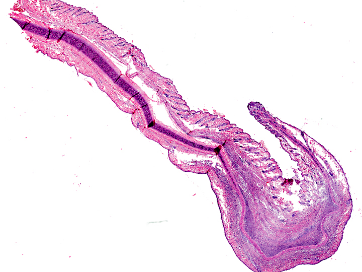
Gross Description:
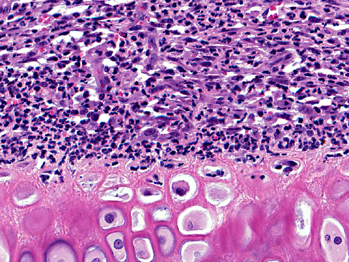
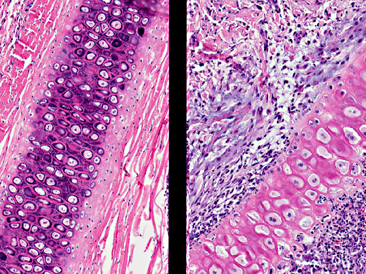
Histopathologic Description:
Morphologic Diagnosis:
Condition:
Contributor Comment:
Sometimes the disease is also called auricular chondritis, because the main gross lesions are ear swelling and discoloration, erythema, pain and curling of one or both ears. However, two case reports describe involvement of additional cartilaginous tissues as reported in people(1, 3).
Histologically, there is degeneration, loss of basophilic staining and necrosis of the ear cartilage. There is also infiltration of mononuclear and polymorphonuclear cells, and perichondrial and perivascular fibrocyte and capillary endothelial cell proliferation. The histological lesions observed in this case are similar to those reported in the cases described in the literature.
JPC Diagnosis:
Conference Comment:
In rats, trauma from cagemates or metal ear tags is suspected as the inciting cause of auricular chondritis, but the disease frequently occurs without history or evidence of trauma. Unlike the feline disease, auricular chondritis in rats is always bilateral, even when the metal ear tag is present in only one ear, and increases in incidence with age(5). The disease presents grossly as firm, multinodular to diffuse thickening of pinnae, and bilateral lesions extend peripherally from the base of the pinnae. Occasionally, pinnae are uniformly thickened rather than having nodular lesions. There is degeneration and lysis of the auricular cartilage plate with granulomatous inflammation and proliferative immature cartilaginous nodules and fibrosis, and osseous metaplasia is characteristic in advanced lesions(5).
Auricular chondritis in rats has been proposed as a model for relapsing polychondritis in humans, which involves several cartilage-containing tissues, including the ear(5). In humans, relapsing polychondritis is associated with auricular chondritis, inflamed cartilage in other sites, and antibodies to type II collagen, IgG and C3 complement. Antibodies to type II collagen have not been demonstrated in the spontaneous auricular chondropathy of rats, and unlike relapsing polychondritis in humans, only the auricular cartilage is involved(4, 5).
In mice, it is speculated that metal ions from ear tags, such as copper and iron, incite an autoimmune process via activation of matrix metalloproteinases. These metal ions supply reactive oxygen species that induce inflammation and fibrosis, and oxidation of cartilage collagen renders the collagen fibrils more brittle and prone to mechanical fatigue. Tagged ears have increased amounts of metallothionein (MT-I and MT-II) and increased expression of Th1 cytokines, including interferon-γ, tumor necrosis factor-α, and interleukin-2; it is postulated that the lesion represents a delayed-type allergic contact dermatitis in response to the metal ions(4). There are two proposed mechanisms for the fibrosis and osseous metaplasia seen in advanced lesions. In the first, cartilage degeneration, characterized by chondrolysis and splitting of the pinnal cartilage plate leads to perichondrial fibrous proliferation, which differentiates into fibroadipose tissue and progresses to fibrochondrous and/or osseochondrous tissue. In the second proposed mechanism, there is focal granulomatous inflammation without chondrolysis, and fibroblasts proliferate within the granulomatous inflammation and then differentiate into fibrochondrous tissue with subsequent chondrous and osseous differentiation(5).
References: