Signalment:
Adult, female,
European rabbit (
Oryctolagus cuniculus).A
member of the public found the collapsed rabbit in a field close to their home
and presented it to the local veterinary practice where the rabbit was
euthanized on humane grounds.
Gross Description:
The
eyelids were bilaterally severely swollen with an exudate lightly adhered to
the surface. The nostrils had a bilateral discharge. The vulva was severely
swollen.
Histopathologic Description:
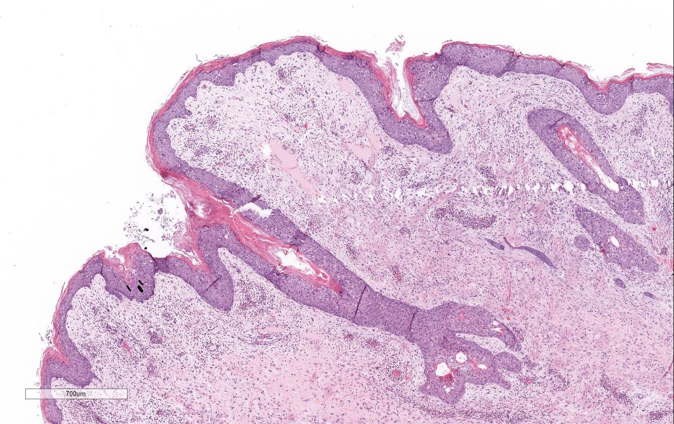
The
slide consists of a section of haired skin and vulva including mucocutaneous
junction. There is diffuse moderate to severe hyperplasia of the mucosal
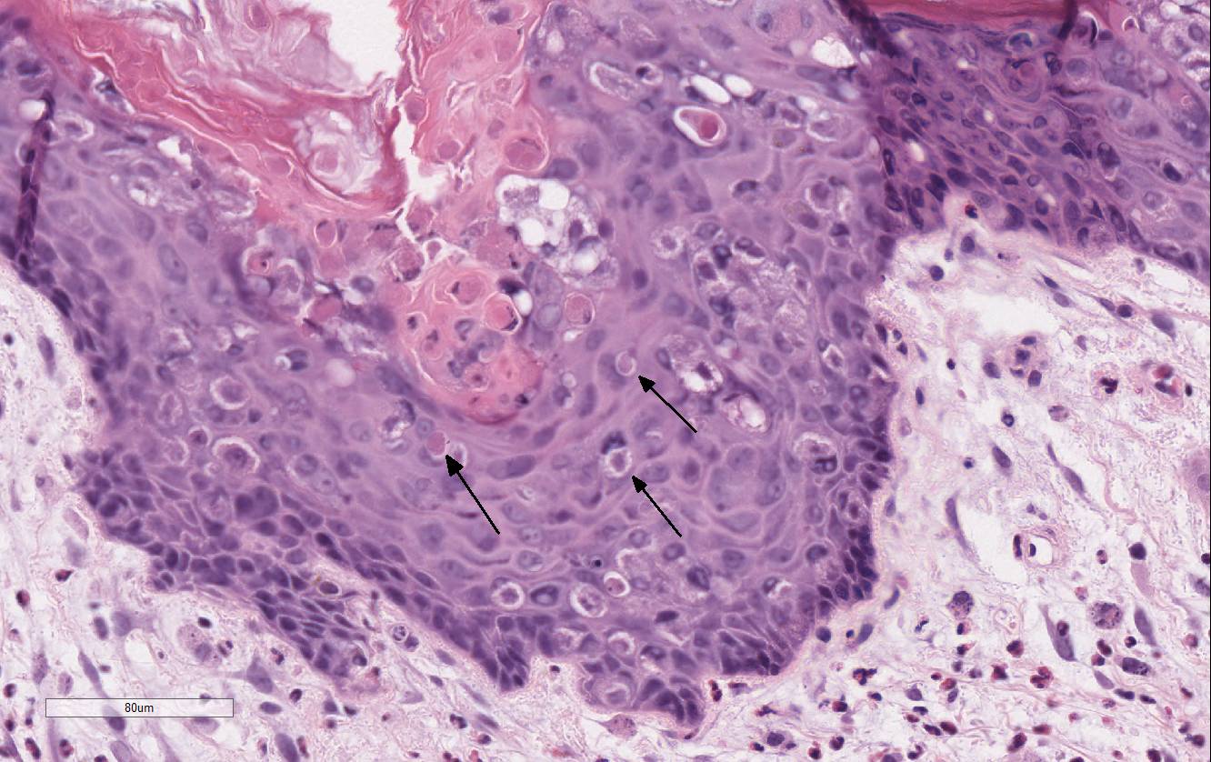
epithelium. Multifocally epithelial cells throughout all the layers of the
epithelium are enlarged, often degenerate, with clear intracytoplasmic spaces
(intra-cellular edema). Often, epidermal cells
contain large, approximately 15 to 20 μm diameter, round to oval,
homogenous, brightly eosinophilic cyto-plasmic inclusions. The same inclusions
may also be observed in sloughed epithelial cells and within the thickened
stratum corneum showing moderate orthokeratotic hyperkeratosis. Randomly
scattered throughout the epidermis there are occasional keratinocytes with
hyper-eosinophilic cytoplasm and karyorrhectic nuclei (single cell necrosis).
There are diffusely scattered basophilic granules within keratinocytes
(keratohyaline granules) of the stratum granulosum and rarely deep within the
stratum basale (dyskeratosis). Multifocally, the epithelial cells of the
vaginal mucosa are expanded by a single large clear vacuole (intracellular
edema / ballooning degeneration).
Diffusely
the lamina propria is characterized by a loosely arranged slightly basophilic
myxoid matrix admixed with edematous areas. Multifocally in the dermis there
are plump elongated to polygonal fibroblasts with stellate processes. These
cells have finely granular basophilic cytoplasm, a single distinct, round to
elongate and centrally placed large nucleus with finely stippled chromatin and
a single evident nucleolus. Anisocytosis and anisokaryosis are moderate and
mitotic figures are rare.
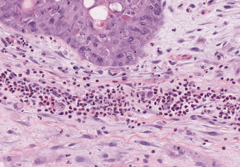
Within the
superficial dermis and surrounding the blood vessels in the deeper dermis there
are moderate, multifocal to coalescing aggregates of lymphocytes, plasma cells,
heterophils, and macrophages. Occasionally, in the superficial dermis, the inflammatory
process is predominately heterophilic.
Morphologic Diagnosis:
1. Vulva, mucocutaneous junction: Diffuse, subacute,
moderate to severe epidermal hyperplasia with intracellular edema and
intracytoplasmic viral inclusions.
2. Vulva,
mucocutaneous junction: Multifocal to coalescing, subacute, moderate
lymphoplasmacytic and heterophilic proliferative dermatitis and sub-mucosal
proliferation with diffuse myxoid changes and edema.
Lab Results:
N/A
Condition:
Vulvar myxomatosis/Myxomatosis virus (Leporipoxvirus)
Contributor Comment:
Myxomatosis is a common, worldwide distribution disease of
rabbits and is now endemic in the wild rabbit population in Europe. Hares may
be carriers of the infection but very rarely exhibit clinical signs.
2
Originally myxo-matosis was introduced in Europe to control the wild rabbit
population.
9 Initially the disease had a very high mortality rate,
but due to natural selection the wild rabbit population began to develop
immunity to the disease and mortality rate is now approximately 25% in the
European rabbits.
9
Myxomatosis
is caused by the Myxoma virus, a leporipoxvirus from the family of poxviruses.
6
Transmission of the virus requires a mosquito or flea vector, but it is
possible for the virus to spread by direct contact. Due to transmission routes,
domestic rabbits, especially the ones kept outdoors are at risk of contracting
the virus from the wild rabbit population and therefore a vaccine has been
developed.
There are two recognized forms of the disease: the classical,
nodular form (as in this case); and the respiratory, amyxomatous form of the
disease. The nodular form is often transmitted by vector route and causes the
classical swelling of the eyelids, nodules over the skin, and edema of the
genitalia. Usually, there is ocular and nasal discharge associated with the
disease. The infection causes a severe depression of the hosts immune system
causing secondary infections to take hold and often the secondary infections
are the ultimate cause of death. The amyxomatous form is usually due to a mild
or attenuated strain of the virus;
6 this causes respiratory signs
and rarely nodular lesions.
Other
skin disease that may be confused for myxomatosis is Shope fibroma caused by
the rabbit Shope fibroma virus (SFV, Leporipoxvirus). Shope fibroma virus
induces discrete fibromas usually restricted to the distal limbs but
occasionally may be found in the head.
JPC Diagnosis:
Mucocutaneous
junction, vulva Atypical mesenchymal proliferation,
diffuse, moderate, with epithelial and epidermal hyperplasia, ballooning
degeneration, lymphoplasmacytic and heterophilic dermatitis, and epithelial
intracytoplasmic eosinophilic inclusions, European rabbit,
Oryctolagus
cuniculus.
Conference Comment:
Poxviruses are a large family of epitheliotropic double-stranded DNA viruses
that cause several important cutaneous and systemic lesions in wild and
domestic mammals, birds, and humans.
8 Most poxviruses cause a mild
localized cutaneous lesion, but some can cause generalized systemic and fatal
disease. An example of the latter is the rabbit myxoma virus, a member of the
genus
Leporipoxvirus, which can cause up to 90% mortality in naïve
susceptible strains of wild rabbits.
2,8,9 However, through natural
selection, genetically resistant strains of wild rabbits now have only about
25% mortality when infected with virulent strains of the virus in endemic
areas.
9 Readers are encouraged to review 2012 for a brief review of the fascinating history of this virus, and its use during
attempted European rabbit (
Oryctolagus cuniculus) eradication programs
in Australia and France in the early 1950s.
Following
inoculation, typically by an arthropod vector, susceptible rabbits develop
localized skin tumors resembling fibromas caused by the rabbit (Shope) fibroma
virus discussed above by the contributor.
3,9 Other characteristic
gross lesions are pronounced gelatinous subcutaneous edema surrounding
mucocutaneous junctions. In this case, the contributor noted severe swelling
around the eyelids and vulva with nasal discharge grossly.
9
While the
microscopic staining of the tissue section of vulva is somewhat pale, this case
nicely illustrates the classic histologic poxviral epithelial intracytoplasmic
in-clusions and ballooning degeneration.
8 In addition, there is a
subepithelial proliferation of large stellate mesenchymal cells within a
homogenous mucinous matrix. Conference participants also noted a moderate
lympho-plasmacytic inflammatory infiltrate admixed with the atypical
mesenchymal cells. In addition to being epitheliotropic, the myxoma virus is
T-lymphocytotrophic and systemic spread occurs via lymphocytes and monocytes to
draining lymph nodes.
9 Myxoma stellate cells are typically present
in lymph nodes, bone marrow, spleen, and centrilobular areas of the liver.
Degenerative and necrotizing lesions are usually confined to the lymphoid
tissue in lymph nodes, lungs, and spleen with lymphoid depletion, particularly
in the T-cell zones.
9
Recently, there has been a great deal of interest in the rabbit
myxoma virus as one of the promising new oncolytic viruses used in virotherapy
for human cancer. Oncolytic viruses are engineered to preferentially infect and
kill cancer cells while sparing normal host cells.
4,7 Several other
oncolytic viruses originated from human pathogens (herpes simplex-1, measles,
etc) and still retain some ability to replicate in normal host tissue. Myxoma
virus is attractive to researchers because its pathogenicity is restricted to
lagomorphs and thus it will not replicate or kill normal human host cells; but
the virus does have significant oncolytic potential for a large variety of
neoplasms in several animal species and humans.
4,7
References:
1. Bangari DS,
Miller MA, Stevenson GW, et. al. Cutaneous and systemic poxviral disease in red
(
Tamiasciurus hudsonicus) and gray (
Sciurus carolinensis)
squirrels.
Vet Patho.l 2009; 46:667-672.
2. Barlow A., et
al. Confirmation of myxomatosis in a European brown hare in Great Britain
. Vet
Rec. 2014; 175(3):75-6.
3. Berto-Moran A,
Pacios I, Serrano E, Moreno S, Rouco C. Coccidian and nematode infections
influence prevalence of antibody to myxoma and rabbit hemorrhagic disease
viruses in European rabbits.
J Wildl Dis. 2013; 49(1):10-17.
4. Chan WM,
McFadden G. Oncolytic poxviruses.
Annu Rev Virol. 2014; 1:119-141.
5. DiGiacomo RF,
Mare CJ. Viral diseases. In: Manning PJ, Ringler DH, Newcomer CE, eds.
The
Biology of the Laboratory Rabbit. 2nd ed. San Diego, CA: Academic Press;
1994:178-180.
6. Kerr, P.J., et
al., Myxoma virus and the Leporipoxviruses: An evolutionary paradigm
. Viruses.
2015;
7(3):1020-61.
7. Kinn VG,
Hilgenberg VA, MacNeill AL. Myxoma virus therapy for human embryonal rhabdo-myosarcoma
in a nude mouse model.
Oncolytic Virother. 2016; 5:59-71.
8. Mauldin E,
Peters-Kennedy J. Integumentary system. In: Maxie MG, ed.
Jubb, Kennedy, and
Palmers Pathology of Domestic Animals. Vol 1. 6th ed. Philadelphia,
PA:Elsevier; 2016:616-625.
9. Percy DH,
Barthold SW.
Pathology of Laboratory Rodents and Rabbits. rd ed. Ames,
IA: Blackwell Publishing; 2016:261-263.