Signalment:  

13-year-old, Gelding, Quarter Horse, Equus caballus2 weeks prior to euthanasia, the horse exhibited signs of colic, was dehydrated and had a temperature of 102.8, CBC and serum chemistries were normal. Horse was treated with Banamine and mineral oil. His temperature and temperament returned to normal. 10 days later, the horse became anorexic, had difficulty breathing, was very weak and developed dependent ventral edema. Severe pleural effusion found on physical exam. The horse treated with lasix. There was no improvement. On 05/23/05, one fourth of a gallon of straw colored fluid was drained from the chest. The horse was euthanized and submitted to the diagnostic laboratory. Necropsy was performed on the same day.


Gross Description:  


Subacute, severe fibrinous serosanguinous thoracic and peritoneal effusion
Subacute, severe, proliferative, fibrinohemorrhagic pericarditis
Subacute, severe, bilateral pulmonary congestion and edema
Subacute, severe, focally extensive, ventral subcutaneous edema


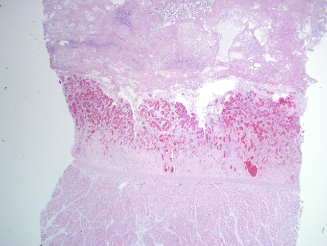
Histopathologic Description:

The epicardium is diffusely congested, hyperplastic and inflamed. The epicardial surface is covered by edematous, well vascularized fibroblastic tissue which contains a marked infiltrate of degenerative neutrophils and scattered macrophages. This proliferative tissue is covered by laminations of fibrin which contains degenerative neutrophils. Within this material scattered clusters of gram negative coccobacilli are detected in replicate tissue sections stained with Brown and Brenn.


Morphologic Diagnosis:  

Subacute, severe, proliferative fibrinosuppurative epicarditis with bacteria.


Lab Results:  

Actinobacillus spp. was isolated in pure culture from a swab of the pericardial sac contents.


Condition:  

Actinobacillus sp.


Contributor Comment:  

The severe pleural effusion noted at necropsy is attributed to heart failure secondary to severe, restrictive, fibrinous pericarditis. Fibrinous pericarditis is a fairly uncommon condition in horses.3 This condition can result from hematogenous bacteria, extension of inflammation from the surrounding tissues via lymphatic spread or by direct inoculation of the pericardial sac by a puncture wound.3,4 Historically, streptococci have been incriminated in equine cases.4 However, a retrospective study of cases of terminal equine pericarditis associated with the mare reproductive loss syndrome during the spring and summer of 2001 confirmed that Actinobacillus species played a significant role in this disease.1


JPC Diagnosis:  

Heart, epicardium: Epicarditis, fibrinous, chronic-active, diffuse, severe (Fig. 1-1), with abundant granulation tissue, Quarter Horse (Equus caballus), equine.


Conference Comment:  

There are three forms of pericarditis: effusive, fibrinous, and constrictive.6,7 Fibrinous pericarditis usually occurs via hematogenous spread of infectious agents.4,7 The fibrin exudate covers the epicardium and pericardium and forms gray-white shabby projections when the two are pulled apart (bread and butter pericarditis).4 Suppurative or purulent pericarditis occurs in the presence of pyogenic bacteria.4 It is seen mainly in cattle with hardware disease, and occasionally in cats and horses with pyothorax. In dogs it can be associated with migrating grass awns.4 Constrictive pericarditis occurs following extensive fibrous proliferation and adhesions forming between the pericardium and epicardium.7 Blunt dissection is usually not sufficient to break down the adhesions formed. The lesions obliterate the pericardial space and impair diastolic filling often leading to right sided heart failure.4,7

Conditions potentially associated with fibrinous pericarditis Table extracted from Maxie et al.4 and Van Vleet et al.7

Cattle: Pasteurellosis (Mannheimia haemolytica and Pasteurella multocida), blackleg (Clostridium chauvoei), sporadic bovine encephalomyelitis (Chlamydophila pecorum), contagious bovine pleuropneumonia (Mycoplasma mycoides mycoides small colony type), clostridial hemoglobinuria (Clostridium haemolyticum), neonatal coliform infections (via umbilicus)
Swine: Glasser's disease (Haemophilus suis), pasteurellosis (Pasteurella multocida and Mannheimia haemolytica), porcine enzootic pneumonia (Mycoplasma hyopneumoniae and other agents), salmonellosis, streptococcal infection of piglets
Sheep: Pasteurellosis (Mannheimia haemolytica and Pasteurella trehalosi)
Lambs: Pasteurellosis, streptococci
Horses: Mycoplasma felis, streptococcal polyarthritis with pericarditis, mare reproductive loss syndrome
Cats (rare): Feline infectious peritonitis
Birds: Pssiticosis (Chlamydia psittaci)

Recent articles concerning an epidemic of fibrinous pericarditis, primarily caused by Actinobacillus spp., indicate a strong relationship with mare reproductive loss syndrome (MRLS).1,2,6 MRLS is a syndrome of abortion in horses that occurred in Kentucky in 2001 and 2002. Features of the syndrome include little to no signs of premonitory illness in the mare, hemorrhages in the chorion, amnion, and amniotic segment of the umbilical cord, pleura, and heart.5 Non-β-hemolytic Streptococcus spp. and/or Actinobacillus spp. were isolated in 50% and 20% of the cultured specimens.5 MRLS has been associated with the Eastern tent caterpillar (Malacosoma americanum), specifically the worm exoskeleton and attached setae.2,5 The exact relationship between exposure to the Eastern tent caterpillar, MRLS, and fibrinous pericarditis is not known.1,2,5,6


References:

1. Bolin D, Donahue J, Vickers M, Harrison L, Sells S, Giles R, Hong C, Poonacha K, Roberts J, Sebastian M, Swerczek T, Tramontin R, Williams N: Microbiologic and pathologic findings in an epidemic of equine pericarditis. J Vet Diagn Invest 17:38-44, 2005
2. Donahue JM, Sells SF, Bolin DC: Classification of Actinobacillus spp. isolates from horses involved in mare reproductive loss syndrome. Am J Vet Res 67:1426-1432, 2006
3. Knottenbelt DC, Pascoe RR: Disorders of the Cardiovascular System. In: Color Atlas of Diseases and Disorders of the Horse, pp. 170-171. Mosby, New York, New York, 2003
4. Maxie MG, Robinson WF: Cardiovascular system. In: Jubb, Kennedy, and Palmers Pathology of Domestic Animals, ed. Maxie MG, 5th ed., vol. 3, pp. 22-30. Elsevier Limited, St. Louis, MO, 2007
5. Schlafer DH, Miller RB: Female genital system. In: Jubb, Kennedy, and Palmers Pathology of Domestic Animals, ed. Maxie MG, 5th ed., vol. 3, pp. 506-507. Elsevier Limited, St. Louis, MO, 2007
6. Seahorn JL, Slovis NM, Reimer JM, Carey VJ, Donahue JG, Cohen ND: Case-control study of factors associated with fibrinous pericarditis among horses in central Kentucky during spring 2001. J Am Vet Med Assoc 223:832-838, 2003
7. Van Vleet, JF, Ferrans VJ: Cardiovascular system. In: Pathologic Basis of Veterinary Disease, eds. McGavin MD, Zachary JF, 4th ed., pp. 575-578. Elsevier, St. Louis, MO, 2007

A virtual slide is not available for this case.


Fig. 1-1 Heart


Back | VP Home | Contact Us |