CASE IV:
Signalment:
9-year-old, spayed female, Labrador mixed breed dog (Canis lupus familiaris)
History: The dog developed non-pruritic periocular and dorsal trunk crusting and alopecia approximately 6 weeks prior. A skin scrape of the affected skin was negative. The dog was treated with Cytopoint, Bravecto, and a two week course of Clavamox. Over the next two weeks, the alopecia and crusting spread around her eyes and down to the nose. Periocular areas were treated with topical neo/poly/dex eye ointment. On recheck, the skin around the nasal mucosa and lips was depigmented, with evidence of uveitis.
Gross Pathology:
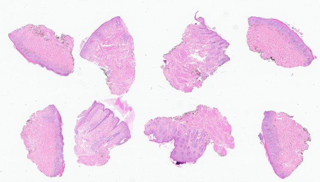
Five punch biopsies of the affected skin around the nose and lip are submitted for histopathology. The epidermal surface of the biopsies is multifocally ulcerated, mottled tan to black, and contains sparse hair. A soft to firm, approximately 2.5 cm subcutaneous mass from the left cranial thorax is submitted alongside the punch biopsies.
Laboratory Results:
Skin culture: Staphylococcus spp.
Microscopic Description:
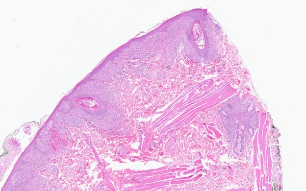
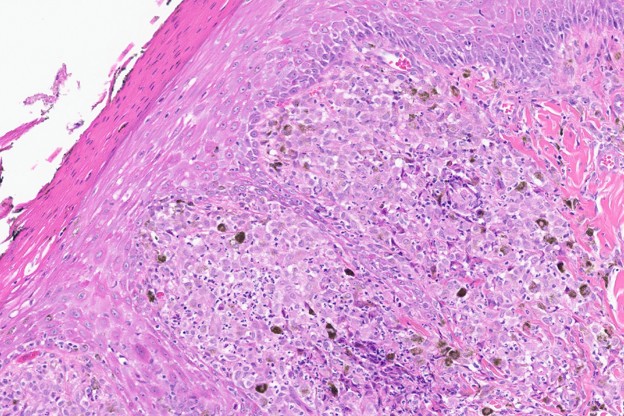
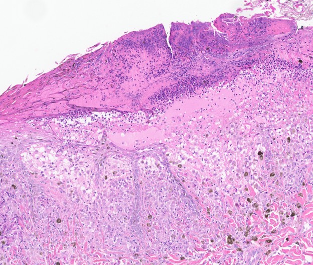
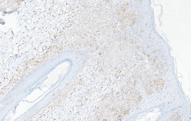
Facial skin (mucocutaneous junction, haired skin, and lips). The mucosal, submucosal, and dermal-epidermal interface is infiltrated by a marked inflammatory infiltrate consisting of macrophages and neutrophils with fewer lymphocytes and plasma cells. Similar inflammation is present in periadnexal areas and occasionally perivascularly. Macrophages containing fine to large, dark brown, cytoplasmic pigment (melanophages) infiltrate the superficial submucosa and are scattered along areas of inflammation (pigmentary incontinence). Rete pegs multifocally extend into the superficial mucosa (mucosal hyperplasia), and the mucosal surface is covered in a moderate amount of nucleated keratin (parakeratotic hyperkeratosis). The superficial mucosal surface is multifocally ulcerated and replaced by serocellular crust composed of degenerate neutrophils and karyorrhectic cellular debris mixed with occasional colonies of basophilic cocci. The epidermal surface is covered in a moderate amount of anucleate keratin (orthokeratotic hyperkeratosis).
Other histopathology findings that are not included in the provided slide:
Left cranial thorax: Soft Tissue Sarcoma (Grade 2, Medium Grade)
Contributor's Morphologic Diagnosis:
- Facial skin: Pyogranulomatous lichenoid dermatitis with pigmentary incontinence
Contributor's Comment:
The histopathological features of the facial skin and clinical history are compatible with uveodermatologic syndrome (UDS). Uveodermatologic syndrome is a rare, suspected immune-mediated skin disease characterized by granulomatous uveitis and facial depigmentation.3 In humans, a similar disease entity, "Vogt-Koyanagi-Harada," results in comparable skin lesions in addition to meningitis with occasional auditory involvement; neurological disease is exceptionally rare in dogs; thus, this disease is referred to as UDS instead of VKH in dogs.1,6 The pathogenesis of VKH in humans demonstrates T cell-mediated autoimmunity against melanin and related proteins; however, this pathogenesis has not been demonstrated in the dog.3 Numerous genetic mutations related to immunity and aberrant activation of Th1 and Th17 pathways have been discovered in humans who develop VKH.8 Studies in Akitas with UDS suggest a role of certain dog leukocyte antigen (DLA) class II gene alleles in the pathogenesis of this disease,1,4,6,7 which is similar to studies in humans that have found an association with human leukocyte antigen (HLA) class II alleles in the formation of VKH.2
Arctic breeds such as Siberian Huskies, Alaskan Malamutes, Akitas, Samoyeds, Chow Chows, and related mixed breed dogs are more commonly affected. This disease has also been diagnosed in the Irish setter, golden retriever, Old English sheepdog, Saint Bernard, Shetland sheepdog, Dachshund, fox terrier, Basset hound, Brazilian fila, Jack Russell terrier, rat terrier, and miniature poodle.3,5,6 The most common ages affected are between 6 months and 6 years, and there is no known sex predilection.3 Ocular signs include anterior and posterior uveitis that presents as photophobia, blepharospasm, conjunctivitis, and corneal edema.3,6,7 Skin signs include bilaterally symmetric facial depigmentation with variable amounts of crusting and erythema, predominantly along the muzzle, periorbital region, nasal planum, and lips.3,4 Lesions may also appear on the external genitalia including the scrotum and vulva, inside the oral cavity, or become more generalized throughout the body.3,4 These lesions may or may not be pruritic. The ocular form of this disease often precedes skin lesions, and affected individuals may not have skin lesions at all; however, concurrent skin disease is an important indicator of the ocular form.3,7
Histologically, the epidermis is hyperplastic, with variable amounts of crusting, ulceration, parakeratosis, and leukocyte exocytosis.3 Oftentimes, the stratum basale has decreased or absent melanin.3 Basal cell degeneration is rare, which helps distinguish it from other immune-mediated causes of dermatitis such as discoid lupus erythematosus.3 There is perivascular to lichenoid dermatitis composed of predominantly large macrophages with fewer neutrophils, lymphocytes, and plasma cells that may extend to periadnexal areas.3,4 Frequently, macrophages will contain fine "dust-like", granular or clumped melanin pigment signifying pigmentary incontinence.3,4,6 Inflammation composed predominantly of macrophages with fine melanin pigment is another key feature in distinguishing this disease from discoid lupus erythematosus.3,6 In chronic lesions, dermal inflammation may be mild.3
The preferred sites of biopsy include areas of depigmentation, crusting, and erythema around the lips and dorsal muzzle.3 It is best to avoid areas of trauma, as secondary infections may obscure important histopathologic lesions.3 In this individual's case, infection was superficial and did not impact histopathologic diagnosis of UDS.
Contributing Institution:
University of Illinois at Urbana-Champaign, Veterinary Diagnostic Laboratory
https://vdl.vetmed.illinois.edu/
JPC Diagnoses:
- Haired skin: Dermatitis, lichenoid, histiocytic, chronic, diffuse, marked with pigmentary incontinence.
- Mucosal membrane: Mucositis, lichenoid, histiocytic, chronic, diffuse, marked with pigmentary incontinence.
JPC Comment:
In humans, autoantibodies against tyrosinase and related melanocytic proteins drive Vogt-Koyanagi-Harada (VKH) syndrome. The tissue distribution of melanocytes accounts for its characteristic presentation of lesions across the uvea, skin, inner ear, and meninges. Canine uveodermatologic syndrome, the disease counterpart in dogs, shares this focus on tissues rich in native melanocytes; however, in dogs, the presentation is almost exclusively limited to ocular and dermatologic lesions. Notably, a recent case report described uveodermatologic syndrome in a dog presenting with ocular lesions and aseptic meningitis without cutaneous involvement. In that patient, the ocular lesions preceded the neurologic signs and became severe enough to require bilateral enucleation to preserve comfort.
During the conference, speakers noted that Akita appear to have a stronger predisposition for immune-mediated diseases than many other breeds. While purebred dogs generally carry a higher risk of inheriting genetic conditions due to selective breeding and occasional practices that neglect outbreeding or genetic testing, the American Akita faces an even greater risk. As the contributor described, this elevated susceptibility likely stems from a unique combination of DLA class II genes that have remained conserved within a small population with limited genetic diversity.
The American Akita is a remarkable breed with historical roots in Japan. Early ancestors of the breed were smaller, close to the size of a fox, yet known for steadfast loyalty. This loyalty is best known through the true story of Hachikō, an Akita owned by a university professor. Each day, Hachikō met his owner at the train station after work. Following the professor's sudden death at the university, Hachikō continued returning to the station daily, waiting faithfully until his own death years later. Today, a bronze statue at the station honors his memory, and families across Japan continue to share his story as an enduring symbol of devotion. In the United States, Helen Keller owned one. The Japanese government gifted her a pair after she expressed deep admiration for Hachikō during a visit to Japan.
References:
- Angles JM, Famula TR, Pedersen NC. Uveodermatologic (VKH-like) syndrome in American Akita dogs is associated with an increased frequency of DQAl *00201. Tissue Antigens. 2005; 66:656-665.
- Anukul N et al. HLA-DRBl *04:05 and HLA-DQBl *04:01: Alleles potentially associated with Vogt-Koyanagi-Harada in Northern Thai Patients. Ocul Immunol Inflamm. 2021; 29(2):260-263.
- Gross TL, Ihrke PJ, Walder EJ, et al. Lichenoid diseases of the dermis. In: Skin Diseases of the Dog and Cat. 2nd Edition. 9600 Garsington Road, Oxford OX4 2DQ, UK: Blackwell Science; 2005.
- Hargis AM, Myers S. Integumentary system. In: Zachary JF, Pathologic Basis of Veterinary Disease. 6th ed. St. Louis, MO: Mosby Inc; 2016: 1104.
- Kang MH, Lim CY, Park HM. Uveodermatologic syndrome concurrent with keratoconjunctivitis sicca in a miniature poodle dog. Can Vet J. 2014;55(6):585-8.
- Mauldin EA, Peters-Kennedy J. Integumentary system. In: Maxie MG, ed. Jubb, Kennedy, and Palmer's Pathology of Domestic Animals. Vol 2. 6th St. Louis, MO: Elsevier; 2016.
- Zarfoss MZ, et al. Clinical findings and outcomes for dogs with uveodermatologic syndrome. J Am Vet Med Assoc. 2018; 252(10): 1263-1271.
- Zhou C, et al. Genetic association of PRKCD and CARD9 polymorphisms with Vogt-Koyanagi-Harada disease in the Chinese Han population. Hum Genomics. 2023; 17(9):1-8.
- Lam JPM, Rozsa B, Paulin MV, et al. Uveodermatologic syndrome presenting with concurrent aseptic meningoencephalitis in a dog. Can Vet J. 2025;66(2):130-137.
- Yamaki K, Gocho K, Hayakawa K, Kondo I, Sakuragi S. Tyrosinase family proteins are antigens specific to Vogt-Koyanagi-Harada disease. J Immunol. 2000;165(12):7323-7329.
- Flaim D. Akita History: Hachik? & the Revival of the Devoted Japanese Breed. American Kennel Club. September 20, 2023. Accessed September 2, 2026. https://www.akc.org/expert-advice/dog-breeds/akita-history-hachiko-japanese-breed/