CASE I: J1251 (JPC 4116584)
Signalment:
Sixteen year old neutered female cat (Felis vulgaris)
History:
This animal was admitted to a local veterinarian with symptoms of chronic vomiting and polyuria and polydipsia. The owner was not motivated to do further diagnostic work and opted for euthanasia. The animal was brought to the veterinary faculty to be necropsied for educational purposes.
Gross Pathology:
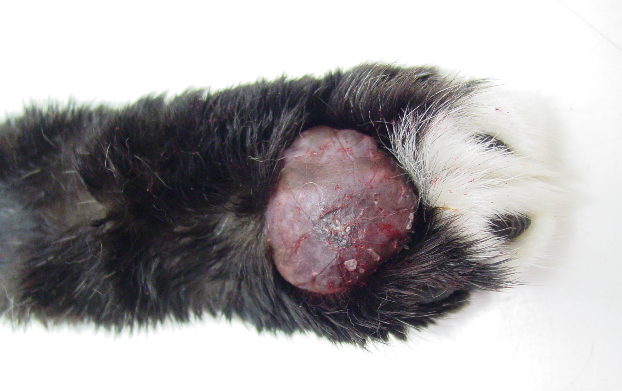
On post-mortem examination, the four footpads of the cat were markedly swollen and very soft. There was mild hyperkeratosis and the footpads were dark red to cyanotic. On cut-section, the tissue within the footpad was diffusely mildly hemorrhagic and very soft in consistency.
Laboratory results:
No blood or other fluids were taken for further analysis.
Microscopic Description:
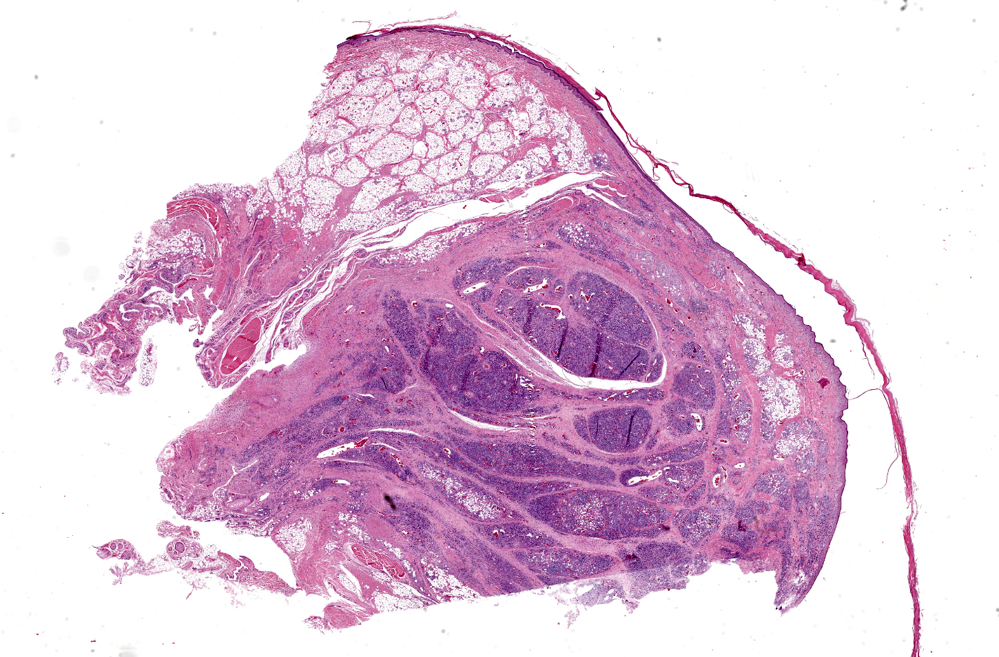
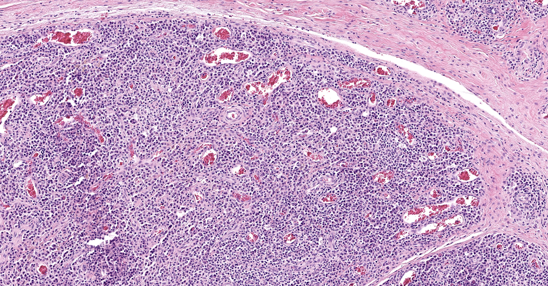
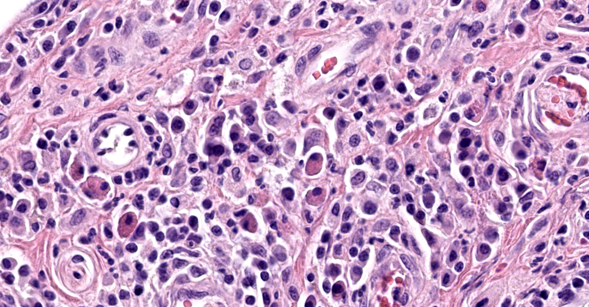
Footpad - The dermis is markedly expanded by multiple nodules and diffuse aggregates of inflammatory cells which are inconspicuously oriented around larger blood vessels and reach the epidermis and infiltrate in lobules of adipocytes. These infiltrates consist of large numbers of plasma cells which are oval-shaped, possess a moderate amount of basophilic cytoplasm and an eccentric small round nucleus (giving a 'fried-egg' appearance). Scattered among those plasma cells are many plasma cells which are swollen by a large amount of brightly eosinophilic globules (Russell bodies) in the cytoplasm, which pushes the nucleus to the periphery (Mott cells). Admixed with the plasma cells are moderate numbers of lymphocytes and macrophages. Within the dermis, there are irregular foci of necrosis with disruption of collagen fibers, cellular debris and karyorrhectic fragments. Scattered melanocytes are found in the superficial dermis (pigmentary incontinence). The epidermis is covered with multiple layers of orthokeratotic keratin.
Contributor's
Morphologic Diagnoses:
Footpad;
Plasmacytic pododermatitis, severe, multinodular, chronic with mild
orthokeratotic hyperkeratosis
Contributor's Comment
Identifying the tissue as a footpad without prior knowledge of where the tissue was taken, might be challenging, but the characteristic histological properties of this entity together with the presence of non-haired skin should guide one to the paws. Feline plasma cell pododermatitis (FPP) is a rare condition with unknown etiology. An immune-mediated pathogenesis seems plausible, due to resolution of lesions and clinical symptoms after administration of immunosuppressive drugs and marked hypergammaglobulinemia.6,8 Some patients require life-long therapy.8 Food hypersensitivity was suspected to be part of the underlying cause in some cats, as patients improved with hyposensitive diets. Seasonal waxing and waning has also been described.8 A large proportion of affected cats test positive for FIV, suggesting a possible role in the development of this entity.6 Some cats exhibit concurrent plasmacytic stomatitis, renal amyloidosis or immune-mediated glomerulonephritis.6 Unfortunately, the kidneys were not histologically examined, but had no gross abnormalities. There is no breed, sex or age predisposition.5,6,8
Affected footpads are markedly swollen and filled with a soft hemorrhagic tissue. There may be mild hyperkeratosis present. All four paws may be affected, but not necessarily. The gross and histologic appearance is practically pathognomonic. An interesting case of a different presentation of likely similar pathogenesis was found in a 7-month-old cat with a firm, haired rounded swelling on the bridge of the nose. Histologically, the lesion closely resembled FPP with perivascular to diffuse infiltrates of plasma cells. This animal did not show any signs of FPP.4
Contributing Institution:
Department of Pathology, Bacteriology and Poultry diseases
Faculty of Veterinary Medicine ? Ghent University
Salisburylaan 133
9820 Merelbeke
Belgium
http://www.ugent.be/di/en/departments?ugentid=DI05
JPC Diagnosis:
Footpad: Pododermatitis, plasmacytic, multinodular, severe.
JPC Comment:
The contributor provides a good overview of feline plasma cell pododermatitis associated literature associated with this rare condition with an unknown underlying cause. As noted within the contributor's description, this condition is often associated with Mott cells and Russell bodies, which are both known to occur in reactive and neoplastic plasma cell disorders.1,2,6
The term "Mott cell" is derived from a surgeon, F.W. Mott, who described "morular cells" (Latin: morus "mulberry) with spherical inclusions packed into the cytoplasm while describing sections of brain tissue from monkeys experimentally infected with various species of genus Trypanosoma while investigating "sleeping sickness" in 1905.7 He recognized the cells were plasma cells and indicative of chronic inflammation.7 However, the first description of these cells is most likely attributed to William Russell. Russell was a pathologist at the School of Medicine at the Royal Infirmary in Edinburgh. Thinking he had discovered the organism that caused cancer, he addressed Pathological Society of London in December 1890 and described large spherical inclusions within cells that often occupied the entire cytoplasm and compressed the nucleus. We now know these inclusions, which may occur in both plasma cell neoplasms and reactive conditions, are derived from immunoglobulin.1,2
The term "Russell body" is inconsistently used between countries and even within countries, with some pathologists using the term to only refer to a single large spherical inclusion displacing the nucleus whereas others use the term to refer to the multiple inclusions within Mott cells. Hand-drawn illustrations of Russell's article reveals he observed multiple spherical inclusions within single cells. Mott cells therefore contain Russell bodies, as reflected in the contributor's microscopic description in this case.1,2
"Immunomodulatory-responsive lymphocytic-plasmacytic pododermatits" has been proposed to denote a similar condition in domestic canines, with a report describing the condition in 20 adult dogs of various breeds. There was chronic (≥ 6 months) inflammation confined to the pedal skin in each case, with lesions present on all four feet in 18/20 cases. Lesions were histopathologically characterized by epidermal hyperplasia, hyperkeratosis, spongiosis, dermal edema, and perivascular aggregates of lymphocytes and plasma cells. Affected dogs had significantly elevated serum IgG and IgM concentrations. None of the dogs responded to antimicrobial therapy administered over an 8 week period, had evidence of ectoparasitism, satisfied criteria for atopic dermatitis, nor responded to a dietary trial using a novel protein source. Each dog responded to immunosuppressive doses of prednisone or cyclosporine.3
During the conference, the moderator noted areas within the section with scant refractile anisotropic material associated with epithelioid macrophages and rare multinucleated giant cells. Although advanced diagnostics are required for confirmation, this is a common observation in cases of feline plasma cell pododermatitis and is consistent with a local foreign body reaction to embedded cat litter.
References:
1. Bain BJ. Russell bodies. Am J Hematol. 2009;84(7):439.
2. Bain BJ. Russell bodies and Mott cells. Am J Hematol. 2009;84(8):516.
3. Breathnach RM, Baker KP, Quinn PJ, McGeady TA, Aherne CM, Jones BR. Clinical, immunological and histopathological findings in a subpopulation of dogs with pododermatitis. Vet Dermatol. 2005;16(6):364-372.
4. Declerq J, Debosschere H, Nasal swelling due to plasma cell infiltrate in a cat without plasma cell pododermatitis. Veterinary Dermatology; 2010;21;412-4
5. Dias Pereira P, Faustino AM, Feline plasma cell pododermatitis: a study of 8 cases. Veterinary Dermatology; 2003;14(6);333-7.
6. Mauldin EA, Peters-Kennedy J. Integumentary system. In: Maxie MG ed. Jubb, Kennedy, and Palmer's Pathology of Domestic Animals. Vol 1. 6th ed. Philadelphia, PA: Elsevier Ltd. 2016: 613-614.
7. Mott FW. Observations on the brains of men and animals infected with various forms of trypanosomes. Preliminary note. Proceedings of the Royal Society of London Series B-Containing Papers of a Biological Character. 1905;76:235?242.
8. Werner A, Zetwo A, Feline Plasma Cell Pododermatitis, Clinican's Brief; 2015