Signalment:
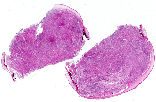
Gross Description:
Histopathologic Description:
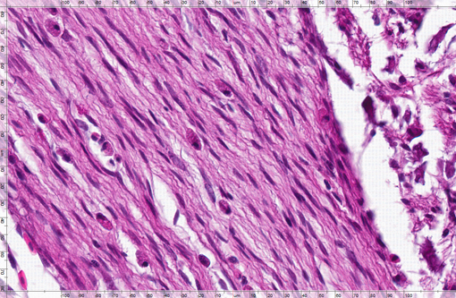
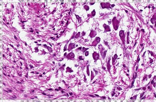
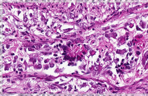
109.006781 Right eye: One section of completely excised tissue is examined. The architecture of the iris, ciliary body and lens is diffusely effaced by a poorly circumscribed population of neoplastic cells that fills the vitreous body and protrudes into the anterior chamber. Neoplastic cells are dimorphic: the first population is spindloid with tapered cell borders, a moderate amount of fibrillar eosinophilic cytoplasm and a large elongate nucleus with stippled chromatin. These neoplastic cells are arranged in thick interlacing fascicles. Interspersed within this population of neoplastic spindle cells, there is a subpopulation of polyhedral to stellate neoplastic cells with abundant eosinophilic cytoplasm, a large eccentric nucleus with a fine chromatin pattern and prominent eosinophilic nucleolus. Mitotic figures average three per ten high power fields for all neoplastic cell types. Neoplastic cells invade the lens capsule and disrupt the lens, and there is moderate histiocytic and lymphoplasmacytic inflammation surrounding the lens fragments.
Morphologic Diagnosis:
Condition:
Contributor Comment:
Immunohistochemical staining with GFAP and vimentin was performed. Few foci of cellular processes within the spindloid cell population in the neoplasm were positive for GFAP, and there was positive internal control staining within the optic nerve. Vimentin staining, however, was negative for neuronal and spindle cells and there was no positive internal control staining within the optic nerve or elsewhere in the tissue.
JPC Diagnosis:
Conference Comment:
Conference participants felt that there were three distinct cell populations in this particular neoplasm, including the spindle cells, ganglion cells, and neuroblast cells with carrot-shaped nuclei that formed scattered rosettes. For this reason, we favor a diagnosis of ganglioneuroblastoma. Ganglioneuroblastomas exhibit histologic features of both well-differentiated neurons and neuroblastic cells(4). Conference participants also felt this neoplasm was malignant due to its locally invasive nature.
Additional differential diagnoses considered for this neoplasm include medulloepithelioma, melanoma, and retinoblastoma. Retinoblastomas present with numerous neuroblasts arranged in rosettes and can be easily confused with medulloepitheliomas in fish, except they are generally not as invasive and do not form heteroplastic elements such as undifferentiated mesenchymal cells, bone, or cartilage(7).
There was some variation among sections due to the submission of both eyes, and some conference participants had evidence of retinal detachment and atrophy, as well as a lenticular cataract with Morgagnian globules in the lens. There were also areas of woven bone surrounding some sections of the cornea, and conference participants hypothesized that this could represent osseous metaplasia from the neoplasm or from exposed lens fibers.
S-100, neurofilament protein (NFP), synaptophysin, chromagranin A, and glial fibrillary acidic protein (GFAP) immunohistochemical stains were performed on this case, but all were non-contributory, which is likely due to the fact that the immunohistochemical stains at the Joint Pathology Center laboratory are not optimized for piscine tissues.
References:
2 Hawkins WE, Overstreet RM, Fournie JW, Walker WW. Development of aquarium fish models for environmental carcinogenesis: tumor induction in seven species. J Appl Toxicol. 5: 261-264, 1985.
3 Hawkins WE, Fournie JW, Overstreet RM, Walker WW. Intraocular neoplasms induced by methylazoxymethanol acetate in Japanses medaka (Oryzias latipes.) J Natnl Cancer Inst. 76: 453-65, 1986.
4 Maxie MG, Youssef S. Nervous system. In: Maxie, MG, ed. Jubb, Kennedy, and Palmers Pathology of Domestic Animals. 5th ed., Philadelphia, PA: Elsevier; 2007:372, 453-455.
5 Porter BF, Storts RW, Payne HR, Edwards JF. Colonic ganglioneuromatosis in a horse. Vet Pathol 44: 207-210, 2007.
6 Roberts RJ. In: Fish Pathology, 2nd ed., p 169. Bailliere Tindall, London, England, 1989.
7 Roberts RJ. Neoplasia of teleosts. In: Roberts RJ, ed. Fish Pathology. 3rd ed. Edinburgh, Scotland: Saunders; 2003:166-7.