Signalment:
Three-year-old male rabbit (
Oryctolagus cuniculus).The animal presented two days of anorexia and lack of defecation.
At physical examination, a single mass was detected by palpation at the right
lumbar area. The presence of a round-shaped mass associated to the caudal pole
of the right kidney was confirmed by ultrasonography. Clinicians suspected a
neoplastic process, and a right nephrectomy was performed.
Gross Description:
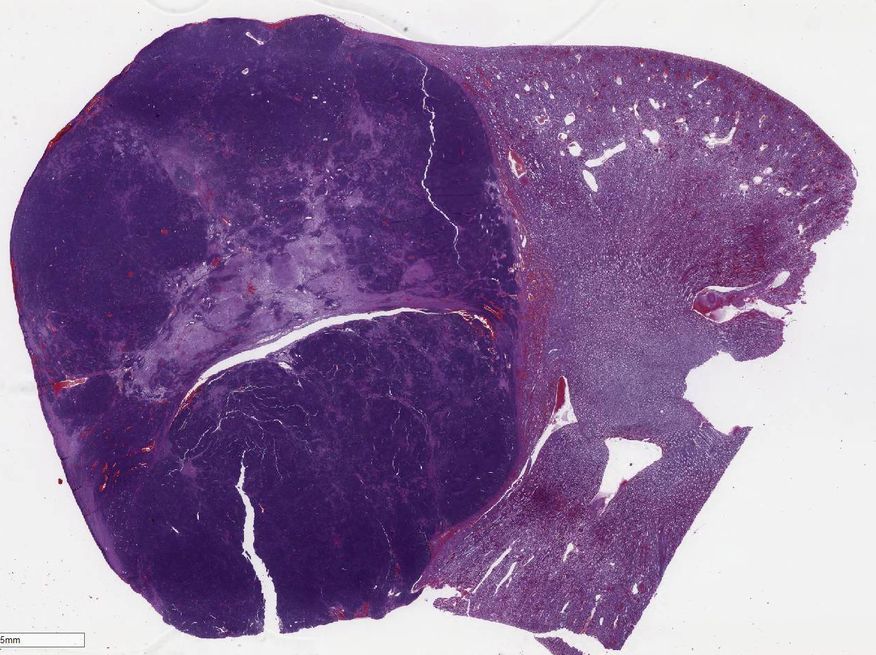
The complete right kidney was submitted
for histopathologic evaluation. A well-circumscribed, oval white mass, 2x1.5 cm
in diameter was observed at the caudal pole of the kidney. On section, its
consistency was soft, and the cut surface was white and homogeneous.
Histopathologic Description:
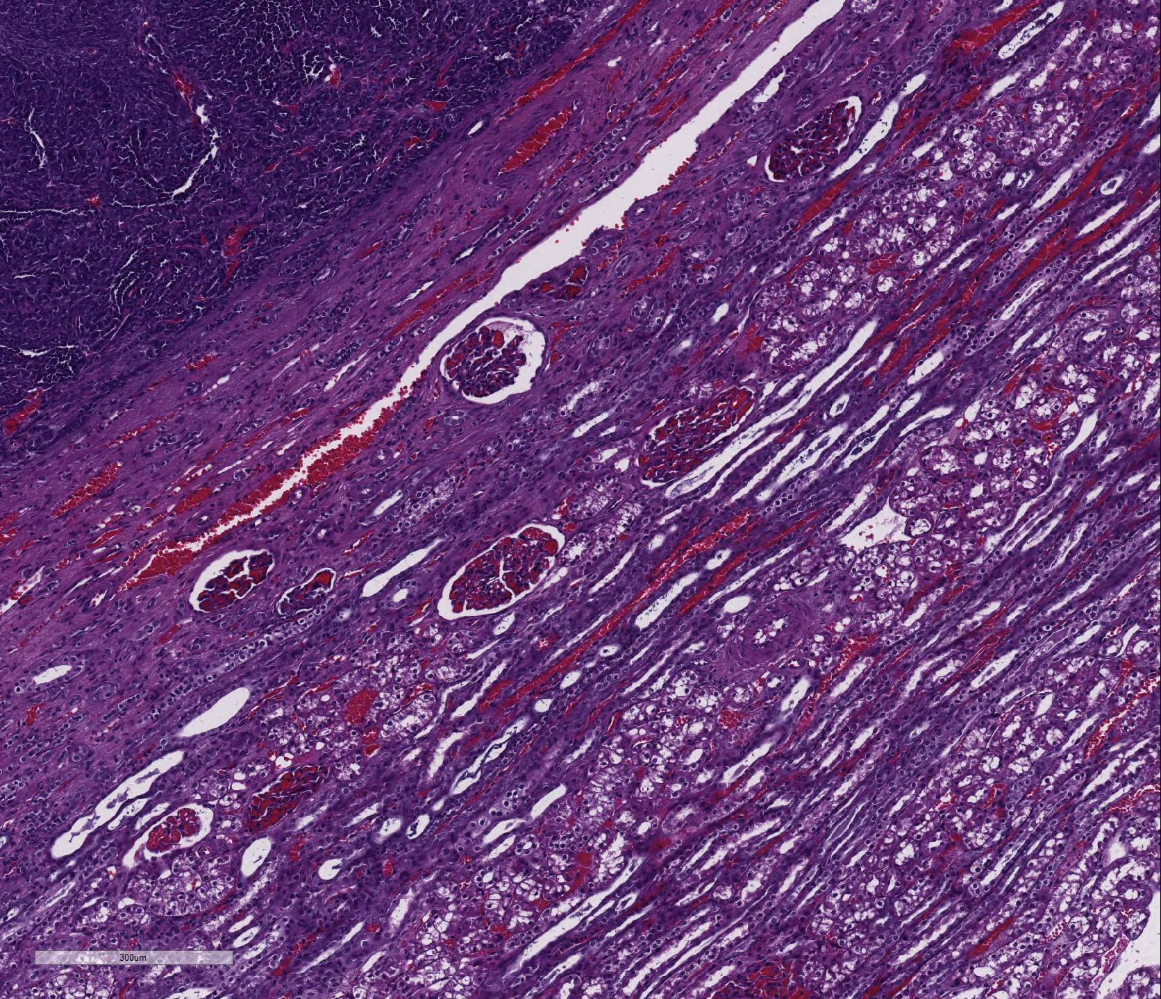
Kidney:
Replacing 50-75% of the renal tissue, there is a densely cellular, partially encapsulated
but infiltrative nodular neoplasm which compresses adjacent parenchyma. The
neoplasm is composed of a disorganized mixture of epithelial, mesenchymal, and
blastemal components supported by an abundant fibrovascular stroma which, in
some areas, is embedded in a loose, pale eosinophilic, collagenous matrix. The
mesenchymal and blastemal components are predominant over the epithelial.
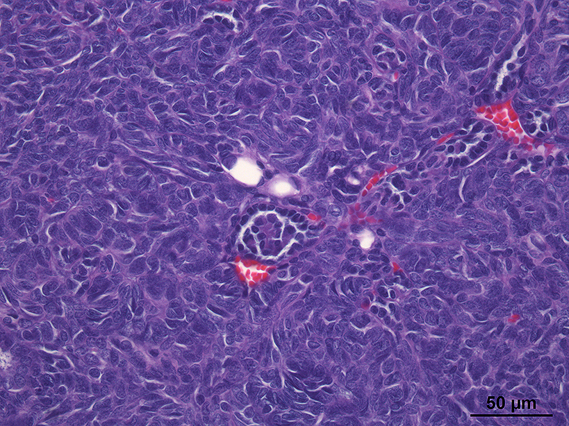
Epithelial neoplastic cells are cuboidal to cylindrical and arranged in a
tubular pattern. Occasionally, there are very scant tuft invaginations into the
tubular lumen lined by flat epithelial cells (primitive glomerular structures).
Those cells present well-defined cytoplasmic borders and moderate amount of
bright eosinophilic cytoplasm. Nuclei are round, irregularly shaped, mostly
basally located, with grossly stippled chromatin and less frequently, finely
stippled chromatin with one basophilic nucleolus. There is low-grade
anisokaryosis and anisocytosis, and low mitotic index (1/40X HPF). The
mesenchymal component is arranged in a storiform pattern or disorganized
bundles. Cells are fusiform, with an indistinct cytoplasmic border, scant
eosinophilic cytoplasm, and fusiform centrally located nuclei with grossly
stippled chromatin. There is a mild anisokaryosis and aniso-cytosis, and a low
mitotic index (0-1/40X HPF). Some of these cells, organized in nest or ribbons,
which are poorly differentiated are identified as possible blastemal cells. Multifocally, in the
neoplastic mass there are areas of hypereosinophilia and loss of cytoplasmic
and nuclear detail (coagulative necrosis) and mild lymphoplasmacytic
inflammatory infiltrate is observed among the neoplasia. The adjacent renal
parenchyma shows a moderate interstitial fibrosis, and intense congestion.
Morphologic Diagnosis:
Renal nephroblastoma.
Lab Results:
None
Condition:
Nephroblastoma
Contributor Comment:
Nephroblastoma, or Wilms´ tumor, is the most common primary renal
tumor of pigs and chickens, and occurs far less often in calves and dogs. It is
frequently observed in young animals, although it may be seen in mature sows,
and is more common in adult dogs than in pups. Clinically, it is usually an incidental
finding except in dogs, in which some animals present with spinal dysfunction
as result of compression of the spinal cord associated with neoplastic
infiltration into the vertebral canal. Macroscopically, it is usually a solitary
unilateral mass located at one pole of the kidney, but it can be multiple and/or
bilateral. Nephroblastoma is a true embryonal tumor that arises from primitive
metanephric blastema and exhibits blastemal, epithelial, and stromal components
in variable proportions. Histologically, it is characterized by the presence of
primitive glomeruli, abortive tubules, and loose spindle-cell stroma that may
differentiate into a variety of mesen-chymal tissues.
Tubular and glomerular differentiation indicate a good prognosis,
whereas anaplasia and sarcomatous stroma are associated with metastasis and a
poor prognosis. In this case, the epithelial component is scant and poorly
differentiated, and there is a clear predominance of undifferentiated mesen-chymal
cells. These features, in combination with the neoplasms invasive growth pattern,
suggest a poor prognosis. The differential diagnosis for renal neoplasia in a rabbit
includes renal carcinoma and renal lymphosarcoma. To
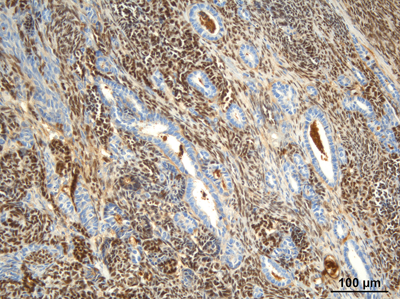
confirm the diagnosis of nephroblastoma, immuno-histochemistry was performed
against Wilm's tumor suppressor (WT1),
which has proven efficacious in dogs. This immunohistochemistry was kindly
provided by Dr. Joan Carles Ferreres, head of the Pathology Department at the
Parc Taulí Sabadell University Hospital. In this case, positive nuclear immunoreactivity
was observed in the mesenchymal and blastemal components, as well as
endothelial cells.
JPC Diagnosis:
Kidney: Nephroblastoma, rabbit,
Oryctolagus cuniculus.
Conference Comment:
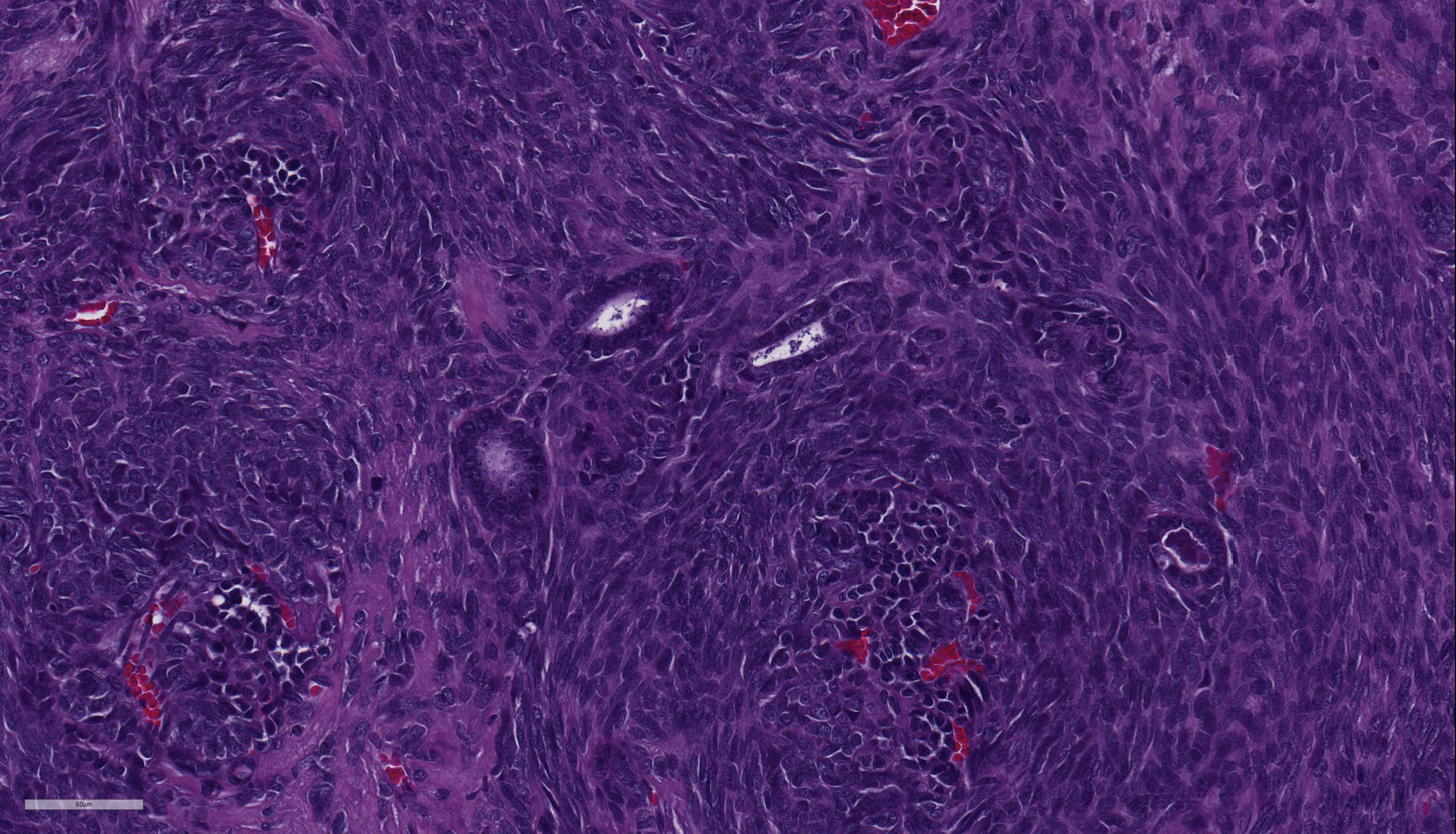
This
case nicely demonstrates the blastemal, epithelial, and stromal components
histologically characteristic for nephroblastomas. Conference participants
readily identified the presence of scattered primitive glomeruli (formed by
infolded papillary projections of polygonal cells within tubule lumina), as
well as low numbers of tortuous, abortive tubules admixed with numerous
blastemal cells and surrounded by an abundant loose spindle-cell stroma. Additionally,
attendees noted several individualized well-differentiated renal tubules
scattered throughout the neoplasm. Conference participants agreed that these
likely represent entrapped non-neoplastic renal tubules within the neoplastic
cell population rather than part of the epithelial component of the neoplasm. There
is moderate slide variability, affecting the proportion of the three components
present. When all three cell types are present in equivalent proportions, the
neoplasm is referred to as triphasic or mixed;
7 however, in this case
there appears to be a preponderance of spindle-cell stromal and blastemal components.
Although not a prominent feature in this case, the spindle-cell stroma
can occasionally differentiate into various types of mesenchymal tissue, such
as striated muscle, collagen, adipose tissue, bone, or cartilage.
6,7
Nephroblastoma rarely causes
clinical signs, although polycythemia is an infrequently reported paraneoplastic
syndrome in rabbits.
2 The vast majority of nephro-blastomas are considered
incidental findings and are noted during necropsy as a solitary (or
occasionally bilateral) renal masses.
1 However, as mentioned by the
contributor, when this neoplasm is characterized predominantly by a poorly
differentiated sarcomatous stromal component, as in this case, it tends to metastasize
to the lung and liver, portending an overall poor prognosis.
7
While nephroblastomas are a
relatively uncommon spontaneous neoplasm in pet rabbits, they can be rapidly
induced at a high frequency in laboratory rabbits and rats after exposure to
direct-acting alkylating carcinogens, such as dimethylnitrosamine.
6
Dimethylnitrosamine has also been reported to experimentally induce triphasic nephro-blastomas in rainbow trout, similar to Wilms tumor in humans.
3
Other than lymphoma, nephroblastomas are the most common primary renal neoplasm
in a variety of species of freshwater fish, with the Japanese eel
over-represented. Research is ongoing to determine if potential carcinogens in
the water may be inducing nephroblastomas in wild fish, with a particular focus
on fish commonly consumed by people.
3
References:
1. Cianciolo RE, Mohr FC. The urinary system. In: Maxie MG ed.
Jubb
Kennedy and Palmer's Pathology of Domestic Animals. Vol 2. 6th ed.
Philadelphia, PA: Elsevier Saunders; 2016:446-447.
2. Cooper TK, Griffith JW, et al. Spontaneous lung lesions in aging
laboratory rabbits (
Oryctolagus cuniculus).
Vet Pathol. 2017;
54(1):178-187.
3. De Lorenzi D, Baroni M, Mandara MT. A
true "triphasic" pattern: Thoracolumbar spinal tumor in a young dog.
Vet
Clin Pathol. 2007; 36:200-203.
4. Hassan J, Katic N, et al. Treatment of nephroblastoma with poly-cythemia
by nephrectomy in a rabbit.
Vet Rec. 2012; 170:465.
5. Lombardini ED, Hard GC, Hashbarger JC. Neoplasms in the urinary
tract in fish.
Vet Pathol. 2014; 51(5):1000-1012.
6. Meuten DJ. Tumours of the urinary system. In: Meuten DJ, ed.
Tumors
in Domestic Animals. Ames, IA: Iowa State Press; 2002:519-520.
7. Newman SJ. The urinary system. In: McGavin MD, Zachary JF, ed.
Pathologic
Basis of Veterinary Disease. 5th ed. St Louis, MO: Elsevier Mosby; 2012:
643.