Signalment:
8-year-old neutered male Cairn terrier, (
Canis familiars).Surgical extirpation of the bulbus, further clinical data not available.
Gross Description:
None
Histopathologic Description:
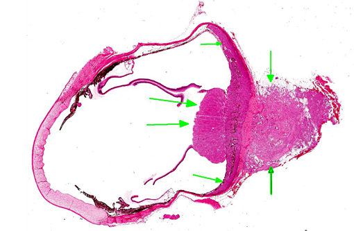
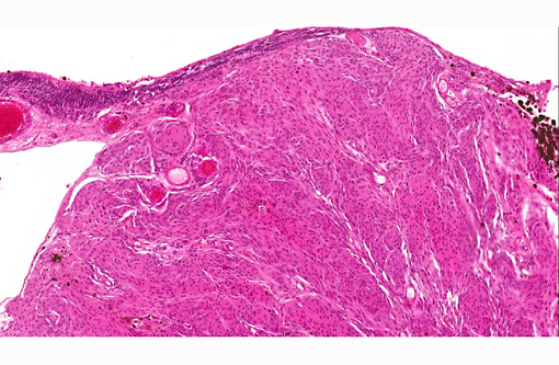
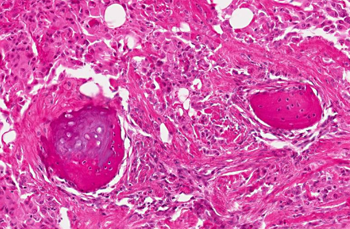
Replacing, infiltrating and expanding the optic nerve disc and proximal parts of the optic nerve origin is a moderately circumscribed, densely cellular neoplasm extending bilaterally into the choroid and the retrobulbar adipose tissue. The mass is separated by a thin fibrovascular stroma into nests of closely packed predominantly epithelioid cells or spindle to polygonal cells arranged in loosely interlacing streams and whorls. In areas extending into the choroid variable numbers of melanin-containing cells are intermingled in the stroma. Neoplastic cells have variably distinct borders with varying amounts of eosinophilic cytoplasm, oval to spindle-shaped and occasionally vesicular nuclei with finely stippled chromatin and predominantly one distinct nucleolus. Epithelioid cells forming small nests often show abundant eosinophilic cytoplasm and eccentric nuclei. Mitotic rate is less than one mitotic figure per high power field. Multifocally, especially in extrabulbar parts of the mass are foci of myxoid, cartilaginous or osseous metaplasia. Multifocally, mainly perivascularly, there are few lymphocytes, plasma cells, neutrophils and intermingled with some pigment (hemosiderin) laden macrophages.
Additionally, segments of the retina adjacent to the tumor are mildly atrophic.
Morphologic Diagnosis:
Eye, optic nerve: Meningioma, optic nerve type, canine.
Condition:
Optic nerve meningioma
Contributor Comment:
In dogs and cats, meningiomas are the most common primary tumor arising in the nervous system.(7) In the dog they occur more commonly in the brain than in the spinal cord(4,7) Those arising in the brain are often localized over the convexities, attached to the falx or the tentorium cerebelli or inside the ventricular system. Rarely they occur retrobulbarly.(3,7) In general intracranial meningiomas are slow growing, discrete, expansile neoplasms and malignant behavior or extracranial metastases are only rarely reported.(1,6) Due to the embryonic origin of the meninges from both mesoderm and neural crest, they can undergo mesenchymal and epithelial differentiation and show highly variable morphological patterns.(7) The current World Health Organization (WHO) classification of tumors of the nervous system in domestic animals(2) describes nine histological patterns: meningothelial, fibrous (fibrobroblastic), transitional (mixed), psammomatous, angiomatous, papillary, granular cell, myxoid and anaplastic (malignant). Montoliu et al. reviewed 30 cases of meningiomas and divided into transitional, meningothelial, psammomatous, anaplastic, fibroblastic, angioblastic, papillary, microcystic and those types arising from the optic nerve.(5) The latter were characterized predominantly by meningothelial or transitional patterns and contained multiple areas of myxoid, cartilaginous and osseous metaplasia. Because of this distinctive morphology they suggested to include optic nerve meningiomas into the classification as a separate entity. The case presented here qualifies for this type due to localization and histological appearance.
Immunohistochemically, most of the cases stain positive for vimentin, less often also for cytokeratin, pointing to a more mesenchymal and less epithelial differentiation. Expression of S-100 protein and neuron-specific enolase (NSE) is inconstant and rarely a predominant feature.(5)
JPC Diagnosis:
Eye, optic nerve: Meningioma with acute neural retinal detachment.
Conference Comment:
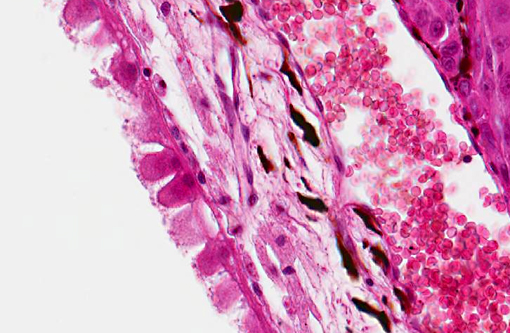
The contributor provides an excellent summary of canine intra- and extracranial meningioma. In addition to the distinct neoplastic features described above, participants observed a striking separation of the retinal pigment epithelium (RPE) from the photoreceptors (i.e., neural retina) with diffuse, marked hypertrophy (tombstoning) and multifocal cystic degeneration of the RPE, as well as pockets of proteinaceous fluid and few neutrophils within the subneural retinal space. There is also mild retinal atrophy, as noted by the contributor. These histopathological findings support an additional diagnosis of neural retinal detachment, which refers specifically to a separation between the neural retina and the RPE. Neural retinal detachment was likely secondary to locally infiltrative neoplastic cells cleaving photoreceptors from their interdigitations with the RPE. Conversely, artifactual retinal separation is a common sequela of formalin fixation, and is distinguished from pathologic retinal detachment by the absence of RPE hypertrophy and absence of fluid and/or inflammatory cells in the subneural retinal space.(8,9)
References:
1. Dugan SJ, Schwarz PD, Roberts SM, Ching SV. Primary optic-nerve meningioma and pulmonary metastasis in a dog. Journal of the American Animal Hospital Association. 1993;29:11-16.
2. Koestner A, Bilzer T, Fatzer R, Schulman FY, Summers BA, van Winkle TJ. World Health Organization: Histological Classification of Tumors of the Nervous System of Domestic Animals. Washington, DC: Armed Forces Institute of Pathology; 1999.
3. Mauldin EA, Deehr AJ, Hertzke D, Dubielzig RR. Canine orbital meningiomas: a review of 22 cases. Vet Ophthalmol. 2000;3:11-16.
4. Meuten DJ. Tumors in Domestic Animals. 4th ed. Ames, IA: Iowa State University Press; 2002:717-723, 753.
5. Montoliu P, Anor S, Vidal E, Pumarola M. Histological and immunohistochemical study of 30 cases of canine meningioma. J Comp Pathol. 2006;135:200-207.
6. Schulman FY, Ribas JL, Carpenter JL, Sisson AF, LeCouteur RA. Intracranial meningioma with pulmonary metastasis in three dogs. Vet Pathol. 1992;29:196-202.
7. Summers BA, Cummings JF, de Lahunta A. Veterinary neuropathology. St. Louis, MO: Mosby; 1995:355-362.
8. Wilcock BP. Eye and ear. In: Maxie MG, ed. Jubb, Kennedy and Palmers Pathology of Domestic Animals. Vol 1. 5th ed. Philadelphia, PA: Elsevier; 2007:518.
9. Yanoff M, Sassani JW, eds. Ocular Pathology. 6th ed. London, UK: Mosby Elsevier; 2009:395, 462-463.