Signalment:
Gross Description:
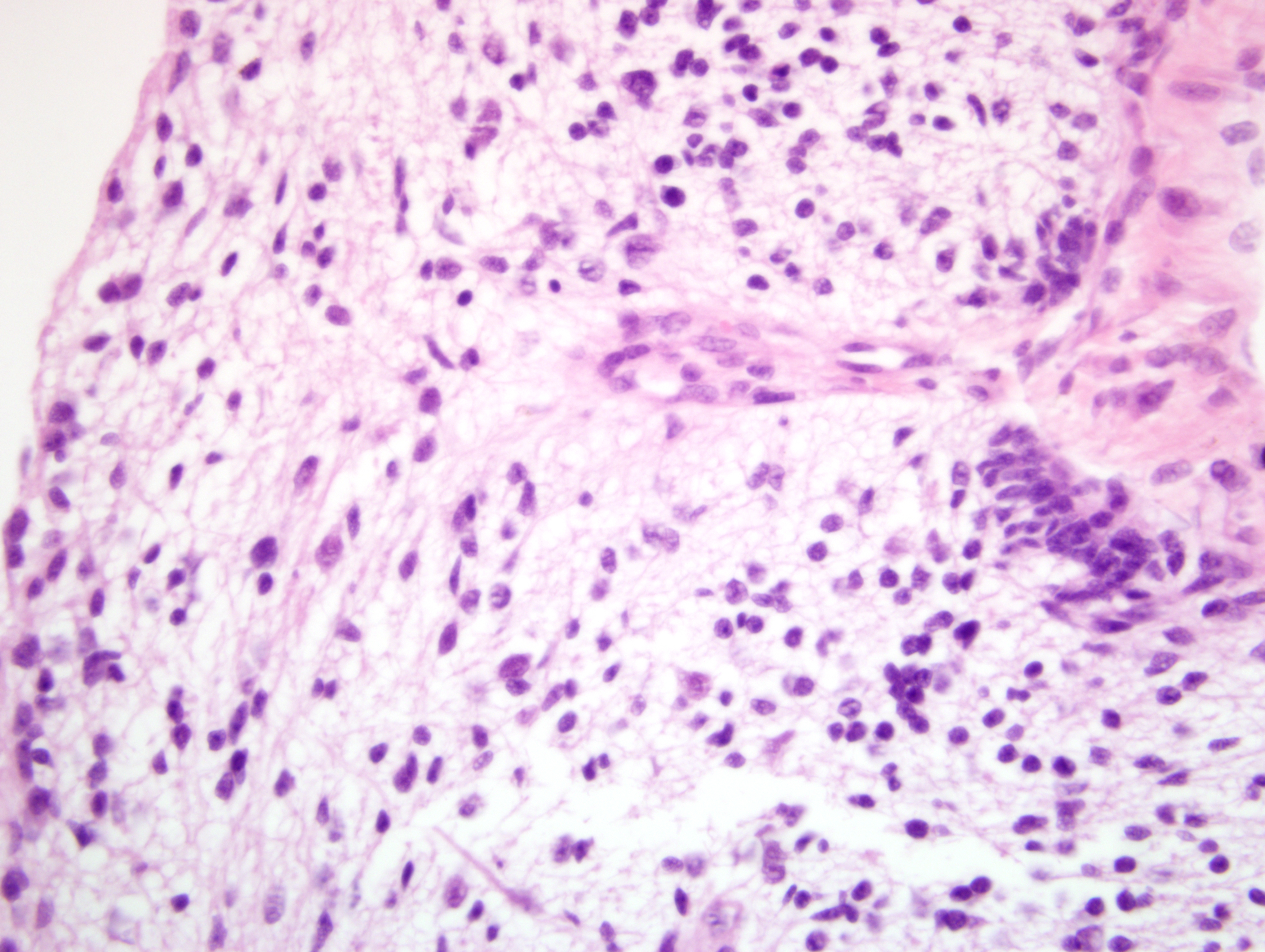
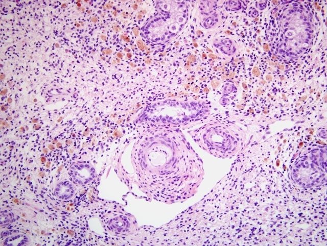
Histopathologic Description:
Morphologic Diagnosis:
Condition:
Contributor Comment:
Teratomas of the testis are the most frequently reported testicular tumor in young horses and are more commonly found in cryptorchid testes.(1) To the contributors knowledge, there are two published reports in horses less than a year old, one in a cryptorchid, neonatal 3 day old foal(5) and the other a cryptorchid 4-day-old foal,(4) in which the spermatic cord entrapped the small colon resulting in signs of abdominal pain. It is thought that testicular teratomas may be congenital(4) and that their presence in a fetal testis may prevent normal descent,(1,4) although this may depend on the size of the tumor relative to the size of the inguinal canal at the time of testicular descent.(4) There have been no reports, to the contributors knowledge, of bilateral testicular teratomas in horses.
Grossly, teratomas may be single or multiple and often have a cystic or multilobular structure. Hair and mucoid or sebaceous-like secretions are often seen on cut section (and these are sometimes referred to as dermoid cysts) as well as yellow-white masses with fibrous, adipose, cartilaginous, and bony tissue.(1) Histologically, teratomas are composed of structures derived from all embryonic germ layers, including ectodermal, neuroectodermal, endodermal, or mesodermal.(1) The presence of nervous and adipose tissue is very common.
The remainder of the testicular parenchyma in this foal is normal for the age of the horse. The seminiferous tubules are immature, as spermatogenesis is not initiated until approximately 2 years of age.(2) From a fetal gestational age of 155 days to 1 year old, the seminiferous epithelium consists of gonocytes and Sertoli cells. Another interesting feature of these testes was the dark-brown discoloration grossly, and the abundant pigmented interstitial cells present around immature seminiferous tubules histologically. This is also a normal feature for testes from a foal of this age (3 months). These pigmented cells are not present between fetal gestational ages 155 days to 248 days, but are present in large numbers in the neonatal 3-day-old testis. They increase to reach a maximum number at 2 months of age, and then gradually diminish and disappear by 3 years of age.(2) A morphologic study on these pigmented cells of the horse testis found that ultrastructurally, they were characterized by the presence of abundant residual bodies consisting of secondary lysosomes, having variable internal structures, and an eccentrically located nucleus.(3) In combination with histochemical staining, it was suggested that the pigmented granules found within the pigmented cells have some characteristics of ceroid. It is hypothesized that these pigmented cells may be derived from macrophages which are phagocytizing the degenerating fetal type of interstitial cells, and that they gradually increase in volume by storing digested materials as ceroid-like pigment.(2,3)
JPC Diagnosis:
Conference Comment:
While gonadal teratomas are uncommon, still rarer are the extragonadal teratomas, the most common form of which is the dentigerous cyst, usually found at the base of the ear in horses. Extragonadal teratomas are well-documented in humans, and sporadically reported in other domestic, wild, and laboratory species. Specifically, adrenal teratomas have been reported in humans, ferrets, an ox, and a rat. Interestingly, extragonadal teratomas are thought to arise from diploid pluripotent progentior cells that escape embryonal organizers during maturation, in divergence from the histogenesis of gonadal teratomas described by the contributor.(7)
Of equal or perhaps greater interest are the pigmented interstitial cells, which garnered the bulk of the discussion during the conference, and remain fairly enigmatic. What little is known about these cells is well-summarized by the contributor; from a diagnostic standpoint, it is important to recognize them as normal structures in the neonatal foal testis.
References:
2. Hondo E, Murabayashi H, Hoshiba H, Kitamura N, Yamanouchi K, Nambo Y, Kobayashi T, Kurohmaru M, Yamada J: Morphological studies on testicular development in the horse. J Reprod Dev 44:377-383, 1998
3. Murabayashi H, Hondo E, Kitamura N, Furuoka H, Taguchi K, Nambo Y, Yamada J: Morphological study on pigmented cells in the horse testis. J Vet Med Sci 61:1183-1186, 1999
4. Parks AH, Wyn-Jones G, Cox JE, Newsholme BJ: Partial obstruction of the small colon associated with an abdominal testicular teratoma in a foal. Equine Vet J 18:342-343, 1986
5. Pollock PJ, Prendergast M, Callanan JJ, Skelly C: Testicular teratoma in a three-day-old foal. Vet Rec 150:348-350, 2002
6. Schlafer DH, Miller RB: Female genital system. In: Jubb, Kennedy, and Palmer's Pathology of Domestic Animals, ed. Maxie MG, 5th ed., vol. 3, pp. 453-454. Saunders Elsevier, Philadelphia, PA, 2007
7. Williams BH, Yantis LD, Craig SL, Geske RS, Li X, Nye R: Adrenal teratoma in four domestic ferrets (Mustela putorius furo). Vet Pathol 38:328-331, 2001