CASE II:
Signalment
adult, female, water buffalo (Bubalus bubalis)
History
One of three water buffaloes found dead peracutely in a herd of 14 animals was submitted for necropsy.
Gross Pathology
Tongue: Severe, multifocal to coalescing erosions and ulcerations affecting approximately 30% of the dorsal lingual mucosa. Rumen: Severe, multifocal to coalescing erosions and ulcerations, predominantly affecting the ruminal pillars. Heart: Multifocal, pale, streaky discoloration of the myocardium. Other: Generalized congestion, cyanosis, and pulmonary edema.
Laboratory Results
PCR: BTV, BVDV, BHV-1, OvHV-2 negative FMDV positive
Microscopic Description
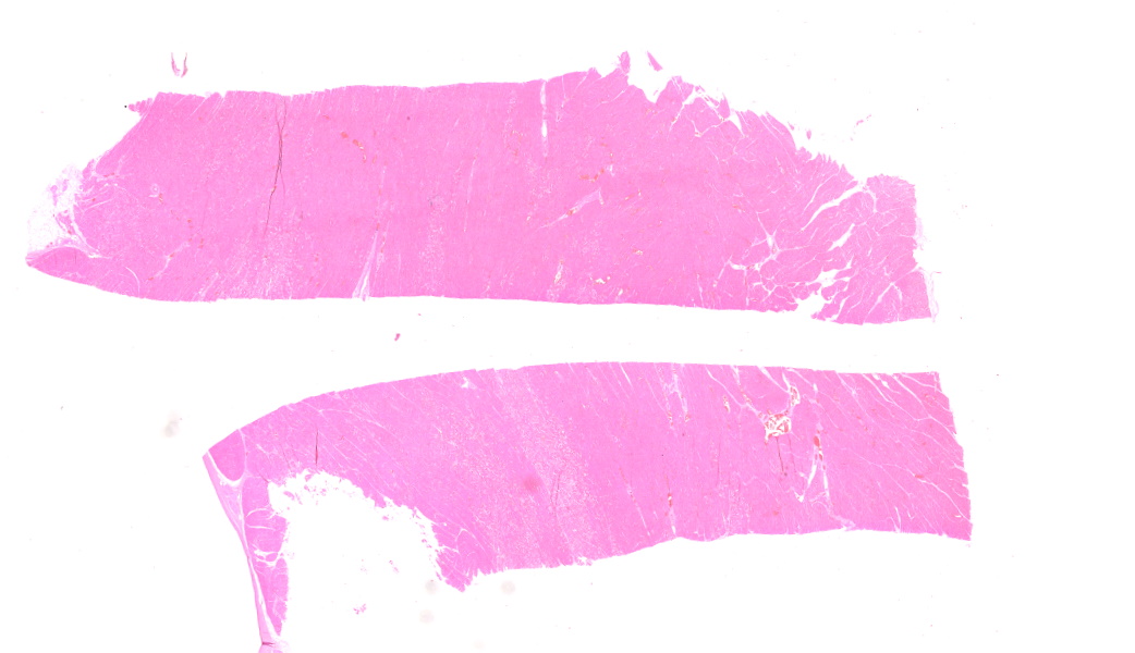
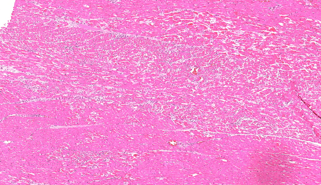
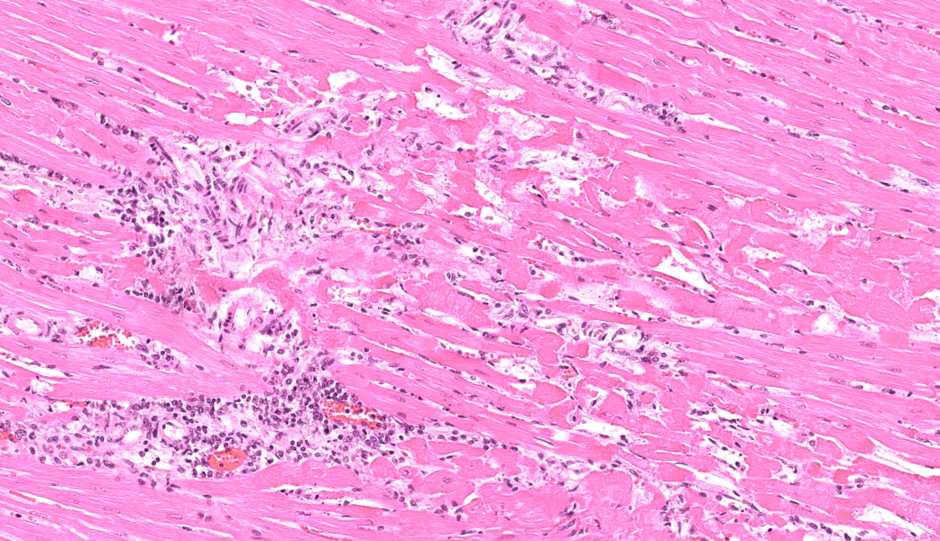
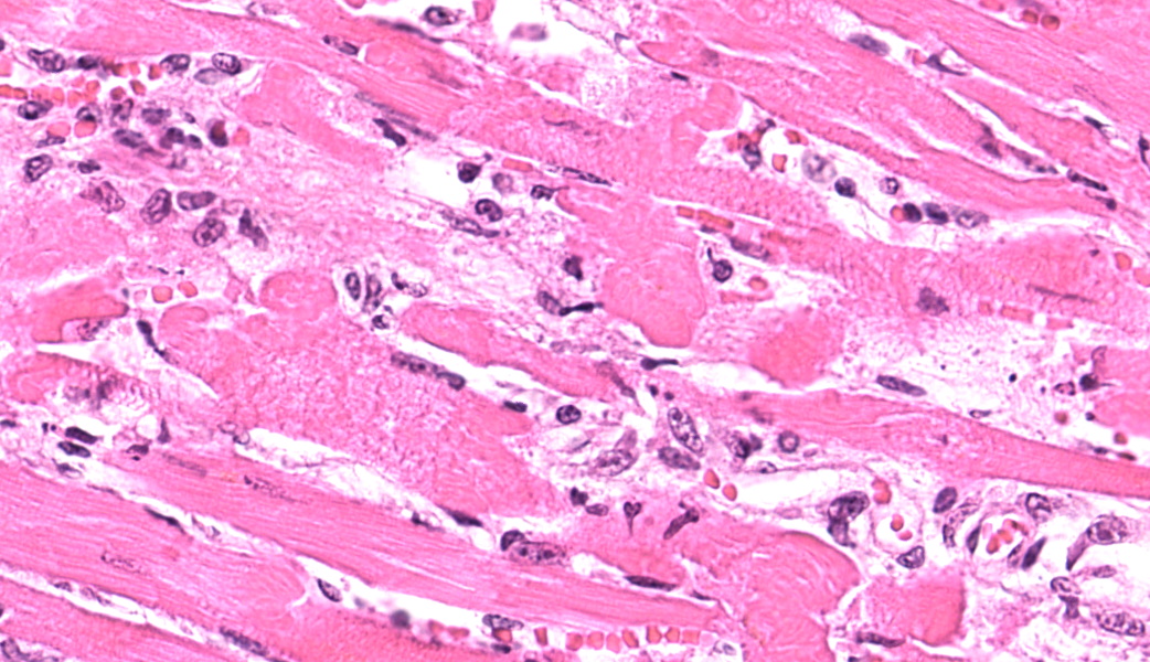
At low magnification, multifocal areas of pallor correspond to the grossly observed streaks within the myocardium. At higher magnification, there is multifocal degeneration and necrosis of cardiomyocytes, characterized by hypereosinophilia, loss of cross striations, and occasional fragmentation. Associated with necrotic myocardial cells, there is mild to moderate inflammatory infiltration composed mainly of histiocytes and fewer lymphocytes. The myocardial interstitium is mildly expanded by edema. Immunohistochemistry reveals intracytoplasmic labeling of cardiomyocytes for FMDV antigen.
Contributor's Morphologic Diagnoses:
Myocardial degeneration and necrosis, acute, multifocal, with mild lymphohistiocytic myocarditis
Contributor's Comment:
The foot-and-mouth disease virus (FMDV), a member of the genus Aphthovirus within the family Picornaviridae, causes a highly contagious disease of cloven-hoofed animals and solipeds. Seven different serotypes are recognized: O, A, C, Asia 1, SAT1, SAT2, and SAT3.1
Transmission most commonly occurs through direct contact between infected and susceptible animals, but can also occur via contaminated materials, including feed.2 Clinical signs vary depending on age and species; however, characteristic findings include fever and the formation of vesicles (aphthae) that progress to erosions and ulcerations on the oral mucosa, tongue, hooves, and udder.3
In addition to epithelial lesions, FMDV can cause degeneration and necrosis of cardiomyocytes due to viral replication within these cells. This may result in the characteristic "tiger heart" appearance, caused by streaks of pale myocardial fibers, as observed grossly in this case. Myocardial involvement is most common in young animals but may also occur in adults and can lead to peracute death without preceding clinical signs. The pathogenesis of FMDV-associated myotropism is not fully understood but is thought to involve a combination of viral and host factors, including age-related susceptibility.3 For in vivo diagnosis, measurement of serum cardiac troponin I levels can be useful, as this biomarker is specific for myocardial cell injury.4 Histopathologically, myocardial lesions vary with age and disease progression. In young animals (e.g., calves, piglets, and neonatal lambs), cardiomyocyte degeneration and necrosis often occur with minimal inflammatory response. In contrast, older animals more commonly develop myocarditis characterized by lymphohistiocytic inflammatory infiltrates, as seen in this case.5
Differential diagnoses for myocardial degeneration and necrosis in ruminants include nutritional myodegeneration (vitamin E/selenium deficiency), toxic cardiomyopathies, and infectious causes. Bacterial agents (e.g., Clostridium spp., Histophilus somni) and parasitic infections (e.g., Sarcocystis spp., Neospora caninum) are more common, whereas viral myocarditis is comparatively rare. In addition to FMDV, encephalomyocarditis virus should also be considered.5
Contributing Institution
Department of Experimental Facilities and Biosafety, Friedrich-Loeffler-Institute https://www.fli.de/en/institutes/department-of-experimental-animal-facilities-and-biorisk-management-atb/
JPC Diagnoses
Heart: Cardiomyocyte degeneration and necrosis, subacute, multifocal to coalescing, with mild multifocal lymphohistiocytic myocarditis.
JPC Comment
The trifecta of foot and mouth disease, a water buffalo, and our longstanding contributor, the Fredrich Loeffler Institute may seem like an unusual combination, so a bit of explanation is in order. In 2024, three individuals in a herd of 14 water buffalo in Brandenburg, Germany died within 48 hours.6 The owner transported one carcass to the state diagnostic lab, at which loss of approximately 1/3 of the lingual epithelium, necrotizing myocarditis and pulmonary edema were identified, and tissues were positive for FMD RNA by real-time PCR (and were corroborated by the FRG National Reference Laboratory. Immediately following these results, the remainder of the herd and 261 susceptible animals (245 pigs, 13 sheep and 3 goats) in three nearby farms and 61 additional susceptible animals (3 cattle, 39 sheep, 19 goats) at a farm that had received hay from the infected farm were culled and tested. The European Union Animal Disease Information System (EUADIS) and the World Animal Health Information System (WAHIS) were notified and the World Organization for Animal Health (WOAH) suspended Germany's FMD-free status.6
The initial tested buffalo had no anti-FMDV antibodies but the remainder of the animals in the herd did. Nine of the remaining buffaloes demonstrated oral lesions, and two had pedal lesions. FMDV antigen and viral genome was identified in the myocardium of the initially necropsied individual. Based on the coding region of the viral capsid protein VP1 (633 nucleotides), the virus isolated from this buffalo was identified as FMDV serotype O, topotype ME-SA and lineage 115 SA-2018. The route of introduction of this outbreak was not identified, and modeling suggested minimal farm connectivity and a low potential for additional transmission. Response to an O/PanAsia-2 vaccine strain was promising, but ultimate the decision not to vaccinate was made by the authorities.6
Water buffaloes are an important part of agriculture in much of the world, especially in tropical and subtropical countries. Buffalo farming has gained popularity in these areas, as in addition to being an effective draft animal, they also provide high-quality red meat and milk. In Bangladesh, farmed buffalo exceed 1.5 million.7
While the literature on FMD infection in water buffaloes is somewhat sparse (but growing, as their popularity increases), the disease is like that seen in cattle. While not associated with high mortality in adults, the lesions result in significant loss of feed conversion (not to mention the economic impact of depopulation.) Lesions in both species are characterized by extensive loss of mucosal epithelium within the gastrointestinal tract and in areas of friction such as the interdigital space and coronary band. In both species, the main lesion in calves less than 6 months of age is necrotizing myocarditis (as seen in this case), which, in buffaloes may range up to 50% in calves less than 1 year and up to 100% in suckling animals.8 Other risk factors for FMD in buffaloes include co-grazing with cattle, exposure to bushy environments, inadequate deworming and vaccination practices, and insufficient isolation facilities.8
References
- Church ME, Estrada M, Leutenegger CM, Dela Cruz FN, Pesavento PA, Woolard KD. BRD4 is associated with raccoon polyomavirus genome and mediates viral gene transcription and maintenance of a stem cell state in neuroglial tumour cells. J Gen Virol. 2016;97:2939-2948.
- Coackley W, Maker D, Smith VW. A possible bovine polyomavirus. Arch Virol. 1980;66:161-166.
- Costa C, Cavallo R. Polyomavirus-associated nephropathy. World J Transplant. 2012;2:84-94.
- Dalianis T, Hirsch HH. Human polyomaviruses in disease and cancer. Virology. 2013;437:63-72.
- Dela Cruz FN, Giannitti F, Li L, et al. Novel polyomavirus associated with brain tumors in free-ranging raccoons, western United States. Emerg Infect Dis. 2013;19:77-84.
- Feng H, Shuda M, Chang Y, Moore PS. Clonal integration of a polyomavirus in human Merkel cell carcinoma. Science. 2008;319:1096-1100.
- Giakoumelou S, Wheelhouse N, Cuschieri K, Entrican G, Howie SEM, Horne AW. The role of infection in miscarriage. Hum Reprod Update. 2016;22:116-133.
- Giannitti F, da Silva Silveira C, Bullock H, et al. Bovine polyomavirus-1 (Epsilonpolyomavirus bovis): An emerging fetal pathogen of cattle that causes renal lesions resembling polyomavirus-associated nephropathy of humans. Viruses. 2022;14:2042.
- Giannitti F, Higgins RJ, Pesavento PA, et al. Temporal and geographic clustering of polyomavirus-associated olfactory tumors in 10 free-ranging raccoons (Procyon lotor). Vet Pathol. 2014;51:832-845.
- Hierweger MM, Koch MC, Seuberlich T. Bovine polyomavirus 2 is a probable cause of non-suppurative encephalitis in cattle. Pathogens. 2020;9:620.
- Imperiale MJ, Jiang M. Polyomavirus persistence. Annu Rev Virol. 2016;3:517-532.
- Moens U, Krumbholz A, Ehlers B, et al. Biology, evolution, and medical importance of polyomaviruses: An update. Infect Genet Evol. 2017;54:18-38.
- Parry JV, Gardner SD. Human exposure to bovine polyomavirus: A zoonosis? Arch Virol. 1986;87:287-296.
- Parry JV, Lucas MH, Richmond JE, Gardner SD. Evidence for a bovine origin of the polyomavirus detected in foetal rhesus monkey cells, FRhK-4 and-6. Arch Virol. 1983;78:151-165.
- Parry JV, Richmond JE, Gardner SD. Polyomavirus in fetal rhesus monkey kidney cell lines used to grow hepatitis A virus. Lancet. 1983;321:994.
- Peretti A, FitzGerald PC, Bliskovsky V, Buck CB, Pastrana DV. Hamburger polyomaviruses. J Gen Virol. 2015;96:833-839.
- Ramos E, Drachenberg CB, Wali R, Hirsch HH. The decade of polyomavirus BK-associated nephropathy: State of affairs. Transplantation. 2009;87:621-630.
- Reissig M, Kelly TJ, Daniel RW, Rangan SR, Shah KV. Identification of the stumptailed macaque virus as a new papovavirus. Infect Immun. 1976;14:225-231.
- Schuurman R, Sol C, van der Noordaa J. The complete nucleotide sequence of bovine polyomavirus. J Gen Virol. 1990;71:1723-1735.
- Wognum AW, Sol CJA, van der Noordaa J, van Steenis G, Osterhaus ADME. Isolation and characterization of a papovavirus from cynomolgus macaque kidney cells. Virology. 1984;134:254-257.
- Horvath CJ, Simon MA, Bergsagel DJ et al. Simian Virus 40-induced disease in rhesus monkeys with simian acquired immunodeficiency syndrome. Am J Path.1192 140(6): 1431-1440.
- JPC Wednesday Slide Conference, Conference 14, Case 3, 2015.
- JPC Wednesday Slide Conference, Conference 7, Case 4, 2002.
- JPC Wednesday Slide Conference, Conference 19, Case 2, 2021.