WSC 2023-2024, Conference 11, Case 4
Signalment:
Adult female Zebrafish (Danio rerio)
History:
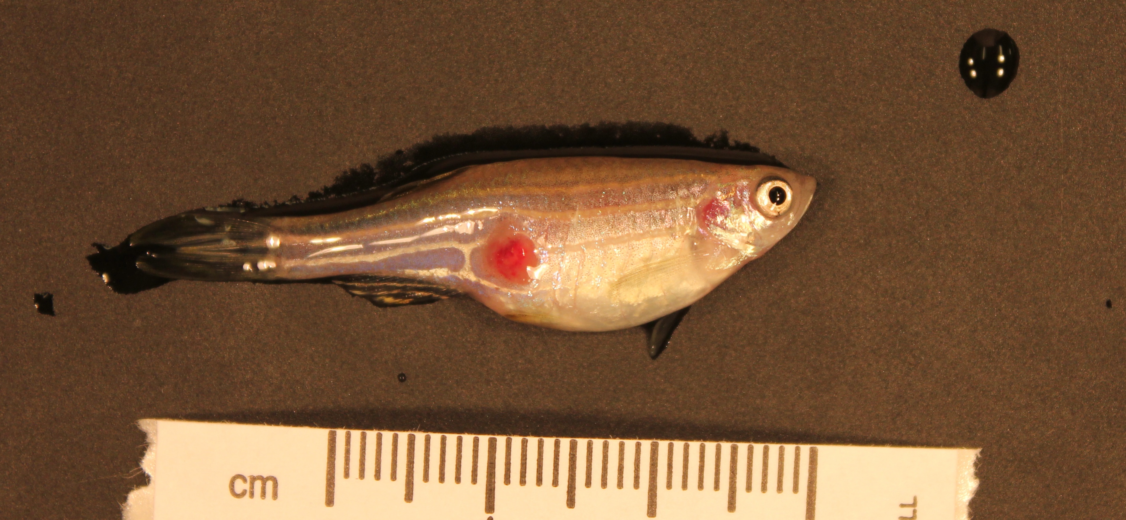
The animal presented with an ulceration on the right caudal abdomen, but appeared otherwise bright, alert, and responsive and was in good body condition.
Gross Pathology:
The skin of the right caudal abdomen contains a single approximately 1.5-2 mm diameter circular pale tan ulceration. Fin clip and gill clip wet mounts are unremarkable.
Laboratory Results:
Aerobic culture of ulcerated region: Mixed bacterial flora including Plesiomonas shigelloides and Pseudomonas putida.
Aerobic and anaerobic culture of kidney: Negative
Microscopic Description:
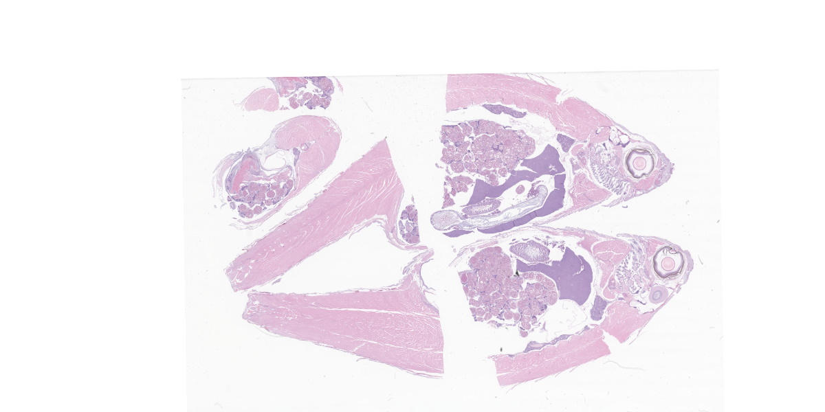
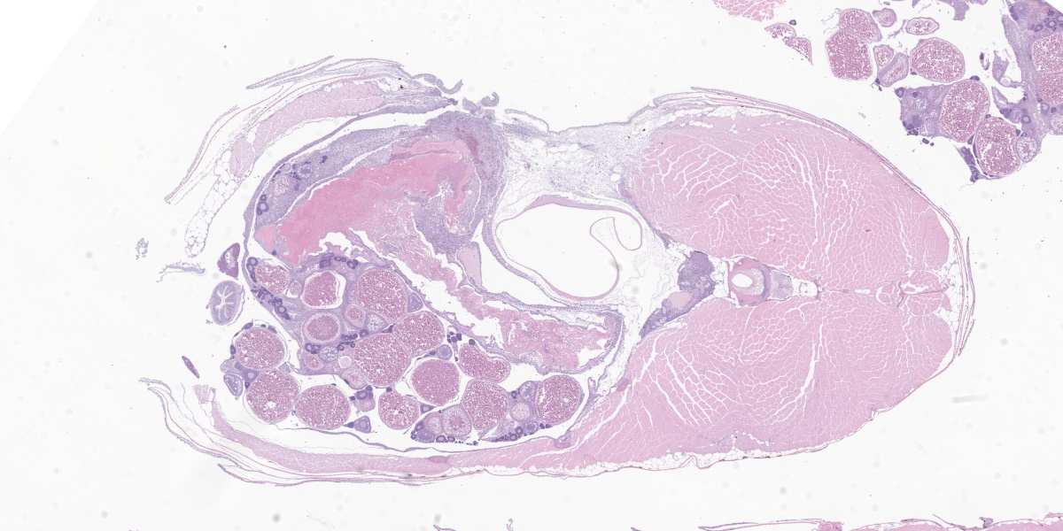
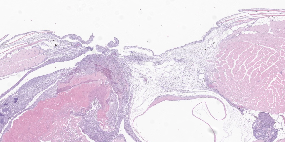
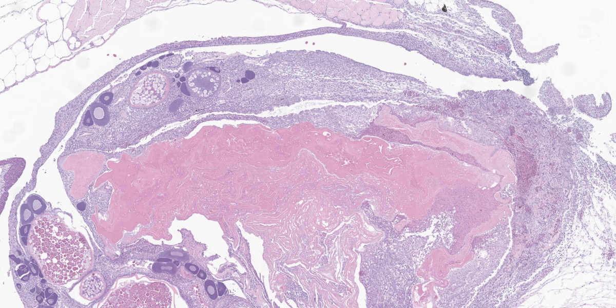
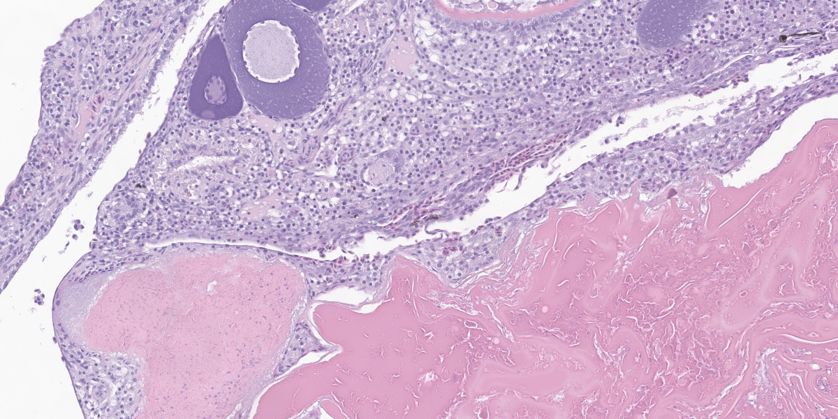
Markedly expanding the coelomic cavity and separating and surrounding viable and degenerate developing eggs within the ovary is a large inflammatory population composed of numerous epithelioid macrophages, granulocytes, and rare multinucleated giant cells. This inflammatory population is admixed with small amounts of necrotic cellular debris, moderate hemorrhage, and fibrin. This inflammatory process extrudes through the extensively ulcerated overlying body wall with loss of all layers including the skin, muscle, and coelomic cavity lining. Few small basophilic rod-shaped bacterial colonies are present near the surface of the ulceration within the degenerate egg material. The myocytes adjacent to this body wall ulceration are multifocally degenerate, with swollen vacuolated sarcoplasm, or necrotic with shrunken, hypereosinophilic, fragmented sarcoplasm. Several macrophages infiltrate between and surround damaged myocytes.
Contributor’s Morphologic Diagnosis:
Ovary and coelom: Severe, multifocal to coalescing, chronic granulomatous and granulocytic oophoritis and coelomitis with hemorrhage, degenerating eggs, myocyte loss, degeneration, and necrosis, and severe full thickness body wall, dermal, and epidermal ulceration with multifocal intralesional rod-shaped bacterial colonies.
Contributor’s Comment:
Chronic ovarian and coelomic inflammation in zebrafish associated with degenerating eggs is a common condition observed in laboratory facilities and is referred to as Egg Associated Inflammation (EAI). The cause of this condition is uncertain with some individuals describing this as a syndrome of egg retention.6 A potential link to Mycobacterial infection has been suggested though no causative agents have been identified currently and it is uncertain that Mycobacterium is a primary pathogen in this disease.2 Acid fast staining of our case was undertaken to rule out Mycobacterial infection and was negative.
Gross examination of these fish reveals a large abdomen that when opened may contain a mass of tissue, possibly adhered to the overlying coelomic cavity lining and body wall. Occasionally, as in our case, a full thickness ulceration may be visible externally, covered or surrounded by a rim of pale tan to white tissue. This ulceration is secondary to the extrusion of tissue through the body wall from the point of underlying coelomic adhesion.
Microscopically, the observed inflammation is often granulomatous with varying numbers of granulocytes and is always associated with various stages of degenerating eggs within the coelom. The inflammatory population may be surrounded and infiltrated by abundant fibroplasia and rarely this may promote the formation of fibromas and fibrosarcomas.6
The rod-shaped bacterial colonies noted on H&E-stained sections highlight as gram negative with gram staining. Aerobic culture of the ulcerated region grew Plesiomonas shigelloides and Pseudomonas putida, both gram negative rod-shaped bacteria. Plesiomonas shigelloides is found commonly within the environment and gut of laboratory and wild zebrafish but has been identified in few cases to be pathogenic to zebrafish.5 Pseudomonas putida is another bacterium widely distributed in soil, water, and on the skin of animals.3 The skin ulceration secondary to EAI likely lead to an optimal situation for these environmental organisms to grow within the damaged tissue.
Contributing Institution:
Laboratory of Comparative Pathology
Memorial Sloan Kettering Cancer Center The Rockefeller University
Weill Cornell Medicine http://www.mskcc.org/research/comparative-medicine-pathology
JPC Diagnosis:
Body wall, ovary, coelom: Granulomatous inflammation with ovarian follicular degeneration, extracellular bacteria, and body wall perforation.
JPC Comment:
As the contributor notes, egg associated inflammation (EAI), also referred to as Egg Associated Inflammation and Fibroplasia (EAIF) is thought to be associated with egg retention in fish that have not had proper opportunity to spawn.1 While mild forms of the condition may be clinically inapparent, in many cases female zebrafish present with an enlarged, ulcerated abdomens and superimposed bacterial infections within the fertile, yolky soil of the affected ovary.6
The histologic features of this case are typical of the condition. The eosinophilic coaglum represents yolk from atretic follicles and the periphery of this material is often characterized by finely granular basophilic material consisting of lysed inflammatory cells, necrotic cellular debris, and visible bacterial colonies.1 This material is usually rimmed by an intense granulomatous reaction with epithelioid macrophages and multinucleated giant cells.1
EAI is currently considered a non-infectious condition, though that assertion is typically heavy with caveats. As the contributor notes, some cases have been associated with mycobacteriosis; however, infectious agents are not usually found in cases of EAI in the absence of body wall defects, and the granulomatous and inflammatory changes seen in zebrafish with mycobacteriosis are typically found in organs other than ovaries, whereas EAI arises, as the name implies, from the ovaries.4
In addition to the detrimental effects of EAI on the individual animal, the condition raises confounding concerns due to the use of zebrafish in experimental interrogations of developmental genetics. Zebrafish are used extensively to investigate the effects of exogenous chemicals, particularly endocrine disrupting compounds, on the vertebrate reproductive system.1 Follicular atresia is a common non-specific effect of several classes of these compounds and research results may be confounded by the chronic oophoritis that is characteristic of EAI.1 In females with subclinical EAI, egg production may be reduced, leading to skewed research results in studies that use fecundity endpoints.1
The appearance of these apparently spontaneous degenerative and inflammatory changes in the ovaries of zebrafish highlight the importance of recognizing baseline variations in the histologic morphology of target organs in research animals.4
The specifics of this condition are largely unknown, leaving scant fodder for conference discussion. Dr. Travis discussed several factors that have been associated with EAI, including small body size, obesity, overcrowding, water quality issues, sex ratio imbalances, stress, and lack of proper environmental cues (e.g., photoperiod, temperature) in cultured fish. Conference participants also considered sequelae of the profound abdominal distention characteristic of the disease, including organ compression and malfunction, prolapse of the ovary through the genital pore, and dystocia.
References:
- Kent ML, Harper C, Wolf JC. Documented and potential research impacts of subclinical diseases in zebrafish. ILAR J. 2012;53(2):126-134.
- Kent ML, Watral VG, Kirchoff NS, Spagnoli ST, Sharpton TJ. Effects of subclinical mycobacterium chelonae infections on fecundity and embryo survival in zebrafish. Zebrafish. 2016;13(S1):S88-95.
- Oh WT, Kim JH, Jun JW, Giri SS, Yun S, Kim HY. Genetic characterization and pathological analysis of a novel bacterial pathogen, Pseudomonas tructae, in rainbow trout (Oncorhynchus mykiss). Microorganisms. 2019;7(10):432.
- Rossteuscher S, Schmidt-Posthaus H, Schafers C, Teigler M, Segner H. Background pathology of the ovary in a laboratory population of zebrafish Danio rerio. Dis Acquat Org. 2008;79:169-172.
- VanderHoek ZK, Browning AM, Washburn Q, Kent ML, Sharpton T, Gaulke CA. Draft genome sequence of Plesiomonas shigelloides Strain zfcc0051 (Phylum Proteobacteria). Microbiol Resour Announc. 2022; 11(7):e0007422.
- Zebrafish International Resource Center. Non-Infectious and Idiopathic Diseases. April 6, 2016. Accessed November 17, 2023. https://zebrafish.org/wiki/health/ disease_manual/noninfectious_and_idio-pathic.