Signalment:
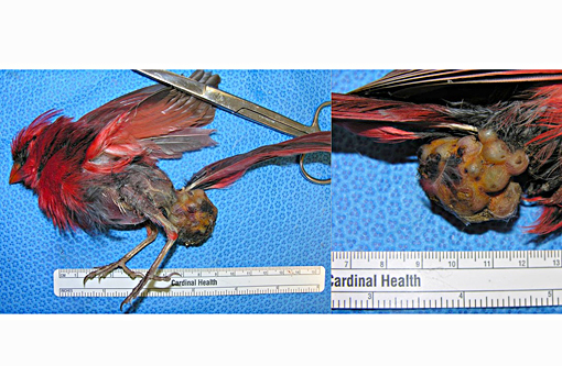
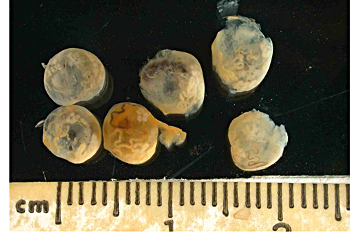
Gross Description:
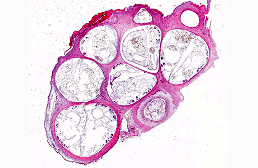
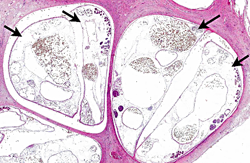
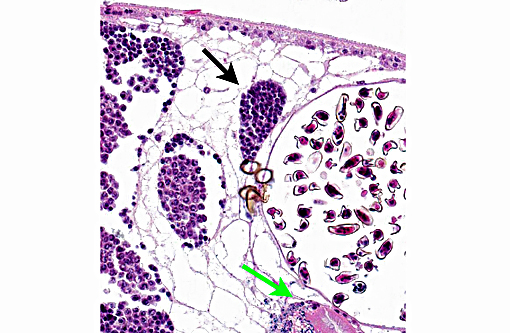
Histopathologic Description:
Blood vessels throughout the section contain large numbers of blood cells parasitized by protozoal organisms (e.g. Leukocytozoon sp. and/or Hemoproteus sp.). In one field, a skeletal muscle myofiber is expanded by a large sarcocyst containing numerous bradyzoites.
Morphologic Diagnosis:
1) Skin and subcutis (dorsal cloacal region): Parasitic pseudocysts, multifocal, with ulcerative and granulocytic dermatitis and intralesional mature adult flukes and fluke ova, etiology consistent with Collyriclum faba
2) Skeletal muscle: Sarcocystosis, mild
3) Protozoal parasitemia, marked
Lab Results:
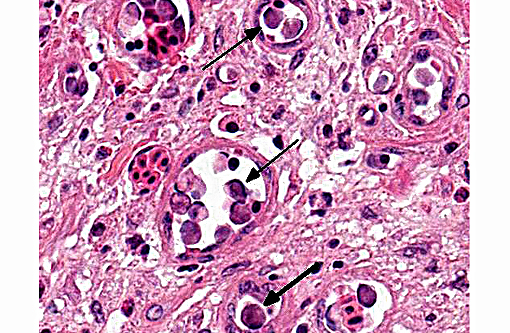
Parasite identification: Trematode parasites extracted from the dermal pseudocysts were identified as Collyriclum faba.
Condition:
Contributor Comment:
The life cycle of this parasite is incompletely understood, however, birds are thought to become infected following ingestion of metacercariae in the putative snail or dragonfly intermediate hosts.(3,7) Fluke eggs are thought to be released from a pore in the dermal pseudocysts of the avian host upon immersion in water where they then infect the intermediate hosts.(3,6) This phenomenon has been demonstrated by rubbing wet gauze over the pseudocysts in live avian patients.(3) Collyriclum faba is not usually associated with clinically significant disease, though it has been associated with cloacal obstruction and emaciation in some cases.(3)
JPC Diagnosis:
1. Glabrous skin: Multiple trematode pseudocysts with mild heterophilic and granulomatous dermatitis.
2. Skeletal muscle: Sarcocysts, multiple (some sections).
3. Circulating erythrocytes: Intracytoplasmic protozoa.
Conference Comment:
There is slide variability with few sarcocysts being present in skeletal muscle in some sections. Sarcocystis spp. protozoan parasites are common incidental findings in various mammalian and avian species. Infective sporocysts are shed in the feces of the definitive host, which are ingested by the intermediate host, where asexual reproduction occurs resulting in the formation of cysts in the skeletal muscle. One common species which infects birds is S. falcatula; the Virginia opossum is the definitive host and many different species of birds serve as intermediate hosts. The birds ingest infective sporocysts in the opossums feces, or by ingesting transport hosts such as cockroaches. In addition to skeletal muscle, sarcocysts may also be found in cardiac muscle and the central nervous system. The definitive host is infected by ingesting a bird which contains sarcocysts. The cysts are digested and zoites invade the intestinal epithelium of the definitive host, developing into gamonts. Fertilization occurs, resulting in the formation of oocysts, which sporulate and are shed as the infective form in the feces. The intermediate host ingests feces with sporocysts; once the sporocysts are ingested, the sporozoites are released in the intestine, where they migrate to blood vessels, eventually developing into meronts. Merozoites are eventually liberated from the meronts and enter circulation, resulting in their migration to cardiac, skeletal muscle and nervous tissue where they develop into sarcocysts, containing metrocytes that produce bradyzoites, which are infective to the definitive host.(2)
The conference histologic description was very similar to the contributors thorough description above. Multiple sections of a small tubular structure, lined by cuboidal to columnar epithelium and surrounded by smooth muscle, are present at the periphery of the section and these structures were tentatively identified as the vas deferens. The excellent gross images submitted with this case were also discussed. Conference participants commented on the nodular and ulcerative nature of the lesion as well as the feather loss and poor body condition, indicated by keel prominence and the absence of subcutaneous fat. The dark brown foci seen grossly overlying the pseudocysts were postulated to be foci of ulceration secondary to rupture and release of eggs. Although difficult to resolve even at high power, the egg shells have small hyalinized spines. Conference participants discussed the different hemoprotozoan genera including Plasmodium, Haemoproteus and Leucocytozoon but agreed it was not possible to definitively determine which organism is present in this case.
References:
1. Blankespoor HD, Wittrock DD, Aho J, Esch GW. Host-parasite interface of the fluke Collyriclum faba (Bremser in Schmalz, 1831) as revealed by light and electron microscopy. Z Parasitenkd. 1982; 68:191-199.
2. Gardiner CH, Fayer R, Dubey JP. An atlas of protozoan parasites in animal tissues. Washington D.C.: Armed Forces institute of pathology; 1998:41.
3. Grove DM, Zajac AM, Spahr J, Duncan RB, Sleeman JM. Combined infection by avian poxvirus and Collyriclum faba in an American crow (Corvus brachyrhynchos). J. Zoo Wildl. Med. 2005;36: 111-114.
4. Heneberg P, Faltynkova A, Bizos J, Mala M, et al. Intermediate hosts of the trematode Collyriclum faba (Plagiochiida: Collyriclidae) identified by an integrated morphological and genetic approach. Parasit Vectors. 2015; 8:85.
5. Literak I, Honza M, Haman A, Pinowska B, Pcola S. Cutaneous trematode Collyriclum faba in wild birds in the central European Carpathians. J. Parasitol. 2003; 89: 412-416.
6. Parker D. What is your diagnosis? J. Avian Med. Surg. 2009; 23:159-161.
7. Taylor MA, Coop RL, Wall RL. Veterinary Parasitology. 3rd ed. Ames, IA: Blackwell Publishing; 2007:521.