Signalment:
Gross Description:
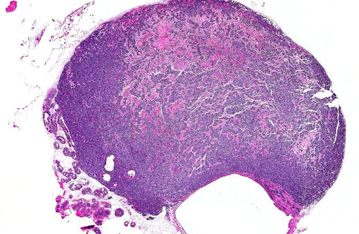
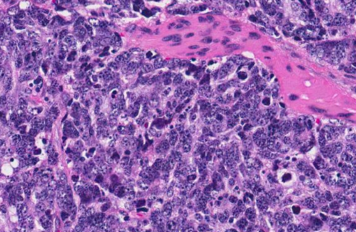
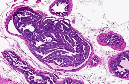
Histopathologic Description:
Morphologic Diagnosis:
1. Prostate gland (anterior lobe), poorly-differentiated prostatic carcinoma.Â
2. Prostate gland (all lobes), high-grade prostatic intraepithelial neoplasia (PIN).
Condition:
Contributor Comment:
The TRAMP model has been based on either C57BL/6 or C57BL/6 TRAMP x FVB hybrid mouse strains.(1,3,4) The prostate tumors observed in male TRAMP mice progress in a stepwise fashion through different preneoplastic and neoplastic lesions, which is a feature of prostate tumors in humans.(1,3) In TRAMP mice, commonly observed lesions include:(1)
- 6- to 12-week old mice: hyperplastic epithelial lesions or prostatic intraepithelial neoplasia (PIN).Â
- 12- to 24-week old mice: well-differentiated prostatic adenocarcinoma.
- >24-week old mice: poorly-differentiated prostatic adenocarcinoma, with development of metastases (commonly iliac lymph nodes and lungs).Â
- 33- to 52-week old mice: death.Â
The classification and grading of lesions in transgenic mouse models of prostatic tumors (including and mainly those observed in TRAMP mice) can be confusing and controversial. The most widely used scheme for classifying such tumors originates from the 2004 Bar Harbor Meeting of the Mouse Models of Human Cancer Consortium Prostate Pathology Committee.(4) This classification scheme refined and stressed the importance of PIN lesions based on a wide body of published work. Recently, a grading scheme was proposed that incorporates some data and concepts developed and published since the 2004 Bar Harbor classification scheme, which in summary includes:(1)
- Grade 0: Normal. Prostate glands lined by a monolayer of cuboidal to columnar epithelium with basally-oriented nuclei.
- Grade 1: Low-grade PIN. Crowding and occasional stratification of prostate epithelial cells, with increased nuclear to cytoplasmic ratio.Â
- Grade 2: Moderate-grade PIN. Similar to low-grade PIN, but there is more frequent stratification.Â
- Grade 3: High-grade PIN. Similar to moderate-grade PIN, but hyperplastic epithelial cells can form papillary projections and/or cribriform patterns.Â
- Grade 4: Phyllodes-like tumor. Consists of papillary projections of loose stroma with loosely-arranged stellate mesenchymal cells.Â
- Grade 5: Well-differentiated adenocarcinoma. Invasive tumor consisting primarily of well-differentiated tubules or acini. There is marked cellular and nuclear atypia, and high mitotic indices.Â
- Grade 6: Moderately-differentiated adenocarcinoma. Invasive tumor that consists of a mass of epithelial cells, some of which form recognizable acini or tubules. There is marked cellular and nuclear atypia, and high mitotic indices.
- Grade 7: Poorly-differentiated carcinoma (neuroendocrine type). Invasive tumors composed of solid sheets of polygonal to elongated cells. There is marked cellular and nuclear atypia, and high mitotic indices.
JPC Diagnosis:
Conference Comment:
Physicians from the Joint Pathology Center (JPC) Genitourinary subspecialty were consulted on this case. These pathologists were impressed by the striking lack of differentiation within this aggressive tumor and noted that the histological features closely approximate the neuroendocrine phenotype described as a grade 7 in the paper previously referenced. Apparently this microscopic appearance is rare in human prostatic carcinomas, which are generally diagnosed and treated well before reaching this stage. JPC pathologists also expressed agreement that the changes in the remaining glands of the prostate gland are consistent with high-grade prostatic intraepithelial neoplasia, which represents an intermediate stage between normal epithelium and invasive malignant carcinoma. In human medicine, PIN is clinically significant in that it provides relatively early identification of patients at risk for malignancy.(2)
References:
2. Brawer MK. Prostatic intraepithelial neoplasia: an overview. Rev Urol. 2005;7(suppl 3);11-18.
3. Chiaverotti T, Couto SS, Donjacour A, et al. Dissociation of epithelial and neuroendocrine carcinoma lineages in the transgenic adenocarcinoma of mouse prostate model of prostate cancer. Am J Pathol. 2008;172:236-246.
4. Schappell SB, Thomas GV, Roberts RL, et al. Prostate pathology of genetically engineered mice: definitions and classification. The consensus report from the Bar Harbor Meeting of the Mouse Models of Human Cancer Consortium Prostate Pathology Committee. Cancer Res. 2004;64:2270-2305.