Signalment:
Sex undetermined juvenile Moorish idol (
Zanclus cornutus)The body of an approximately 6cm juvenile Moorish idol was received in formalin. The fish was one of a
group of recently shipped animals under observation and treatment in quarantine prior to display in a public
aquarium. Constant low level mortalities had occurred from the time of arrival.
Gross Description:
Reported gross findings varied among individuals and included lethargy, emaciation, abdominal
distension, and intestines severely distended by thick yellow contents.
Histopathologic Description:
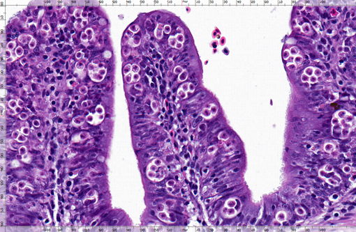
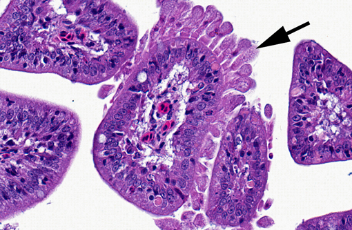
Intestine: Multiple intestinal segments are characterized by plicae
irregularly expanded by variable, mild to moderate, mixed populations of lymphocytes, plasma cells, and
macrophages containing granular to refractile debris. Within affected areas, the normal mucosal architecture is
altered by a combination of mild epithelial hypertrophy, hyperplasia, and pseudostratification, with compression and
distortion of individual cells resulting from the presence of large numbers of approximately 20 μm, ovoid,
intraepithelial parasitic bodies. Individual bodies (pansporoblasts) are defined by a thin pale eosinophilic limiting
membrane and compressed peripheral nucleus, surrounding a vacuolated space typically containing 4-8 ovoid, 5 x 8
μm structures (sporoblasts and accessory cells). The round to ovoid internal structures have moderate amounts of
slightly vacuolated pale eosinophilic cytoplasm and possess 1 or 2 central, condensed, basophilic nuclei, often with a
prominent nucleolus. Similar structures are present within luminal debris. Occasional lifting of the epithelium from
the basement membrane is evident in some areas and pockets of refractile debris are scattered throughout the
epithelium.
Morphologic Diagnosis:
Intestine: Enteritis, segmental, subacute to chronic, moderate, with numerous intralesional myxozoal pansporoblasts
Lab Results:
Intestinal mucosal scrapings were reported to contain numerous signet ring-like structures
with multiple internal spheres.
Condition:
Enteromyxm leei
Contributor Comment:
Microscopic findings are consistent with widespread intestinal infection by the
myxozoan parasite
Enteromyxm (Myxidium) leei. Enteromyxm leei causes severe enteritis and losses, particularly in
sea bream and sea bass mariculture in the Mediterranean area.(6,8) The parasite exhibits an unusually low degree of
host specificity and infections have been transmitted to over 25 marine fish species in a public aquarium.(8) An
alternate host or actinosporean form of the parasite has not been identified and direct transmission has been
demonstrated by cohabitation, exposure to contaminated effluent, and by the feeding of infected tissue.(4) Gross signs
may include emaciation, darkening, lethargy, abdominal distension, pale soft liver, enlarged gallbladder, and a
thickened and hemorrhagic intestinal wall.(6,8) Lesions occur throughout the intestine from the pyloric ceca to rectum,
and occasionally in the gallbladder and intrahepatic bile ducts.(1,6,8) Development occurs between epithelial cells near
the basement membrane. Pansporoblasts, up to 40 μm, consist of an enveloping pericyte or primary cell surrounding
a clear space housing two developing sporoblasts and two or more accompanying accessory cells.(1) Although
Giemsa, Gram, and acid-fast stains were performed, mature spores were not identified. Mature spores, when present,
are arcuate, average 6.9 x 14.7 μm, and have two elongate polar capsules tapering to their distal ends.(3)
Myxozoans are important metazoan parasites of freshwater and marine fish closely related to the Cnidaria. This
large group of obligate parasites includes over 1350 species in 52 genera, involving intracellular and intercellular
histozoic representatives, as well as many coelozoic forms. Life cycles are complex and not fully understood for
many species, although at least 25 are known to involve an invertebrate (oligochaete, polychaete, or bryozoan)
alternative host. Taxonomy is largely based on the morphology of myxospores, characterized by multiple cells
arranged in 1-7 spore valves, 1-2 amoeboid sporoplasms, and 2-7 nematocyst-like polar capsules with coiled
extrudable polar filaments.(2,7)
Additional findings present in some sections include plasmodia of a coelozoic myxozoan associated with mild bile
duct ectasia, epithelial attenuation, and periductular fibrosis. Some intestinal sections also include a focal area with
large numbers of elongate structures with abundant finely granular to vacuolated cytoplasm, one to rarely two small
nuclei, and a broad base of attachment to the mucosal epithelium. The nature of this organism is unknown, but
possibly represents a developmental stage of another myxozoan.
JPC Diagnosis:
Intestine: Enteritis, histiocytic and lymphocytic, segmental, moderate to marked, with mucosal
hypertrophy and hyperplasia and numerous intracytoplasmic myxosporidian pansporoblasts.
Conference Comment:
Conference participants discussed the usual mode of transmission for this myxozoan.
Direct fish-to-fish transmission can occur through the ingestion of exfoliated infected intestinal epithelium(5). The
intestinal epithelium contains autophagic vacuoles, a common finding in fish with gastrointestinal disturbances.
Conference participants also discussed the presence of exocrine pancreatic atrophy, lack of coelomic adipose tissue,
and atrophy and loss of the dorsal skeletal muscle, all of which are consistent with the reported gross findings and
indicate cachexia. Participants attributed areas of myodegeneration in the dorsal skeletal muscle to possible capture
myopathy.
References:
1. Alvarez-Pellitero P, Palenzuela O, and Sitja-Bobadila A. 2008. Histopathology and cellular response in
Enteromyxm leei (Myxozoa) infections of
Diplodus puntazzo (Teleostei). Parasitology International
57:110-120.
2. Bruno DW, Nowak B, and Elliott DG. 2006. Guide to the identification of fish protozoans and metazoan parasites in stained tissue sections. Diseases of Aquatic Organisms 70:1-36.
3. Diamant A, Lom J, and I Dykova. 1994.Â
Myxidium leei n. sp., a pathogenic myxosporean of cultured sea bream Sparus aurata. Diseases of Aquatic Organisms 20:137-141.
4. Diamant A. 1997. Fish-to-fish transmission of a marine myxosporean. Diseases of Aquatic Organisms 30:99-105.
5. Fesit SW, Longshaw M. Phylum myxozoa. In: Woo PTK, ed.Â
Fish Diseases and Disorders. 2nd ed. Vol 1. Cambridge, MA: CABI; 2006:271.
6. Fleurance R, Sauvegrain, Marques A, Le Breton A, Guereaud C, Cherel Y, and Wyers M. 2008. Histopathological changes caused by
Enteromyxm leei infection in farmed sea bream
Sparus aurata. 219-228.
7. Kent ML et al. 2001. Recent advances in or knowledge of the myxozoa. The Journal of Eukaryotic Microbiology 48:395-413.
8. Padr³s F, Palenzuela O, Hispano C, Tosas O, zarza C, Crespo S, and Alvarez-Pellitero P. 2001. Diseases of Aquatic Organisms 47:57-62.