Signalment:
Two-year-old male B6C3F1 mouse (
Mus
musculus).This mouse was
in the control group of a two-year study investigating the carcinogenicity of
kava kava extract. It was sacrificed on Test Day 668, due to moribund
condition. Clinical observations included ruffled fur, lethargy and thin body
condition.
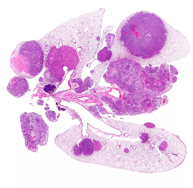
Gross Description:
Gross
lesions described at necropsy included: a single tan mass, approximately
27x17x12 mm, on the left lateral liver lobe; and several white nodules, up to 5
mm diameter, on the visceral pleura, lungs, and mediastinum.
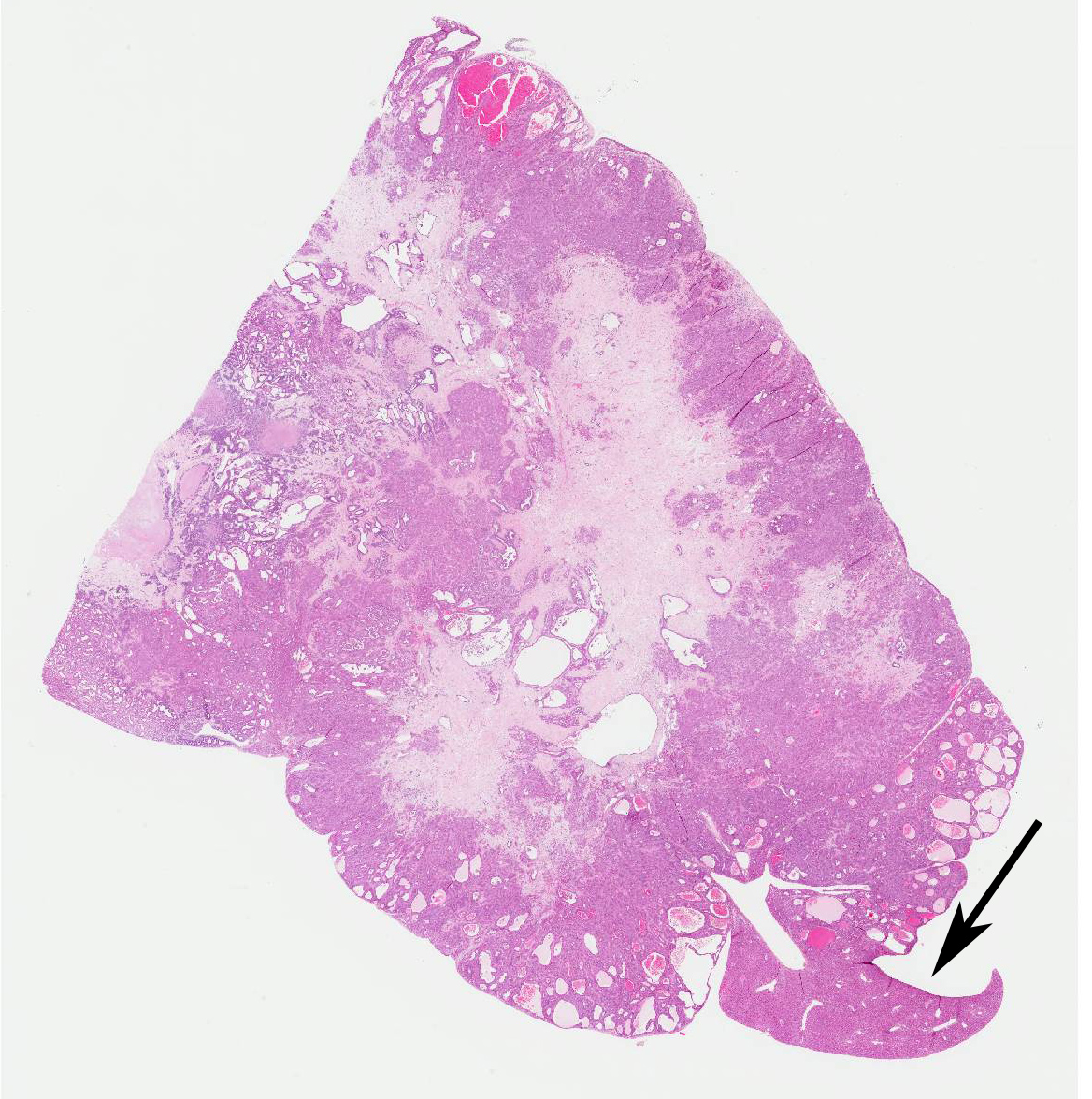
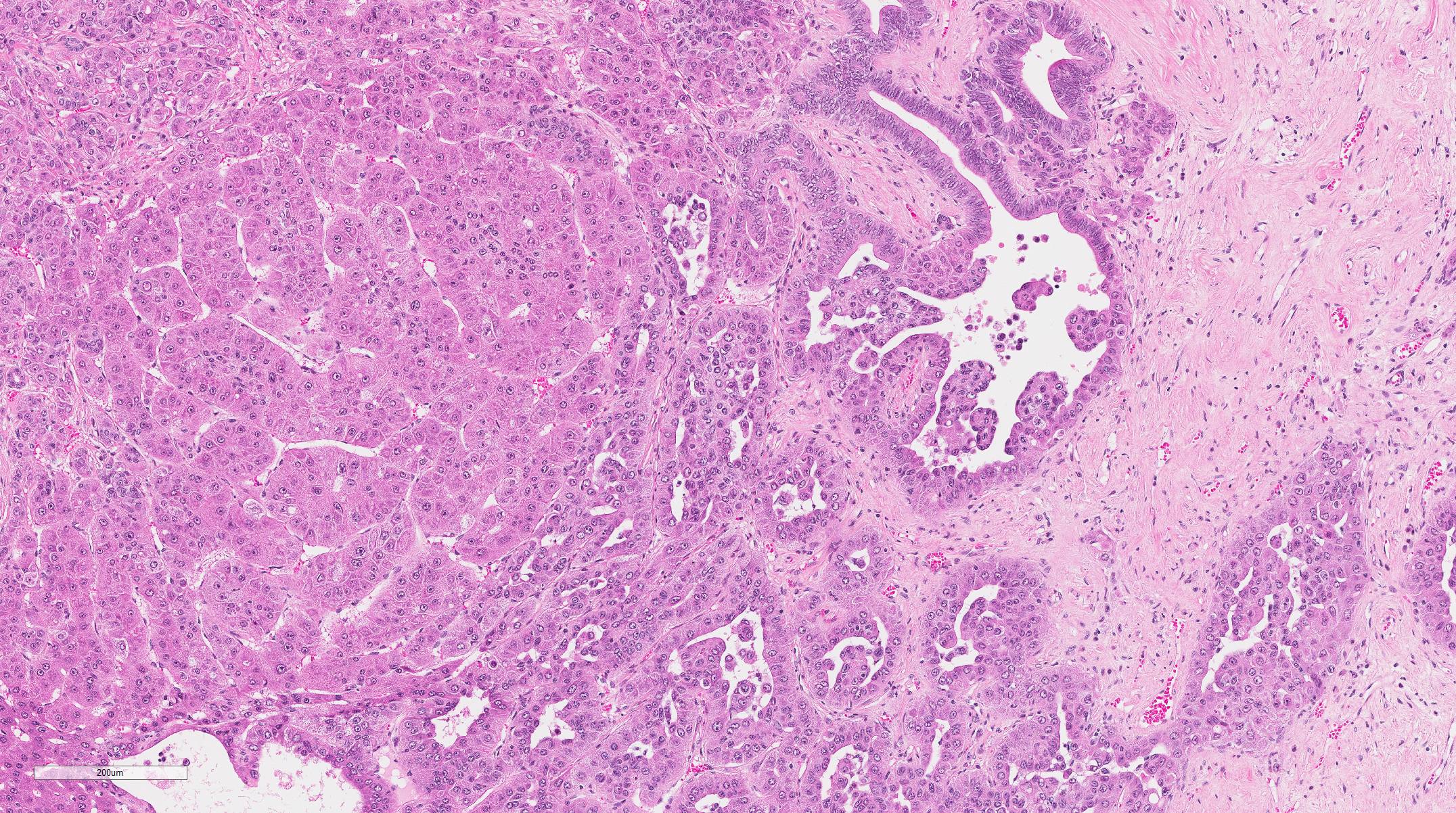
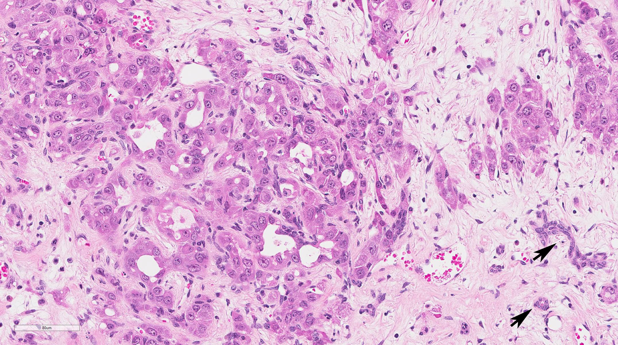
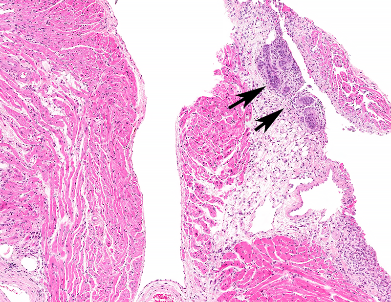
Histopathologic Description:
Most
of this section of liver lobe is replaced by a large mass extending to the
capsular surface. The mass is composed of irregular acini and variably-sized
cystic spaces interspersed with more densely cellular areas and areas of
fibrosis or necrosis. Acini are lined by columnar epithelium with prominent
hyper-chromatic nuclei and scant cytoplasm. More densely
cellular areas are comprised of hepatocytes with a central vesicular nucleus
containing 1 or 2 prominent nucleoli, and abundant cytoplasm. These cells are
arranged in packets, in trabeculae more than 3 cells thick, or around cystic
spaces. The cystic spaces are lined by one or more layers of flattened to plump
hepatocytes, with occasional papillary projections extending into the lumens;
lumens often contain sloughed epithelial cells, lightly stained fibrillar
eosinophilic material, and occasional extravasated erythrocytes. In other
areas, densely-packed, small round to elongated cells with hyperchromatic
nuclei form a sarcomatous pattern, often extending into areas of fibrosis. Occasional
endothelial-lined spaces contain erythrocytes, small clusters of white blood
cells (mainly neutrophils) and single or clusters of neoplastic cells.
Morphologic Diagnosis:
Liver Hepatocholangiocarcinoma. Tissue
not included: Lung Hepatocholangiocarcinoma,
metasta-tic
Lymph node, mediastinal
Hepatocholan-giocarcinoma, metastatic
Thymus Hepatocholangiocarcinoma,
metastatic
Heart, pericardium Hepatocholangio-carcinoma,
metastatic
Lab Results:
None
Condition:
Hepatocholangicarcinoma, mouse
Contributor Comment:
This
hepato-cholangiocarcinoma (HCCC) typically contains both malignant
hepatocellular and malignant biliary components, as well as foci of markedly
undifferentiated sarcoma-like cells, as described in a poster presented by the
National Institute of Environmental Health Sciences (NIEHS) and Experimental
Pathology Laboratories, Inc. (EPL).
4 HCCC is rarely reported in mice
and rats. However, it has been induced by some chemicals tested in the National
Toxicology Program (NTP) studies.
2,4 It is an aggressive neoplasm
that metastasizes readily.
JPC Diagnosis:
Liver: Hepato-cholangiocarcinoma, B6C3F1 mouse,
Mus musculus.
Conference Comment:
Hepato-cholangiocarcinoma
(HCCC) is a rare spontaneous neoplasm with an incidence of less than 0.5% in
B6C3F1 mice, according to the National Toxicology Program (NTP) database.
4,6
HCCC is a primary liver neoplasm comprised of both neoplastic hepatocytes and
neoplastic bile duct epithelial cells.
4,6 The biliary component
forms tubules and acini or small nests, while the hepatocellular component
forms trabeculae, glands, or solidly cellular areas of neoplastic cells. Both
neoplastic hepatocytes and bile duct epithelial cells have many characteristics
of malignancy, such as frequent mitoses, nuclear pleo-morphism, local invasion,
and widespread metastasis.
4,6,7 HCCC often contains large cystic or
necrotic areas within the central portions of the neoplasm, as demonstrated in
this case. Additionally, there are occasional ducts within the neoplasm, lined
by both hepatocytic and biliary epithelial cells, in conjunction with
multifocal areas of poorly differentiated mesenchymal spindloid cells arranged
in short interlacing streams and bundles. Proliferation of sarcomatous cells is
a frequently reported feature of this malignant neoplasm.
6,7 In
previously studies, 84% of mice with HCCC had metastasis to multiple tissues,
similar to what is reported by the contributor here.
6 The histo-morphologic
variability of this neoplasm and widespread metastasis may pose a diagnostic
challenge in determining the site of origin. Metastatic lesions may contain
only one of the three neoplastic populations mentioned above, further
confounding the diagnosis.
6
As
mentioned previously, spontaneous occurrence of HCCC is exceedingly rare in
mice; however, malignant transformation has been experimentally associated with
chemical carcinogens, such as trimethylolpropane tricrylate, benzidine
dihydrochloride, and N-2-acetylaminofluorene.
4
The B6C3F1mouse is a hybrid strain that is the result of a cross between a male
C3H and a female C57BL/6 mouse. This relatively hardy mouse strain has been
used by the NTP for decades as part of carcinogenesis, toxicity, and transplant
studies.
2
References:
1. Adams ET,
Auerbach S, Blackshear PE et al. Proceeding of the 2010 National Toxicology
Program satellite symposium. Toxicol Pathol. 2010; 39:240-246.
2. Gad SC, Clifford
C, Goodman D. The mouse. In Gad SC ed. Animal Models in Toxicology. 3rd
ed. Boca Raton, FL: CRC Press; 2016:76-78.
3. Hailey JR, Nold
JB, Brown RH et al. Biliary proliferative lesions in the Sprague-Dawley rat:
Adverse/non-adverse. Toxicol Pathol. 2014; 42:844-854.
4.Harada T,
Enomoto A, Boorman GA, Maronpot RR. Liver and gallbladder. In: Maronpot RR,
Boorman GA, Gaul BW eds. Pathology of the Mouse. Vienna, IL: Cache River
Press; 1999:153.
5. Maronpot RR.
Rodent Liver - Neoplastic. 2015. http://focusontoxpath.com/rodent-liver-lesions-neoplastic/
6. Moore R,Willson
G, Miller R, Malarkey D, Kissling et al. Hepatocholangiocarcinomas in B6C3F1
Mice in National Toxicology Program (NTP) Studies.
7.
Thoolen
B, Maronpot RR, Harada T et al. Proliferative and nonproliferative lesions of
the rat and mouse hepatobiliary system. Toxicol Pathol. 2011; 38:5S-81S.