WSC 2023-2024, Conference 15, Case 4
Signalment:
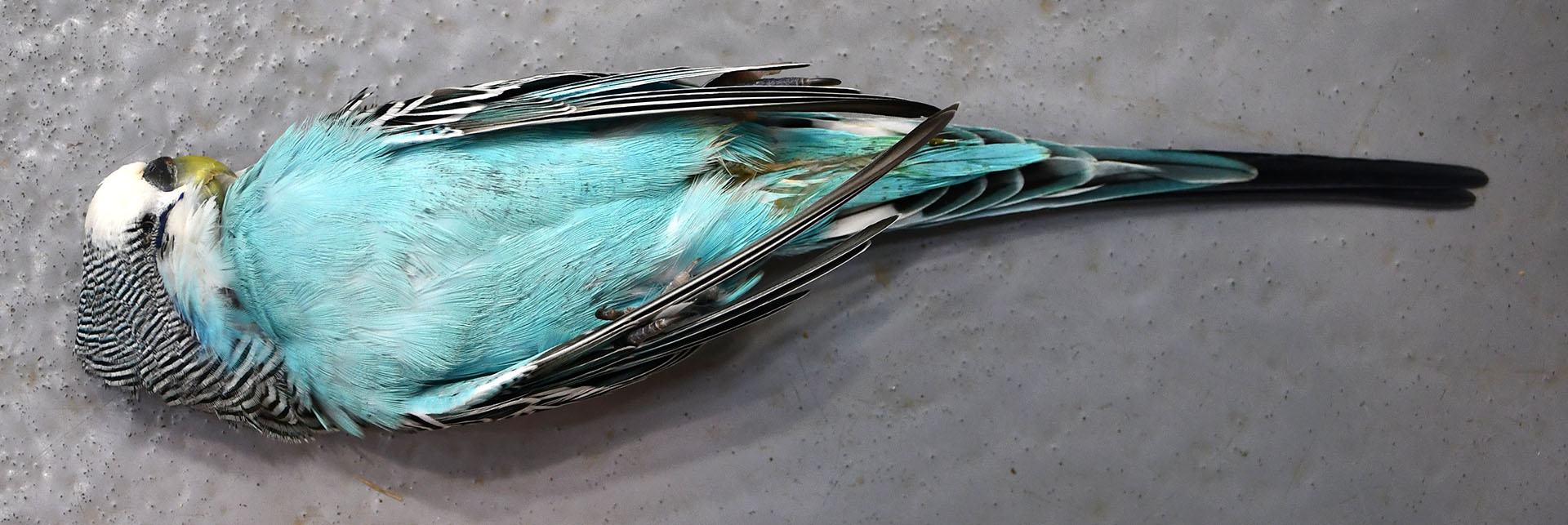
3-year-old, male intact budgerigar (Melopsittacus undulatus)
History:
This animal was a member of a zoological collection and was found dead in its enclosure.
Gross Pathology:
This blue, 39.3g male budgerigar had a dark blue cere. Both testes were enlarged (left was bilobed and 8x5x5mm, right was spherical and 5x5x5mm in diameter).
Microscopic Description:
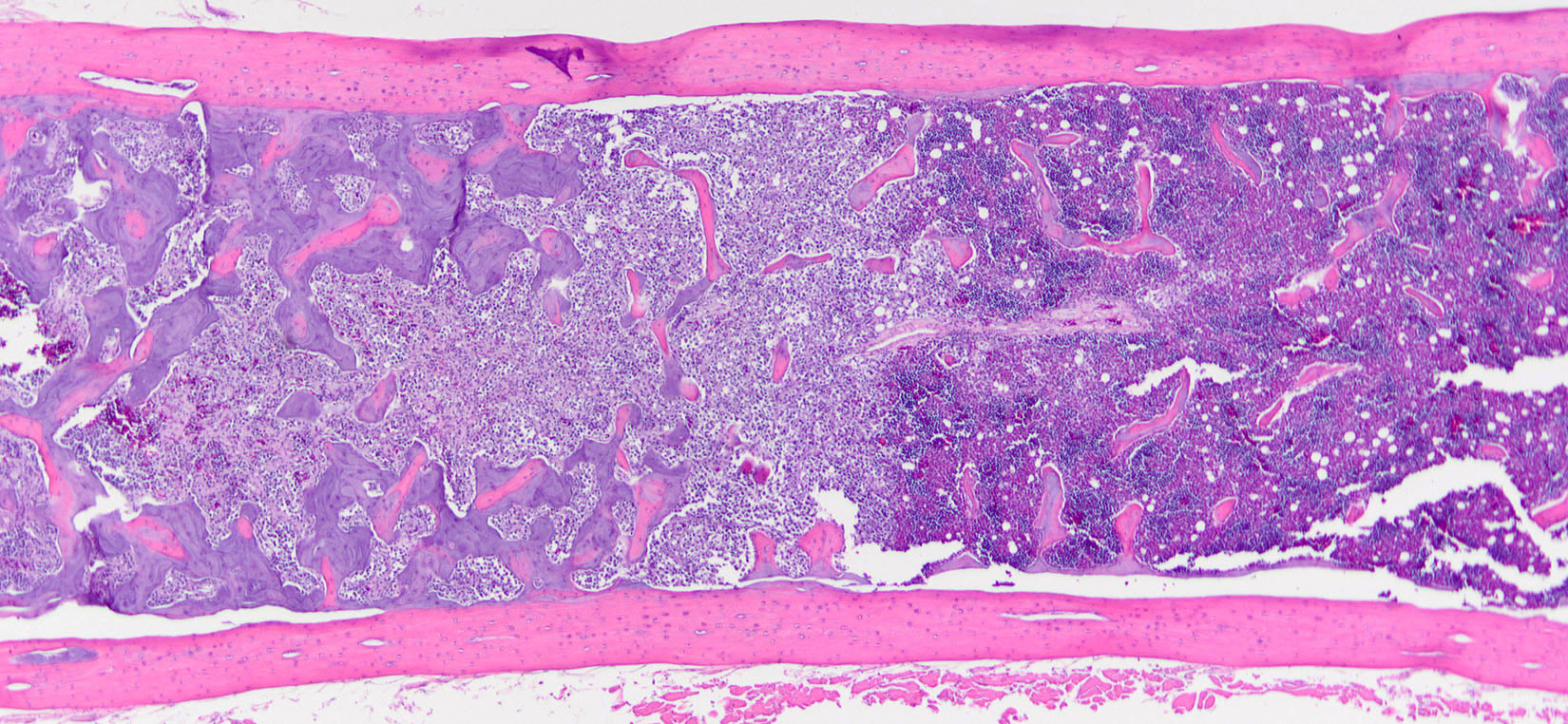
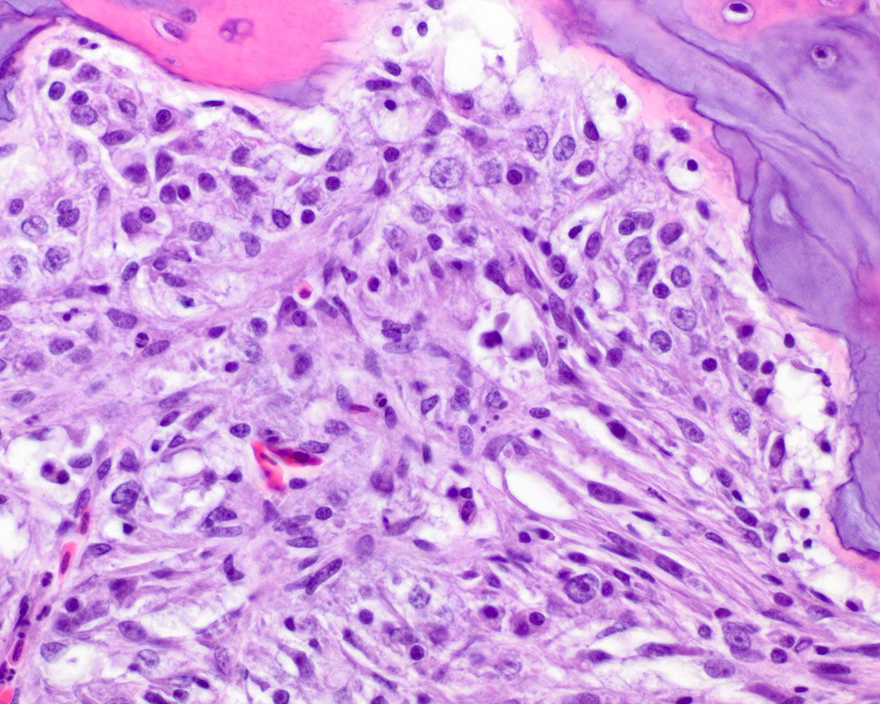
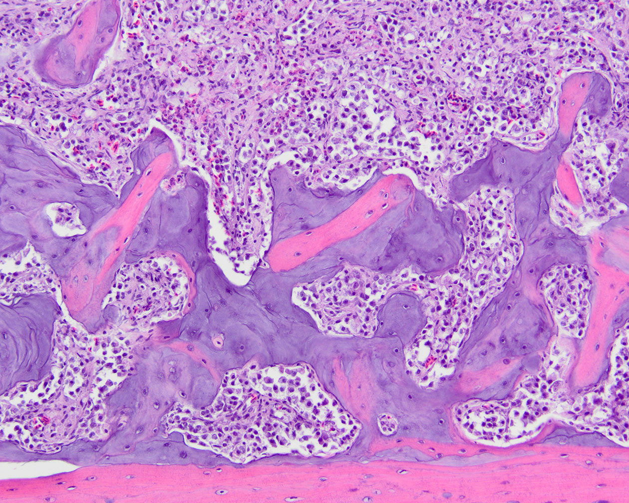
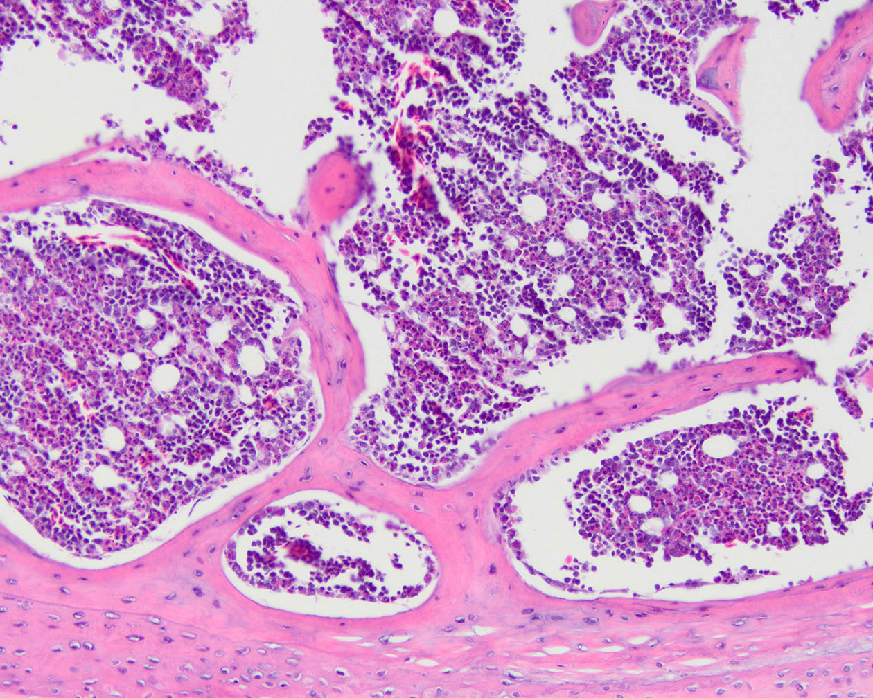
Both testes (slide not provided) contained a neoplasm composed of polygonal cells in packets separated by a dense fibrous stroma. Neoplastic cells had distinct borders, eosinophilic cytoplasm, and round nuclei. Foci of a similar neoplastic population were within the marrow of both femurs. Both femurs and humeri had basophilic, non-birefringent bone lining the medullary trabeculae (medullary bone) that was most pronounced near the metastatic foci.
Contributor’s Morphologic Diagnosis:
Femur: Metastatic Sertoli cell tumor with associated medullary bone.
Contributor’s Comment:
Medullary bone (also known as laying bone) is formed and removed along the endosteal surfaces of bones in female birds during the egg-laying cycle as a source of calcium for eggshells. In females, it typically spares the pneumatic bones, mostly forming in the hematopoietic bones such as the femur and tibiotarsus, but this varies with species.
This femur is from one of 7 male budgerigars in a publication that described medullary bone as a feature of paraneoplastic feminization. The medullary bone deposition was diffuse in 4 cases and multifocal in 3 (including this one) and affected both pneumatic (humeri) and hematopoietic (femurs) bones.2 This was the only case in which bony metastases were detected. Interestingly, the medullary bone in this case is most pronounced near the metastatic foci.
The testicular tumors associated with medullary bone included Sertoli (sustentacular) cell tumors, seminomas, and one mixed (Sertoli-interstitial cell) tumor.2 Although estrogen production is characteristic of Sertoli cells tumors, estrogen production and feminization have been reported in humans and dogs with seminomas.1,3
Female budgerigars normally have a bright pink cere, while males have a light blue cere. Cere color change is another feature of paraneoplastic feminization reported in budgerigars.4 In the published study of 7 budgerigars with testicular tumors and medullary bone, 4 had a blue-brown or red-brown cere, 2 had a light blue cere, and one (this bird, case 3 in reference 2) had a dark blue cere.2
Contributing Institution:
University of Tennessee
College of Veterinary Medicine
Biomedical and Diagnostic Sciences
2407 River Drive, Room A201
Knoxville, TN 37996
https://vetmed.tennessee.edu/academics/biomedical-and-diagnostic-sciences/
JPC Diagnosis:
Long bone: Metastatic sustentacular cell tumor with associated medullary bone.
JPC Comment:
This fascinating case provides an excellent example of medullary bone, a new concept for many conference participants this week. As noted by the contributor, medullary bone is normally produced in the hematopoietic bones of female birds to serve as calcium stores for the arduous task of mineralizing eggs. This sex-specific tissue is produced by female birds from approximately 10 days pre-ovulation until the completely mineralized egg is laid.2 Medullary bone can be distinguished from woven bone by its increased basophilia, due to an increased proteoglycan content compared to woven bone, and by the absence of birefringence under polarized light.2
The formation of medullary bone occurs simultaneously with the maturation of ovarian follicles, and is believed to be due to incompletely understood interactions among the hypothalamus, pituitary gland, and ovaries.2 The ovum produces estrogens upon the binding of luteinizing hormone produced by the pituitary gland, and these estrogens bind estrogen receptors on osteoblasts, leading to the deposition of type I collagen on the endosteal surfaces of hematopoietic bones of female birds.2 Once the egg is ovulated and reaches the shell gland, mineralization-induced hypocalcemia induces parathyroid hormone secretion, which in turn stimulates osteoclasts to resorb medullary bone, releasing calcium stores into the bloodstream for use in egg mineralization.2
The contributor provides an excellent overview of the concept of paraneoplastic feminization, defined as “the acquisition of feminine characteristics as an indirect consequence of a neoplasm, usually due to production of chemical signalling molecules such as hormones.2
In veterinary medicine, this condition is well-described in male dogs with sustentacular (Sertoli) cell tumors, in which estrogen production by neoplastic cells produces female behavior and other characteristic changes such as gynecomastia, alopecia, prostatic squamous metaplasia, and myelosuppression.2 Removal of the neoplastic tissue typical results in normalization of serum estrogen and reversion to more gender-stereotypic behavior and physiology. In male budgerigars with testicular neoplasia, paraneoplastic feminization is typically described as involving medullary bone formation, cere color change, widening of the pelvic inlet, and protrusion of the cloaca.2 The production of medullary bone can be diagnostically useful, as the resulting increase in bone density can be identified radiographically and can serve as an early indication of testicular neoplasia.2
Conference discussion focused on the histologic appearance of medullary bone, including the basophilia and lack of birefringence, as noted above. Conference participants also examined Masson and Movat pentachrome stains, in which the medullary bone stained blue and green, respectively. The moderator noted that medullary bone in male budgerigars can be present with all three testicular tumors though not all produce the estrogen throught responsible for the formation of medullary bone. This curiosity highlights the fact that the physiologic basis for medullary bone deposition remains incompletely understood and is likely multifactorial.
References:
- Duparc C, Boissiere-Veverka G, Le-febvre H, et al. An oestrogen-producing seminoma responsible for gynaecomastia. Horm Metab Res. 2003;35(5):324-329.
- Hoggard NK, Craig LE. Medullary bone in male budgerigars (Melopsittacus undulatus) with testicular neoplasms. Vet Pathol. 2022;59(2):333-339.
- Kim O, Kim KS. Seminoma with hyperesterogenemia in a Yorkshire terrier. J Vet Med Sci. 2005;67(1):121-123.
- Mans C, Pilny A. Use of GnRH-agonists for medical management of reproductive disorders in birds. Vet Clin North Am Exot Anim Pract. 2014;17(1):23–33.