Signalment:
Gross Description:
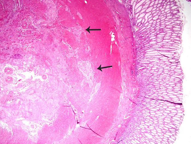
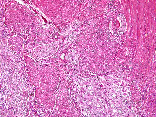
Histopathologic Description:
Morphologic Diagnosis:
Lab Results:
Condition:
Contributor Comment:
Immunohistochemically, some cases of ganglioneuromatosis were shown to be a complex hyperplasia of several peptidergic, cholinergic, and probably adrenergic nerve fibers instead of a selective overgrowth of one type of nerve fibers.4
Some rare cases of intestinal ganglioneuromatosis or ganglioneuromas have been reported, most often in young animals: in a horse1, a steer3, a cat9, and 3 dogs.5,11,13 In all cases, there were clinical signs (e.g. colic, impaction, anorexia, vomiting, diarrhea, rectal prolapse) that led to surgical resection of the masses. Masses were located in the small intestine (3 cases), colon (1 case), colorectum (2 cases) or Vaters papilla (1 case). This is the first reported case of asymptomatic ganglioneuromatosis in a dog.Â
JPC Diagnosis:
Conference Comment:
Intussusceptions are described as having three layers: (1) outer wall of the receiving segment, (2) middle returning segment of invaginated bowel, and (3) inner entering segment. The intussusception seen in this lesion is unusual in that it contains only two of the three layers, a feature that will occur only through the invagination of a blind pouch (in this case, the tip of the cecum). Cecal inversion is another term for such a lesion (Fig. 3-3). Various causes of intussusception may include linear foreign bodies, heavy parasitism, previous intestinal surgery, enteritis, and intramural lesions. It may also develop as a terminal, agonal or postmortem event.2

Fig 3-3
A. Intussusception of tubular section of bowel consisting of three layers: (1) outer wall of the receiving segment, (2) middle returning, segment of invaginated bowel, and (3) inner entering segment.Â
B. Intussusception of a blind pouch consisting of two layers: (1) outer wall of the receiving segment, and (2) the middle returning, segment of invaginated bowel.
We appreciate the assistance from the Departments of Gastrointestinal Pathology, Neuropathology, and Soft Tissue Pathology at the Armed Forces Institute of Pathology in consultation on this case.
References:
2. Brown CC, Baker DC, Barker IK: Alimentary system. In: Jubb, Kennedy, and Palmers Pathology of Domestic Animals, ed. Maxie MG, 5th ed., vol. 2, pp. 96-97. Elsevier Limited, St. Louis, MO, 2007
3. Cole DE, Migaki G, Leipold HW: Colonic ganglioneuromatosis in a steer. Vet Pathol 27:461-462, 1990
4. DAmore ES, Manivel JC, Pettinato G, Niehans GA, Snover DC: Intestinal ganglioneuromatosis: mucosal and transmural types. A clinicopahologic and immunohistochemical study of six cases. Hum Pathol 22:276-286, 1991
5. Fairley RA, McEntee MF: Colorectal ganglioneuromatosis in a young female dog (Lhasa Apso). Vet Pathol 27:206-207, 1990
6. Goyal RK, Hirano I: The enteric nervous system. N Engl J Med 334:1106-1115, 1996
7. Head KW, Else RW, Dubielzig: Tumors of the alimentary tract. In: Tumors in Domestic Animals, ed. Meuten DJ, 4th ed., p. 470. Blackwell Publishing, Ames, IA, 2002
8. Komminoth P: Multiple endocrine neoplasia type 1 and 2: from morphology to molecular pathology. Vehr Dtsch Ges Path 81:125-138, 1997
9. Patnaik AK, Lieberman PH, Johnson GF: Intestinal ganglioneuroma in a kitten a case report and review of literature. J Small Anim Pract 19:735-742, 1978
10. Porter BF, Storts RW, Payne HR, Edwards JF: Colonic ganglioneuromatosis in a horse. Vet Pathol 44:207-210, 2007
11. Ribas JL, Kwapien RP, Pope ET: Immunohistochemistry and ultrastructure of intestinal ganglioneuroma in a dog. Vet Pathol 27:376-379, 1990
12. Shekitka KM, Sobin LH: Ganglioneuromas of the gastrointestinal tract. Relation to Von Recklinghausen disease and other multiple tumor syndromes. Am J Surg Pathol 18:250-257, 1994
13. Van Den Ingh TSGAM, Rothuizen J: Ganglioneuroma of Vaters papilla and extrahepatic cholestasis in a dog. Vet Pathol 21:254-256, 1984