Signalment:
Adult, male, rosy finch (
Leucosticte spp.)A wild-caught research flock of outdoor-housed rosy finches (
Leucosticte spp.) captured in California and white-crowned sparrows (
Zonotrichia leucophrys) captured in eastern Washington state presented during the winter of 2001 with history of periocular ulceration, pododermatitis and death.
Gross Description:
Numerous birds had similar lesions that included proliferative and necrotizing palpebral dermatitis with conjunctivitis and/or fibrinonecrotic and proliferative stomatitis and glossitis. One rosy finch had a large (5-7mm) mass on the right wing. Numerous mites were noted within the feathers.
Histopathologic Description:
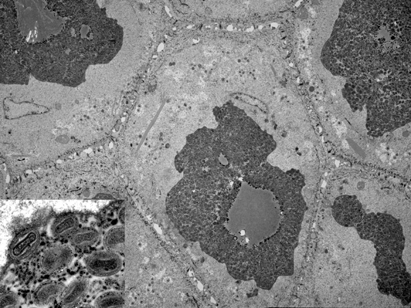
EM Skin of Wing (5,000X)
(Fig. 4-1): Numerous polygonal cells with desmosomes, few discernable organelles consistent with squamous epithelial cells, have displaced and flattened nuclei, loss of organelle detail, granular homogenous cytoplasm and widened intercellular spaces. In each squamous cell, there is a large electron dense cytoplasmic inclusion body that contains abundant virions.Â
Fig. 4-1 Inset (60,000X): Enveloped virions approximately 300nm long with brick-shaped nucleocaspid and biconcave core consistent with pox virions.
Morphologic Diagnosis:
Squamous epithelial degeneration, intra- and intercellular edema with single large intracytoplasmic inclusion bodies composed of virions consistent with pox.Â
Lab Results:
The mites were not further characterized.
Candida albicans and
Staphylococcus aureus were cultured from the tongue.
Condition:
Avian poxvirus
Contributor Comment:
The gross, histological lesions are consistent with avipox infection that was confirmed by electron microscopy. Several strains of poxvirus infect a variety of avian hosts including passerines. Transmission is via direct contact, ingestion or mechanical vectors such as mosquitoes or other insects. Pox infection results in cutaneous necroproliferative lesions (cutaneous form or dry pox) and/or fibrinonecrotic and proliferative lesions affecting the mucous membranes of the upper gastrointestinal and respiratory systems (diphtheritic form or wet pox). Both forms can cause significant morbidity by interfering with bodily functions. Secondary bacterial or fungal infections also increase morbidity and mortality. Songbirds are more commonly affected in the winter months as seen in this epornitic. Diagnosis is via gross and histological findings, electron microscopy, viral isolation serology and PCR. Distinctive microscopic findings include epithelial hyperplasia and enlargement of epithelial cells with the characteristic eosinophilic intracytoplasmic inclusion bodies (Bollinger bodies). Electron microscopy can confirm diagnosis by identifying 250 x 354 nm virions with a distinctive brick or dumbbell shape. The presumptive introduction of poxvirus into this research aviary was through the recently caught white-crowned sparrows. The stress of the capture may have exacerbated the infection and the mechanical vectors such as the mites, feather dander, and mosquitoes in the environment likely resulted in the spread if the infection to the finches. Vaccination of the remainder of the flock with a canary pox vaccine has reduced incidence of new disease.
JPC Diagnosis:
Skin, epithelium (per contributor): Intracytoplasmic inclusions, with mature virions, etiology consistent with poxvirus, rosy finch (
Leucosticte spp.), avian.
Conference Comment:
The Avipoxvirus genus contains many members that are mostly species-specific, although there are some that may cross over species, genus or family barriers.
1 Avipoxvirus strains vary in virulence and protective immunity appears to be strain specific.
4 Characteristic histopathologic lesions of avipoxvirus infection include intracytoplasmic, eosinophilic inclusion bodies (Bollinger bodies) of the epithelial cells in the integument, respiratory tract, and oral cavity.
1 Transmission occurs primarily via innoculation, primarily through mosquitoes, although stable flies and blowflies have also been implicated.
3
Three forms of the disease have been described: cutaneous form (dry pox), diphtheroid form (wet pox), and septicemic form.
1,4 The cutaneous form is most commonly characterized by cutaneous proliferative lesions around the eyes, beak, nares, vent, and distal to the tarsometatarsus. It is the most common form of the disease in raptors and Passeriformes, but not the Psittaciformes.
1,4 The diphtheroid form consists of multifocal to coalescing fibrinous and caseous lesions on the mucosa of the tongue, pharynx and larynx. Grossly, these lesions are similar to lesions caused by vitamin A deficiency, infectious laryngotracheitis,
Trichomonas gallinae, Capillaria sp., and
Candida albicans.
1 The septicemic form occurs most commonly in canaries and canary finch crosses, and is characterized by small pneumonic foci and hemorrhages, with cutaneous lesions occuring only rarely.
1 All three forms may occur simultaneously in the same individual.
1
References:
1. Gerlach H: Viruses. In: Avian Medicine: Principles and Application, eds. Ritchie BW, Harrison GJ, Harrison LR, pp. 865-874. Wingers Publishing Inc., Lake Worth, FL, 1994
2. Hukkanen RR, Richardson M, Wingfield JC, Treuting PM, Brabb T: Avipox in a Grey-crowned Rosy Finch (
Leucosticte tephrocotis) Colony. Comp Medicine 53:548-52, 2003.
3. Pledger A: Avian pox virus infection in a mourning dove. Can Vet J 46:1143-1145, 2005
4. Thiel T, Whiteman NK, Tirap+�-� A, Baquero MI, Cede+�-�o V, Walsh T, Uzc+�-�tegui GJ, Parker PG: Characterization of canarypox-like viruses infecting endemic birds in the Gal+�-�pagos Islands. J Wildl Dis 41:342-353, 2005