Conference #25
Case IV:
Signalment:
27 y.o female Philippine crocodile (Crocodylis mindorensis)
History:
Adult female Philippine crocodile presented with blepharospasm and palpebral edema of left eye. Physical exam with ultrasound revealed retrobulbar mass of soft tissue. Fine needle aspirate cytology report revealed mesenchymal cells assessed as potentially neoplastic. A prolonged course of meloxicam and long-acting antibiotic was initiated, but eye did not improve. Two weeks later, CT revealed mass invading posterior chamber at the eye as well as widespread pulmonary consolidation and fibrosis. Repeated ocular ultrasound confirmed invasion of posterior chamber. Later enucleation of eye with as much retrobulbar tissue as possible was performed, and bulb and adnexa were submitted to pathology as a biopsy. The animal was found dead the morning after enucleation and was submitted to pathology for necropsy.
Gross Pathology:
The dorsomedial aspect of the globe is expanded by a rounded tan soft tissue mass that rises approximately 3 mm from the scleral surface and infiltrates through the fibrous tunic of the eye. Upon sectioning the eye, the entire posterior chamber is occupied by a multinodular tan soft tissue mass with multifocal hemorrhage. The mass anteriorly displaces the lens, and the anterior chamber is partially collapsed.
Philippine crocodile, left globe, bisected. The posterior chamber is filled by a multilobular white mass with multifocal hemorrhage.
Laboratory Results:
Fungal 18S rRNA PCR sequencing identified Purpureocillium lilacinum in tracheal wash samples from this animal
Microscopic Description:
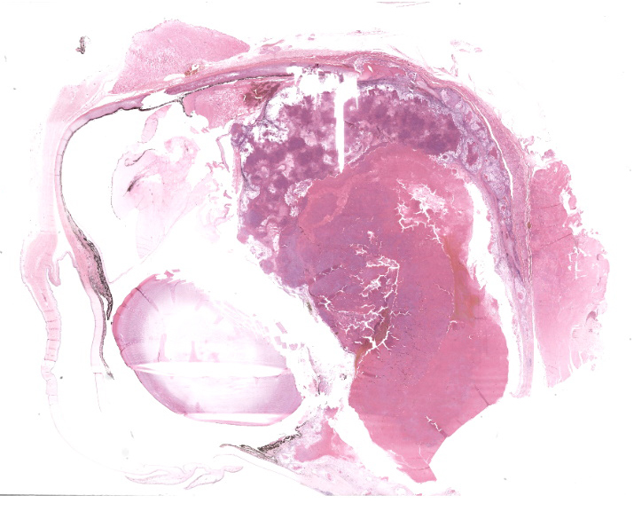
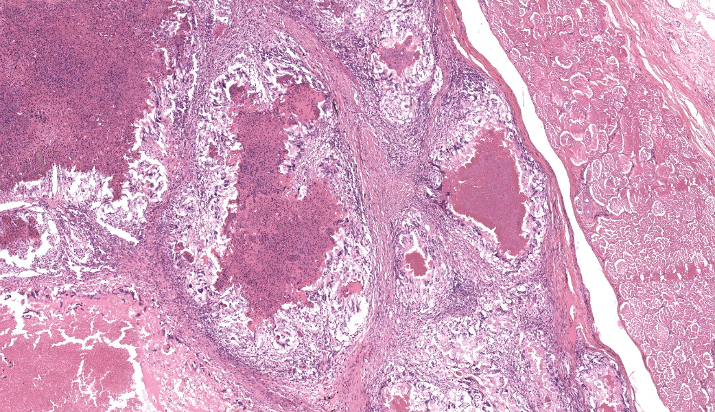
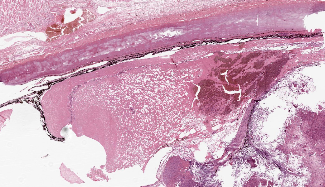
Left eye. Disrupting the fibrocartilaginous tunic of the eye, infiltrating and obliterating the choroid, anteriorly displacing the retina, and filling >80% of the posterior chamber is a multilobular mass of coalescing granulomas, necrotic and cellular debris, and hemorrhage. Granulomas are characterized by central cores of densely eosinophilic protein, necrotic debris, degranulated heterophils, and negative-staining hyphal filaments. These cores are surrounded by abundant multinucleated giant cells and epithelioid to foamy macrophages, many with large, clear vacuoles. Individual lobules are separated by variable amounts of fibrous tissue infiltrated by lymphocytes, plasma cells, and intact heterophils.
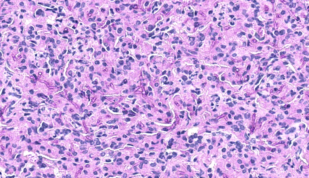
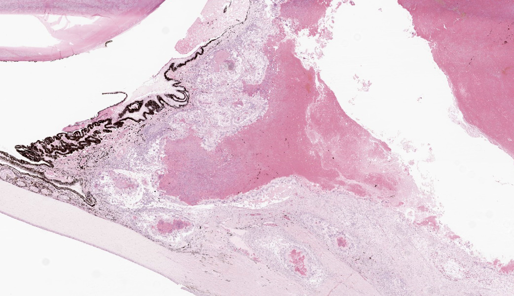
Fungal hyphae are nonpigmented, 2-4 um in diameter, parallel-walled, and septate, with dichotomous branching. Portions of the retina are adhered to the posterior surface of the ciliary body. The remaining retina is fragmented, vacuolated, and infiltrated by heterophils. Granulomas focally extend into anteriorly displace the ciliary body. The remaining vitreous humor is multifocally coagulated and contains heterophils, detached retinal pigmented epithelium and erythrocytes. The anterior surface of the lens capsule is multifocally adered by fibrin to pigmented epithelial cells consistent with those on the posterior surface of the iris and ciliary body, which also display accumulations of fibrin (posterior synechae). The iris is anteriorly displaced, the iridocorneal angle is collapsed, and the anterior chamber is reduced. The iris is infiltrated by low numbers of lymphocytes, macrophages and heterophils. The cornea has reduced stromal separation artifact (corneal edema).
Contributor's Morphologic Diagnosis: Left eye (OS): Panophthalmitis, granulomatous and heterophilic, severe, with abundant nonpigmented fungal hyphae.
Contributor's Comment:
The origin of the intra-ocular mycosis is presumed dissemination of fungi from a granulomatous pneumonia, which was found on necropsy the day following this enucleation. The pulmonary granulomas contained abundant morphologically identical fungal hyphae and widespread Pseudomonas aeruginosa colonies. Tracheal exudate cultured a fungus that was identified via 18S rRNA PCR as Purpureocillium lilacinum (formerly Paecilomyces lilacinus).
The ocular lesions in this case are not particular to a specific fungal organism. Indeed, ocular dissemination is a well-reported manifestation of systemic aspergillosis in German shepherd dogs. Anatomically, the retina of crocodilians is largely avascular, relying almost entirely on the richly vascular choroid layer just below.7 The expansile granulomas visible in this case clearly show that they originated in this vascular layer, displacing the retina anteriorly and deforming and infiltrating the sclera.
Hematogenous spread of fungal pathogens is a potential complication of any angioinvasive fungal infection, and in domestic animal veterinary medicine Aspergillus, Fusarium, and the Mucorales are often implicated.7 In addition to saprophytic fungi such as Aspergillus, in reptiles, systemic mycoses are often caused by entomopathogenic fungi, such as Purpurocilleum or Metarhizium.3,9 P. lilacinum is well reported as a cause of fatal systemic mycoses in turtles, crocodilians, and snakes.3,6,8,9 Rapidly-growing opportunists such as P. lilacinum are a particular danger to reptiles under high stocking density, cold stress, or unsanitary environmental conditions.8 In this case, P. aeruginosa, a bacterium fully capable of acting as a primary pathogen in an immunocompetent host, may have initiated the pneumonia and P. lilacinum was a secondary invader that subsequently seeded the eye. It is also plausible that there was an undetected stressor that resulted in P. lilacinum pneumonia with a secondary Pseudomonas co-infection.
This fungus is not solely a reptile pathogen. P. lilacinum has also been reported as a cause of fungal keratitis in humans, and general or local immunosuppression was a risk factor.2 A recent paper from Louisiana identified 21 fungal pneumonia cases in wild Virginia opossums, a subset of which were successfully identified via fungal isolation and/or PCR as P. lilacinum.5 The genus Purpurocilleum has recently been found to contain another species, P. lavendulum, closely related to P. lilacinum, which has been reported in fibrinous pneumonias of a green tree python and a panther chameleon.10 P. Javendulum replicates more readily below 35°C than P. macinum and is thus of potential importance as a reptile pathogen though of less concern for mammals or birds.
Contributing Institution:
Smithsonian's National Zoo and Conservation Biology Institute
P.O. Box 37012, MRC 5501, Washington, DC 20013-7012
https://nationalzoo.si.edu/animals/veterinary-care
JPC Morphologic Diagnosis:
Globe: Panophthalmitis, heterophilic and granulomatous, chronic, diffuse, severe, with anterior and posterior synechiae, retinal detachment and degeneration, anterior lens luxation, and numerous fungal hyphae.
JPC Comment:
Dr. Holder opened the final case of the year by ranking crocodilians on the "Holder Scale of Spiciness." The Philippine crocodile, she explained, is "low spicy per unit mass", which translates to a contextually polite, manageable reptile compared to, say, the saltwater crocodile, which is "high spicy." The Cuban crocodile, however, she described as "the ultimate spicy." With longer legs set more underneath the body, Cuban crocs can gallop and (kind of) jump, and they do so with a raging bad attitude. With that energy established, the group turned to the eye.
One of the first questions raised during the discussion of this case was the increased cellularity of the lens. Participants wondered whether this represented a degenerative change; however, reptiles and birds possess an annular pad, which is a more densely cellular structure which supports visual accommodation.4 It is not a degenerative change like one might see in other species, but rather is normal and a direct result of reptile evolution.
Participants also noted lymphoid follicles in the conjunctiva. These were initially mistaken by some as small foci of lymphoplasmacytic inflammation, but crocodilians have normal conjunctival lymphoid tissue.4 These follicles were a bit hyperplastic, however, in response to the raging fungal panophthalmitis. On H&E, the fungal hyphae stained negatively, appearing as faint, ghostly outlines that Dr. Holder described as "haunting the slide." A PAS stain brought them out into the light. This led to a broader discussion about fungal diagnostics and how temperature can profoundly affect fungal morphology.9 For this reason, fungi may have different appearances in tissue between mammals and reptiles. And a reminder to all - appearance alone on histology should not be considered definitive for speciation9 - accurate identification requires molecular testing, supported by morphology, for any fungus.
The culprit in this case, Purpureocillium lilacinum, is a saprophytic fungus that thrives in humid environments where meat is used as a food source. Basically, crocodile enclosures are its dream home. Fat from food creates a film on the water, which deposits on the floor of the tank and serves as an organic substrate for the fungus to grow.7 Purpureocillium lilacinum gets its name from the formation of purple to rosy fungal mats, both in culture and in tissue, making it one of few fungi with built-in aesthetics.1 Dr. Holder noted that Purpureocillium spp are also associated with pneumonia in opossums, whose lower body temperature makes them "step-reptiles" in terms of fungal susceptibility.5 She also emphasized that stocking density plays a major role in Purpureocillium outbreaks, and crowded enclosures create ideal conditions for fungal spread.10
References:
- Chen W, Hu Q. Secondary Metabolites of Purpureocilliumlilacinum. Molecules. 2021;27(1):18.
- Chew R, Dorman A, Woods ML. Purpureocillium lilacinum keratitis: a case series and review of the literature. Canadian Journal of Ophthalmology. 2016;1;51(5):382-5.
- Heard DJ, Cantor GH, Jacobson ER, Purich B, Ajello L, Padhye AA. Hyalohyphomycosis caused by Paecilomyces lilacinus in an Aldabra tortoise. JAVMA. 1986;189(9):1143-5.
- Holmberg BJ. Ophthalmology of Exotic Pets. Slatter's Fundamentals of Veterinary Ophthalmology. 2008;427-441.
- Falconnier N, Mitchell MA, et al. Pulmonary lesions in Virginia opossums (Didelphis virginiana): Characterizing fungal pneumonia associated with Purpureocillium lilacinum. Veterinary Pathology. 2025:03009858251338845.
- Lafortune M, Wellehan JF, Terrell SP, Jacobson ER, Heard D, Kimbrough JW. Shell and systemic hyalohyphomycosis in Fly River turtles (Carettochelys insculpta) caused by Paecilomyces lilacinus. Journal of Herpetological Medicine and Surgery. 2005;15(2):15-9.
- Martinez PS, Plummer CE. Ophthalmology of Crocodilia: Alligators, Crocodiles, Caimans, and Gharials. in: Montiani-Ferreira F. Wild and Exotic Animal Ophthalmology: Volume 1: Invertebrates, Fishes, Amphibians, Reptiles, and Birds. Cham: Springer International Publishing AG; 2022.
- Meyer J, Loncaric I, Richter B, Spergser J. Fatal Purpureocillium lilacinum pneumonia in a green tree python. JVDI. 2018;30(2):305-9.
- Pare JA, Conley KJ. Mycotic diseases of reptiles. In: Infectious diseases and pathology of reptiles. CRC Press; 2020.
- Schmidt V, Klasen L, Schneider J, Hubel J, Cramer K. Pulmonary fungal granulomas and fibrinous pneumonia caused by different hypocrealean fungi in reptiles. Veterinary Microbiology. 2018;1;225:58-63.