Signalment:
Gross Description:
Histopathologic Description:
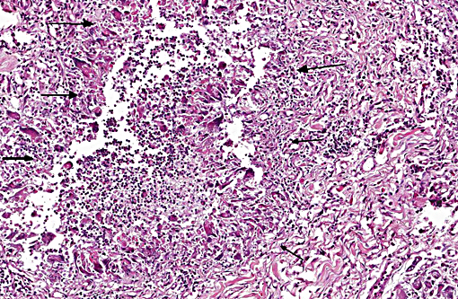
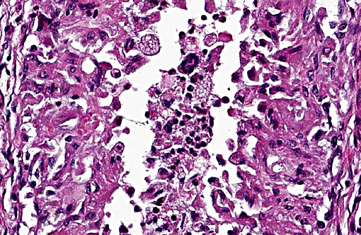
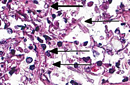
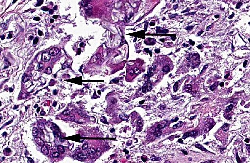
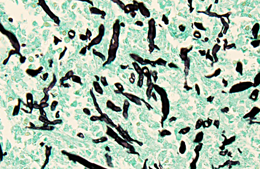
In all tissues, normal architecture is obscured by variably-sized coalescing cellular nodules that are often separated by thick bands of fibrous connective tissue. Nodules often have centers comprised of cellular debris, few to moderate neutrophils and few eosinophils. Macrophages, epithelioid cells, multinucleated giant cells, Langhans giant cells, few to moderate lymphocytes, plasma cells, and fibroblasts surround these necrotic centers. Other nodules lack necrotic centers and are comprised of variable combinations and concen-trations of plasma cells, macrophages, epithelioid cells, multinucleated and Langhans giant cells. Giant cells often contain intracytoplasmic round, 2-4 micron structures with clear centers and thick eosinophilic walls. Within cellular masses, the periodic acid-Schiff stain reveals numerous non-parallel hyphae with thick walls. Hyphae are poorly septate, 6-12 micron wide with occasional non-dichotomous branching (Fig. 1).
Morphologic Diagnosis:
Condition:
Contributor Comment:
Dogs with compromised immune systems are highly susceptible to both conditions. These infections are usually obtained via inhalation or ingestion with primary sites in the respiratory and gastrointestinal systems. Cutaneous infections in both immunosuppressed and immunocompetent dogs are often secondary to open skin wounds. Pythium insidiosum may be present in water, and when animals with open skin wounds enter contaminated bodies of water, they are susceptible to these infections. Skin lesions are typically characterized by nodules that are frequently ulcerated with necrotic centers and granulomatous and or pyogranulomatous inflammation. Hyphae with non-septate or poorly septate, non-parallel walls, and non-dichotomous branches are present in nodule centers. Hyphae are 4-16 micron wide. These features are beneficial in distinguishing Zycomycetes and Pythium from their primary differential diagnoses of aspergillosis and candidiasis. Aspergillus sp. is septate and 2-3 micron wide and Candida sp. is 2-3 micron wide with pseudohyphae.(2) Drugs that are effective against fungal infections are ineffective for treatment of Pythium species, which is not a fungus but a member of the phylum Oomycota.(3)
JPC Diagnosis:
Conference Comment:
Pythium insidiosum and Lagenidum species Oomycetes are dimorphic water molds (previously thought to be fungi) found in tropical or subtropical areas such as the southern United States. Infection is primarily cutaneous or subcutaneous, but systemic, gastrointestinal and vascular sites can occur in domestic animals and humans. Infection occurs when the host comes into contact with standing or stagnant water containing the motile aquatic flagellate zoo-spores; this infectious form of the organism is attracted to injured mammalian tissue. Infections in domestic animals are most commonly reported in the limbs, ventral thorax and abdomen of horses. In horses, the organisms cause large granuloma like mass lesions that may contain a purulent discharge and clinically resemble cutaneous habronemiasis, neoplasia, and exuberant granulation tissue.(1) The cutaneous condition in the dog is less common than the gastrointestinal form, where the condition is also often progressive and refractory to medical therapy; the lesion starts as a dermal nodule that may resemble a lick granuloma and expands into the subcutis, spreads peripherally, may ulcerate and develop draining tracts and is often associated with an eosinophilia. Infection in cats and cattle has been reported, but is less common in these species. Lagenidum species cutane-ous/subcutaneous infection is very similar to pythiosis clinically, and is equally aggressive, but has only been reported in dogs.(1)
Zygomycosis is caused by fungi in the class Zygomycetes which is divided into the two orders, Mucorales and Entomophthorales. The hyphae are described as being infrequently septate and their broad nature allows them to be differentiated from other fungal organisms. Like pythiosis, zygomycosis most commonly occurs in horses and the cutaneous lesions resemble those of pythiosis. However, zygomycotic cutaneous lesions in horses are usually found on the lateral neck, trunk or head, are smaller than those of pythiosis, and often have no association with standing water.(1)
References:
1. Ginn PE, Mansell JEKL, Rakich PM. Skin and appendages. In: Maxie MG, ed. Jubb, Kennedy and Palmers Pathology of Domestic Animals. 5th ed. Philadelphia, PA: Elsevier Saunders; 2007:700-709.
2. Ribes JA, Vanover-Sams CL, Baker CL. Zygomycetes in Human Disease. Clin Microbiol Rev. 2000;13:236-301.
3. Wim G, Len JA, et al. Pythium insidiosum: An Overview. Veterinary Microbiology. 2010; 146:1-16.