Signalment:
Gross Description:
Histopathologic Description:
Morphologic Diagnosis:
Condition:
Contributor Comment:
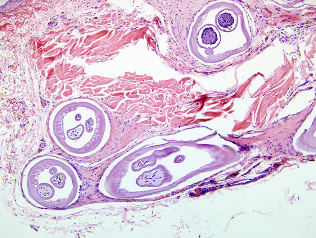
Onchocerca sp. has worldwide distribution and infects ungulates and humans. This tissue is from a series of 9 cases of canine ocular onchoceriasis we received from the Western United States (California, Nevada, Utah, Arizona) since 2004. The typical lesion is an episcleral or conjunctival nodule with lymphogranulomatous inflammation, eosinophils, plasma cells, fibrosis and granuloma formation.
Series of canine cases of episcleral Onchocerca infestation have been reported from Greece, Hungary, and the Western US. Controversy exists as to the Onchocerca species causing canine infection. The Greek and Hungarian isolates are thought to be of the same species; light microscopic study indicates morphologic similarities in U.S. versus Hungarian and Greek parasites. Many previous reports regarding Greek and Hungarian cases incriminate O. lupi with the dog as the definitive host. Other authors believe that aberrant migration of O. lienalis of cattle is responsible for American canine ocular onchocerciasis. While O. lienalis is considered the most likely diagnosis in the cases of American canine onchocerciasis based on widespread geographic incidence of O. lienalis and parasite morphology, experimental infection of dogs with O. lienalis has not been successful, and canine Onchocerca has not been found in cattle-specific locations (gastro-splenic ligament). Furthermore, although the natural host of O. lienalis is cattle, the worms found in dogs have been gravid, suggesting patent infection.
JPC Diagnosis:
Conference Comment:
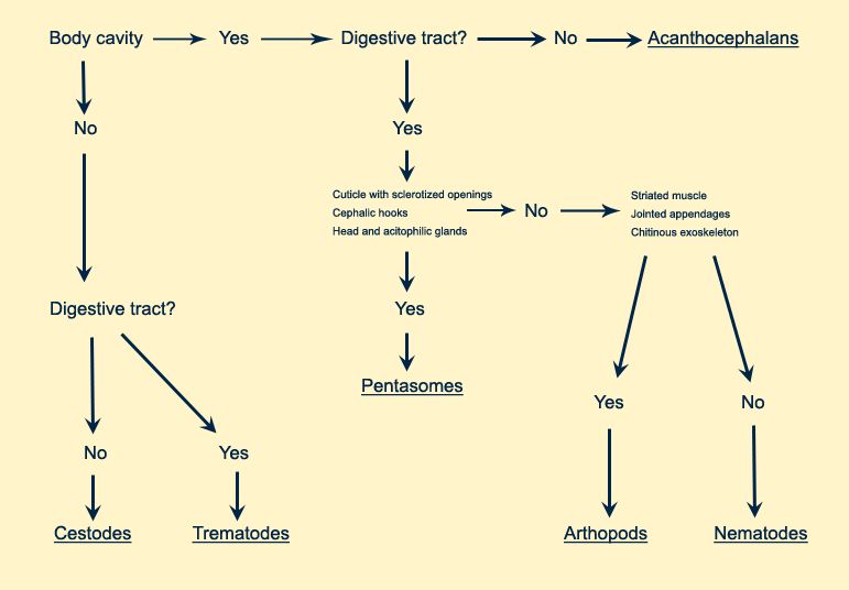
There was slide variation in the presence and severity of associated granulomatous inflammation; in most slides it was minimal. The conference discussion centered largely on the identification of this filarid nematode and the distinguishing characteristics of not only filarids but Onchocerca sp. Filarids are small nematodes that infect a number of different domestic animals. The majority of parasites in this group produce microfilaria, which are very distinctive larvae that when seen in the adult female are helpful in identification.(1) Dr. Chris Gardiner, the AFIP parasitology consultant, has mentioned that these larvae often are very basophilic and look like a bag of nuclei. Filarids have coelomyarian musculature. In Onchocerca, these muscles atrophy and are replaced hypodermal tissue. Even more important than microfilaria in identification of filarids is their tell-tale intestine, which is very small and a key diagnostic feature. These characteristics help differentiate Onchocerca sp. from the spirurid Thelazia sp., another common parasitic scourge of the mammalian eye.(1) The moderator also discussed various features of metazoan parasites in histologic section. A chart used by AFIP residents is included here to aid in the classification of parasites in tissue section.
References:
2. Zarfoss KM, Dubielzig RR, Eberhard ML Schmidt KS: Canine ocular onchocerciasis in the United States: Two new cases and a review of the literature. Vet Ophthal 8:51-57, 2005