CASE III:
Signalment:
A ten-year-old castrated male domestic short-haired cat.
History:
The cat had relapsing icterus, anemia, and lethargy following an episode of similar clinical signs 2 months before presentation. The cat was treated with conservative care and showed minor improvement; its owners elected euthanasia.
Gross Pathology:
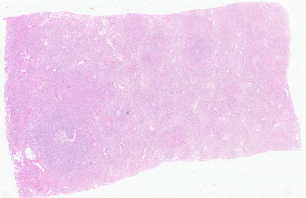
Pale and yellow mucous membranes and severe flea infestation. A small amount of transparent pericardial fluid was noted. The liver is diffusely yellow, has an irregular, rugged surface with multifocal nodules, and a single 2-centimeter, slightly firm, roughly spherical mass in the right central lobe. The adrenal glands are bilaterally symmetrically enlarged. The mesenteric lymph nodes are moderately enlarged but retain normal architecture.
Laboratory Results:
Titer of 1:1024 for antibodies to Toxoplasma gondii
Microscopic Description:
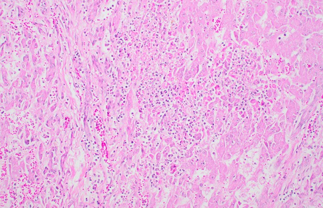
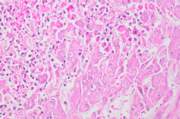
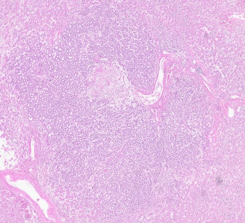
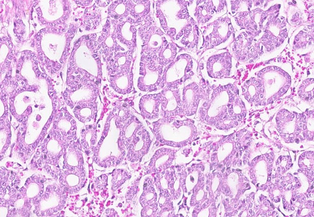
Liver: The liver is expanded by a well-demarcated, unencapsulated, expansile, moderately dense cellular neoplastic cell proliferation. The neoplastic cells are arranged as closely packed tubules and are supported by small amounts of fibrous stroma. The neoplastic cells are cuboidal to polygonal, have variably distinct cell borders, and small to moderate amounts of eosinophilic cytoplasm. The nuclei are intermediate to large, centrally located, have finely stippled chromatin, and 1-2 inconspicuous nucleoli. Anisokaryosis and anisocytosis are mild to moderate. Mitotic index is 10. Within the neoplastic cell proliferation and the adjacent liver, there are multiple randomly distributed foci of coagulative necrosis, admixed with small numbers of neutrophils, macrophages, lymphocytes, and plasma cells (most prominent at the periphery of the necrotic foci). Within the necrotic foci, there are numerous 20-30 micrometer in diameter, round to oval parasitic cysts; each one has a thin 0.5 micrometer basophilic cyst wall and contains numerous 2-3 micrometer, basophilic round to banana-shaped bradyzoites. Occasionally, scattered tachyzoites are present within macrophages and hepatocytes.
Contributor's Morphologic Diagnoses:
Liver:
1) Cholangioma.
2) Severe, multifocal to coalescing acute necrotizing hepatitis with myriad protozoal cysts and zoites (consistent with Toxoplasma).
Contributor's Comment:
Lesions in other organs include severe necrotizing adrenalitis with protozoal cysts, mild lymphocytic myocarditis, and mild interstitial pneumonia with pulmonary edema. Immunohistochemistry for Toxoplasma gondii showed strong positive staining.
Toxoplasma gondii uses felids as definitive hosts and is heteroxenous - cats can be infected by ingesting asexual stages in prey tissue or by ingesting oocysts. In intermediate hosts and in cats, asexual development occurs in many organs and tissues.
Toxoplasma gondii (and Neospora caninum) are unique among protists because of their ability to parasitize a wide range of hosts. Natural infection occurs in birds, primates (including humans), rodents, insectivores, herbivores, and carnivores. Systemic toxoplasmosis occurs most often in immunologically immature or immunocompromised animals. In the cat, intestinal and systemic infection occur almost simultaneously. It can spread via lymphocytes to the lymph nodes and from there to the blood/lymph and portal circulation to the liver.
Focal necrosis is common and appears to be related to rapid replication of tachyzoites. Immune animals develop a chronic or dormant form of toxoplasma infection that is characterized by the formation of cysts containing bradyzoites. In the liver, irregular foci of coagulative necrosis are scattered randomly; usually, only little inflammation is associated with the necrotic area. variable number of tachyzoites may be present in hepatocytes and Kupffer cells, usually at the periphery of the lesions. In the adrenals, vast areas of necrosis are usually seen in the adrenal cortex with minimal inflammation.
The hepatic neoplasm in this case is an incidental finding. We discuss three differentials. diagnoses for it. Cholangiocarcinoma, biliary cystadenoma (the proper term now is congenital biliary cysts), and cholangioma. The small size, demarcation, low cellular pleomorphism, lack of invasion or metastases, lack of substantial desmoplasia, and lack of aggregates of hepatocytes between the neoplastic tubules favored the diagnosis of cholangioma.
Contributing Institution:
Kimron Veterinary Institute
https://www.gov.il/en/government-service-branches/veterinary_institute
JPC Diagnoses:
1. Liver: Hepatitis, necrotizing, subacute, multifocal to coalescing, marked, with intra-hepatocytic, intra-histiocytic, and extracellular apicomplexan zoites.
2. Liver: Biliary adenoma.
JPC Comment:
Conference discussion for this case focused on differentiating biliary hyperplasia, biliary adenoma, and cholangiocarcinoma, as well as the differential diagnoses for feline hepatic necrosis. In the primary liver section evaluated by WSC staff and the conference moderator, the proliferation of irregular bile ducts was predominantly focal, featuring several satellite lobules adjacent to the main lesion. In subsequent sections cut for the resident unknown slide, special stains, and immunohistochemistry, the proliferative mass was diminished in extent, appearing as a single, 3-mm-diameter lobule of relatively well-differentiated bile ducts. For this reason, the presenting resident favored biliary hyperplasia, aligning with her top differential diagnosis for the concurrent hepatic necrosis, which was acetaminophen toxicosis. Although hepatic necrosis, bile duct proliferation, and cholestasis are recognized findings in feline acetaminophen toxicity, three primary features rule out that diagnosis in this case1.
First, although extensive necrosis obscures the architectural pattern, several centrilobular and periportal regions remain unaffected. These spared regions support a multifocal to coalescing, random pattern of necrosis, which is inconsistent with hepatotoxins like acetaminophen that classically cause centrilobular to midzonal necrosis.
Second, the necrotic foci are directly centered on intra- and extracellular apicomplexan zoites. Felids serve as both the definitive and intermediate hosts for Toxoplasma gondii1. In intermediate hosts, primary infection typically transitions to subclinical, chronic carriage of dormant tissue cysts. However, immunosuppression or concurrent disease impairs the CD8+ T-cell and interferon-gamma responses that maintain encystment, which precipitates rupture of tissue cysts and clinical disease4. Although a predisposing cause of immunosuppression was not definitive in this animal's clinical history, common triggers include young age or concurrent immunosuppressive viral infections (e.g., feline leukemia virus, feline immunodeficiency virus, or feline panleukopenia virus)5,6.
Finally, while ductular reaction occurs in subacute feline acetaminophen hepatopathy, marked, solitary focal proliferation is atypical. Acetaminophen-induced ductular change typically presents as a diffuse or multifocal periportal increase in bile duct profiles rather than a discrete focal nodule.
References:
- Jubb, Kennedy and Palmer's Pathology of Domestic Animals Fifth edition vol I p437, vol 2 p270-272, vol 3 p410
- Stacey A. Elmore, Jeffrey L. Jones, Patricia A. Conrad, Sharon Patton, David S. Lindsay, and J.P. Dubay. Toxoplasma gondii: epidemiology, feline clinical aspects, and prevention. 2010.
- Donald J. Meuten. Tumors in domestic animals. 2002;9:493-498
- Durieux MF, Lopez JG, Banjari M, et al. Toxoplasmosis in patients with an autoimmune disease and immunosuppressive agents: A multicenter study and literature review. PLoS Negl Trop Dis. 2022;16(8):e0010691.
- Moore A, Burrows AK, Malik R, Ghubash RM, Last RD, Remaj B. Fatal disseminated toxoplasmosis in a feline immunodeficiency virus-positive cat receiving oclacitinib for feline atopic skin syndrome. Vet Dermatol. 2022;33(5):435-439.
- Bartova E, Sedlak K, Vodlan S, Budikova M, Racka K. Seroprevalence of Toxoplasma gondii and Feline Immunodeficiency Virus in Domestic Cats and Their Associations with Clinical Signs. Acta Parasitol. 2026;71(4):149.