CASE I:
Signalment
6-month-old, intact male, Maine Coon, Felis catus, feline
History
This cat had an acute onset of lethargy and anorexia. The cat was presented to a small animal emergency service for neurologic signs consistent with obtundation. On presentation, the eyes were bilaterally sunken and third eyelids were protruding. Further examination revealed slightly increased bronchovesicular sounds, elevated body temperature, and ambulatory ataxia. During hospitalization, the cat experienced acute respiratory arrest followed by cardiac arrest.
Gross Pathology
At necropsy, the lungs were soft, diffusely mottled red to dark red, and oozed dark red, opaque fluid on cut section. The ileocecal lymph node was moderately enlarged and measured approximately 2.5 cm x 1.5 cm x 1.5 cm. There were no other significant gross lesions observed.
Laboratory Results
Michigan State University Veterinary Diagnostic Laboratory
RT-PCR Avian Influenza virus matrix PCR Nasal swab - Non-negative (AIV 31.91 CT) Avian Influenza Virus H5 PCR Nasal swab - Negative (AIV 32.39 CT) Avian influenza Virus H5 2.3.4.4 PCR Nasal swab - Non-negative (AIV 33.02 CT)
Immunohistochemistry
Cerebrum - AIV Antigen Detected Lung - AIV Antigen Not Detected
Michigan Department of Health and Human Services
Indirect fluorescent antibody test, brain: Rabies negativeNational Veterinary Services Laboratory
RT-PCR - Positive for HPAI A(H5N1) virus, clade 2.3.4.4b, genotype B3.13
Microscopic Description
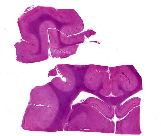
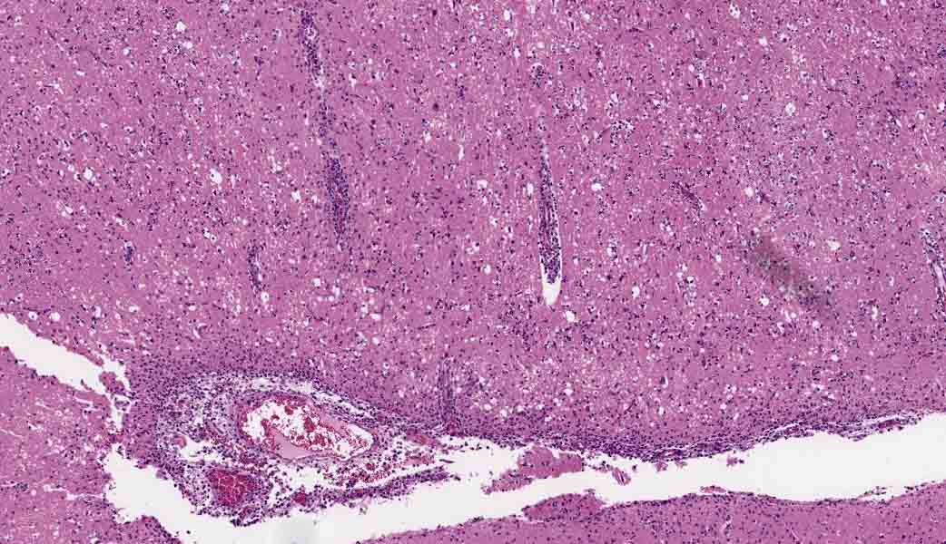
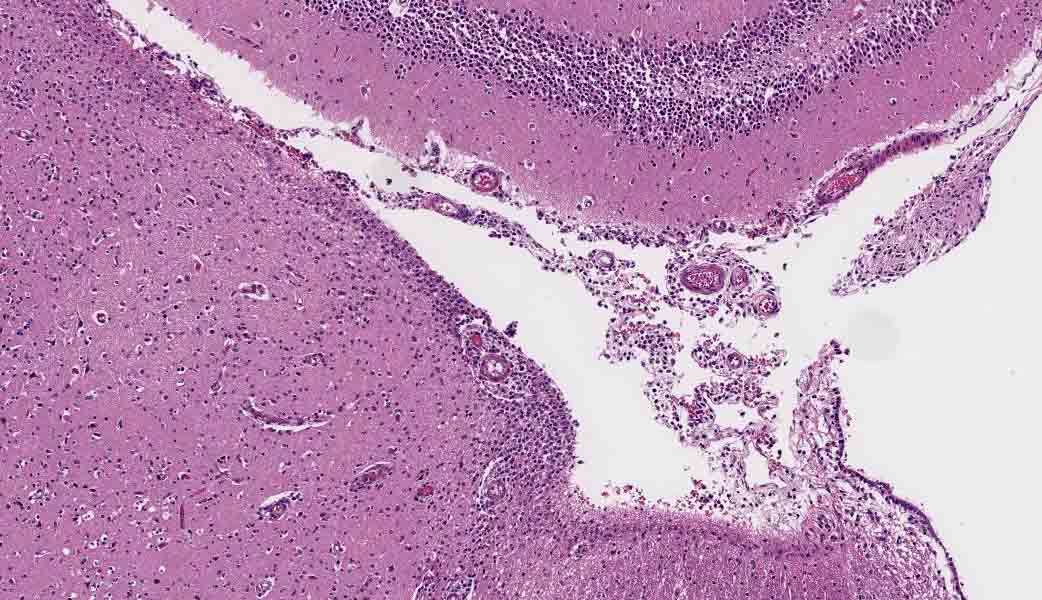
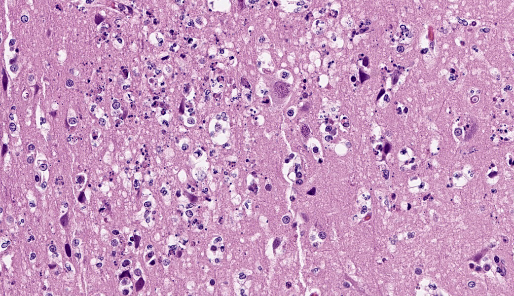
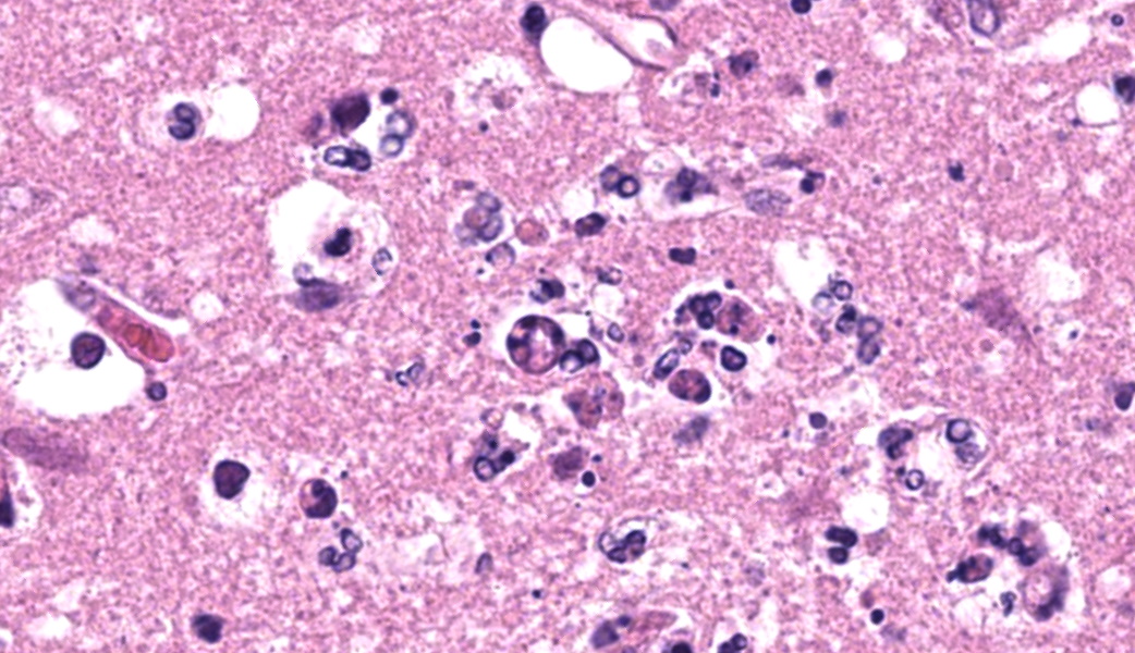
Throughout all examined sections of the cerebrum and within the brainstem, Virchow Robin spaces are mildly to moderately expanded by perivascular cuffs comprised of lymphocytes, histiocytes, and rare neutrophils ranging from 1-4 cell layers thick. The adjacent neuroparenchyma often contains increased numbers of scattered to small aggregates of glial cells admixed with low numbers of scattered lymphocytes, histiocytes, and rare neutrophils. Neuronal cell bodies within these regions and within the hippocampus are multifocally rimmed by increased numbers of glial cells, consistent with satellitosis. Neurons are occasionally shrunken, angular, and hypereosinophilic. The leptomeninges are mildly expanded by low to moderate numbers of lymphocytes and histiocytes, which multifocally infiltrate the surrounding cerebral grey matter. In one section, the adjacent grey matter contains numerous, multifocal small pools of perivascular hemorrhage rimmed by small amounts of karyorrhectic debris.
Contributor's Morphologic Diagnoses:
Cerebrum, brainstem, and meninges: Severe lymphohistiocytic meningoencephalitis with perivascular cuffing, multifocal hemorrhage, satellitosis, and neuronal necrosis
Contributor's Comment:
Over the last few decades, highly pathogenic avian influenza (HPAI) H5N1 has spread across the globe through the migration of numerous wild avian species.7,8 During this time, the H5 lineage has evolved within reservoir hosts, such as migratory waterfowl, via genetic drift and reassortment of viral RNA resulting in transformation of the virus into new strains which have led to devastating outbreaks within the poultry industry with concurrent spillover into an expansive array of mammalian hosts.7,8 Transmission from the reservoir hosts into spillover mammalian species can occur through direct transmission or adaptation of multiple viruses within intermediate hosts.7,8
In general, the efficacy of influenza virus to target intermediate hosts and spillover species depends on the ability of the viral hemagglutinin molecules to bind to sialic acid residues on epithelial cell surface glycoproteins.8 These surface glycoproteins often vary between different tissues within the body and different species resulting in a wide array of clinical presentations dynamically impacting various animals as well as humans.8 While not completely elucidated, transmission of the virus from reservoir or intermediate hosts to other mammals is thought to mainly occur via predator/prey relationships by consumption of infected animals by carnivorous species.7,8 However, recent outbreaks have highlighted the importance of fomite transmission through viral shedding and environmental contamination.1,8
Starting in March 2024, highly pathogenic avian influenza (H5N1) was identified in dairy cattle across the United States originating in Texas and spreading throughout multiple states, including Michigan.1,3,5 This strain of HPAI (H5N1, clade 2.3.4.4b) was detected in the United Sates in late 2021 to early 2022 and has been documented to affect multiple species, including cats.1,2,4,6,7 During the 2024 outbreak, HPAI was found in multiple dairy herds throughout Michigan with concurrent reports of feral or domesticated barn cats afflicted by serious, fatal infections.1 Previous HPAI outbreaks had similar reports with one of the first documented domestic feline HPAI H5N1 clade 2.3.4.4b infection occurring in France in December 2022 characterized by severe, fatal neurologic and respiratory signs which developed after close proximity to a duck farm with confirmed HPAI positive animals.2,6
In this case, the affected cat was an indoor only cat with no history of direct contact with other affected animals or history of drinking unpasteurized milk. Further investigation revealed that one of the members of the family worked with a HPAI positive dairy farm. As such, infection of this animal is presumed to have occurred secondary to fomite transmission of the virus from clothes worn by this individual who worked closely with positive HPAI dairy cattle and reported exposure to unpasteurized milk to the face and clothing.6 Personal protective equipment was not utilized, and the clothing was worn in the household prior to the development of neurologic signs and subsequent acute decline of the affected cat.6 Given the potential of fomite transmission, this case highlights the importance of utilizing personal protective equipment and strict potential post-exposure hygiene practices by veterinary professionals and people with careers where exposure to HPAI positive animals is possible.
Contributing Institution
Michigan State University Veterinary Diagnostic Laboratory 4125 Beaumont Rd, Lansing, MI 48910 https://cvm.msu.edu/vdl
JPC Diagnoses
Cerebrum: Meningoencephalitis, necrotizing and lymphohistiocytic, subacute, multifocal, marked with neuronal necrosis and gliosis
JPC Comment
This week's moderator was Dr. Thomas Cecere, Clinical Professor of Anatomic Pathology in the Department of Biomedical Sciences and Pathobiology, at the Virginia-Maryland College of Veterinary Medicine, a popular moderator at the JPC who has abiding interest in infectious disease and microbial immunology. Highly pathogenic avian influenza has been a frequent visitor to the WSC over the years with 15 entries including a range of poultry, waterfowl, and mammals including pigs, mustelids, and marine mammals, which demonstrates its increasing ability to a variety of avian and mammalian hosts.
A recent and interesting feature of the H5N1, clade 2.3.4.4b (and a potentially very worrisome one) is its ability to infect the mammary gland of dairy cattle. This is not the first documented case of milk-associated virus transmission in ruminants, which has been previously identified in experimental infection of Powassan virus in goats, tick-borne encephalitis virus in cattle and goats, and Middle East Respiratory Coronavirus (MERS-CoV) has been detected in camel milk.9 Obviously however, the current ability of H5N1, clade 2.3.4.4b's ability to infect the mammary gland of domestic cattle ad its ability to infect a wide range of additional hosts (including man) poses a significantly greater health risk than other viruses mentioned above.9 Pasteurization, however, has the ability to inactivate this virus.9
H5N1, clade 2.3.4.4b possesses not only the ability to infect the mammary gland but also can cause mastitis as well as produce heavy viral loads. Older multiparous animals appear to be more severely affected. Secretory epithelial cells in the bovine mammary gland express large amounts of the H5N1 viral entry receptor, ?2,3 sialic acid (while respiratory epithelium appears to express very few of these receptors, explaining why dairy cattle infected with this agent do not demonstrate significant respiratory disease.9 This finding also helps to explain why more severe clinical disease is seen in intramammary inoculated cattle over aerosol-infected animals. The U.S. Department of Agriculture has determined that milk from infected cows is the primary source of virus transmission to other dairy cattle, animals, and humans (Preliminary studies suggest that contaminated milking equipment, as well as infected air and water, may play a role in transmission between cows.9
Feline infection with HPAI is not a new occurrence, but previous infections have been associated with ingestion of infected poultry. In 2004, a variety of domestic cats and large zoo felids in Thailand died following being fed poultry that had died following an HPAI infection.10 Lesions in natural and experimental infections are not confined to the nervous system, and mirror the widespread infection seen in a variety of avian and mammalian species. Degenerative vascular changes and thrombosis are seen in a range of organs, lymphoid depletion and necrosis of epithelial cells in the airways, pancreas, liver, and adrenal gland, and the kidney have been identified in affected felids.11
Molecular analysis of H5N1 clade 2.3.4.4b infections in cats continue to yield clues to neurotropism of this virus in felids. Investigation of a 2024 outbreak which resulted in the deaths of ten cats in the same household with heavy viral loads in the brain demonstrated a number of unique viral mutations which may represent viral adaptation in this host species, including T143A in haemagglutinin, and two novel mutations in polymerase activity proteins (F314L, L342Q).12
Finally, another study demonstrates that brain is an excellent sample when testing for HPAI infection in cats, resulting in the lowest PCR cycle threshold values among sampled tissues. The authors suggest than random samples of Among these cases, brain was the best diagnostic sample, giving the lowest PCR Ct values when multiple sample types were tested. The authors propose that a random sampling of cerebral cortex is likely sufficient for testing given the abundant viral load seen in this organ in infected cats.13
References
- Burrough ER, Magstadt DR, Petersen B, Timmermans SJ, Gauger PC, Zhang J, et al. Highly pathogenic avian influenza A(H5N1) clade 2.3.4.4b virus infection in domestic dairy cattle and cats, United States, 2024. Emerg Infect Dis.
- Briand, F., Souchaud, F., Pierre, I., Beven, V., Hirchaud, E., Herault, F....Grasland, B. (2023). Highly Pathogenic Avian Influenza A(H5N1) Clade 2.3.4.4b Virus in Domestic Cat, France, 2022. Emerging Infectious Diseases, 29(8), 1696-1698.
- Garg S, Reed C, Davis CT, et al. Outbreak of Highly Pathogenic Avian Influenza A(H5N1) Viruses in U.S. Dairy Cattle and Detection of Two Human Cases ? United States, 2024. MMWR Morb Mortal Wkly Rep 2024;73:501-505.
- Graziosi, G., Lupini, C., Catelli, E., & Carnaccini, S. (2024). H5 Clade 2.3.4.4b Virus Infection in Birds and Mammals. Animals. 2024; 14(9):1372.
- Ly, H. (2024). Highly pathogenic avian influenza H5N1 virus infections of dairy and livestock handlers in the United States of America. Virulence, 15(1).
- Naraharisetti R, Weinberg M, Stoddard B, et al. Highly Pathogenic Avian Influenza A(H5N1) Virus Infection of Indoor Domestic Cats Within Dairy Industry Worker Households & Michigan, May 2024. MMWR Morb Mortal Wkly Rep 2025;74:61-65.
- Puryear, W. B., & Runstadler, J. A. (2024). High-pathogenicity avian influenza in wildlife: a changing disease dynamic that is expanding in wild birds and having an increasing impact on a growing number of mammals. Journal of the American Veterinary Medical Association, 262(5), 601-609.
- Kelvin AA, Baker PH, Ghosh S, Schultz-Cherry S, Langel SN. Influenza infection of the mammary gland. J Virol. 2025 Sep 23;99(9):e0194024. doi: 10.1128/jvi.01940-24.
- Runstadler, J. A., & Puryear, W. B. (2024). The virus is out of the barn: the emergence of HPAI as a pathogen of avian and mammalian wildlife around the globe. American Journal of Veterinary Research, 85(5), ajvr.24.01.0018.
- Keawcharoen J, Oraveerakul K, Kuiken T, et al. Avian influenza H5N1 in tigers and leopards. Emerg Infect Dis 2004; 10: 2189-2191.
- Schlachter AD, Bruno-McClung E, Diaz-Delgado J, Odom M, Banyard AC, Nunez A. Feline high pathogenicity avian influenza H5N1 infection: past and present. J Feline Med Surg. 2025 Oct;27(10):1098612X251370695. doi: 10.1177/1098612X251370695.
- Chothe SK, Srinivas S, Misra S, Nallipogu NC, Gilbride E, LaBella L, Mukherjee S, Gauthier CH, Pecoraro HL, Webb BT, Pipas JM, Ramasamy S, Kuchipudi SV. Marked neurotropism and potential adaptation of H5N1 clade 2.3.4.4.b virus in naturally infected domestic cats. Emerg Microbes Infect. 2025 Dec;14(1):2440498. doi: 10.1080/22221751.2024.2440498.
- Sillman SJ, Drozd M, Loy D, Harris SP. Naturally occurring highly pathogenic avian influenza virus H5N1 clade 2.3.4.4b infection in three domestic cats in North America during 2023. J Comp Pathol. 2023 Aug;205:17-23. doi: 10.1016/j.jcpa.2023.07.001. Epub 2023 Aug 14. PMID: 37586267.