Signalment:
Nine-year-old,
female, Papillon (
Canis familiaris).The dog
presented for the mammary masses near the right third nipple and under the left
forth nipple. As a result of the physical examination, the ovarian mass was also found. The ovaries and uterus were surgically with masses were sent to our laboratory for pathological examination.
Gross Description:
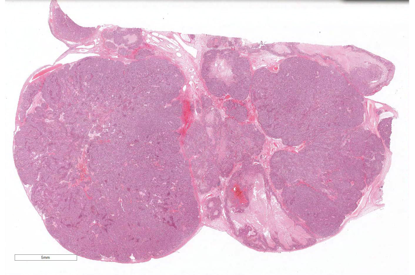
The left ovarian mass after fixed in neutral-buffered formalin was 5.2 x 4.5
x3.2 cm and was soft with milky white smooth to nodular surface. The cut
surface showed white to light yellow solid area with necrosis.
Histopathologic Description:
The
left ovarian mass consists of multiple lobules surrounded by a thin connective
tissue stroma with very few interstitial glands of original ovarian structures.
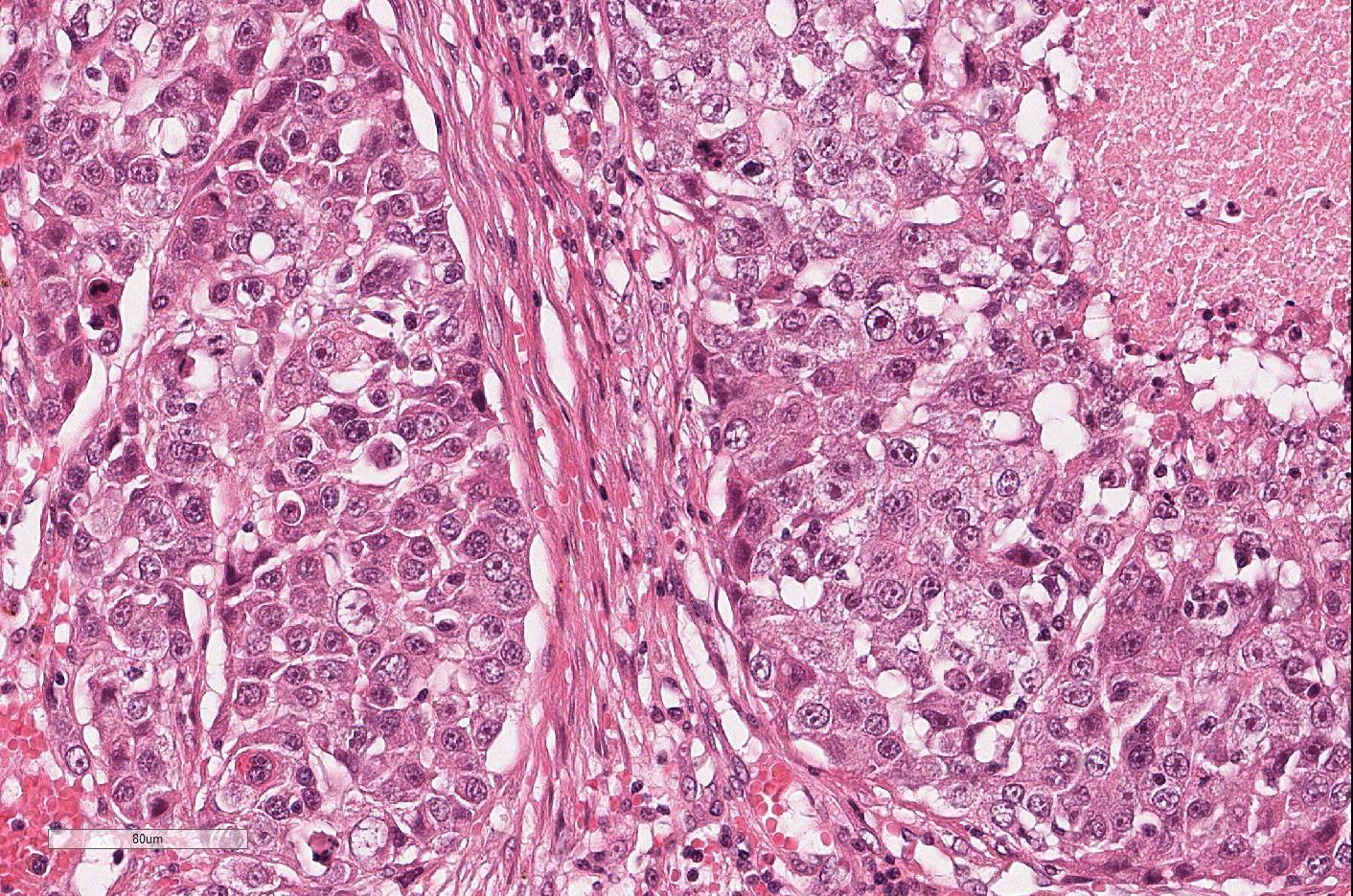
There are occasional multifocal to coalescing areas of necrosis in the mass. Lymphocytes
and plasma cells slightly infiltrated in stroma around tumor cells. Each lobule
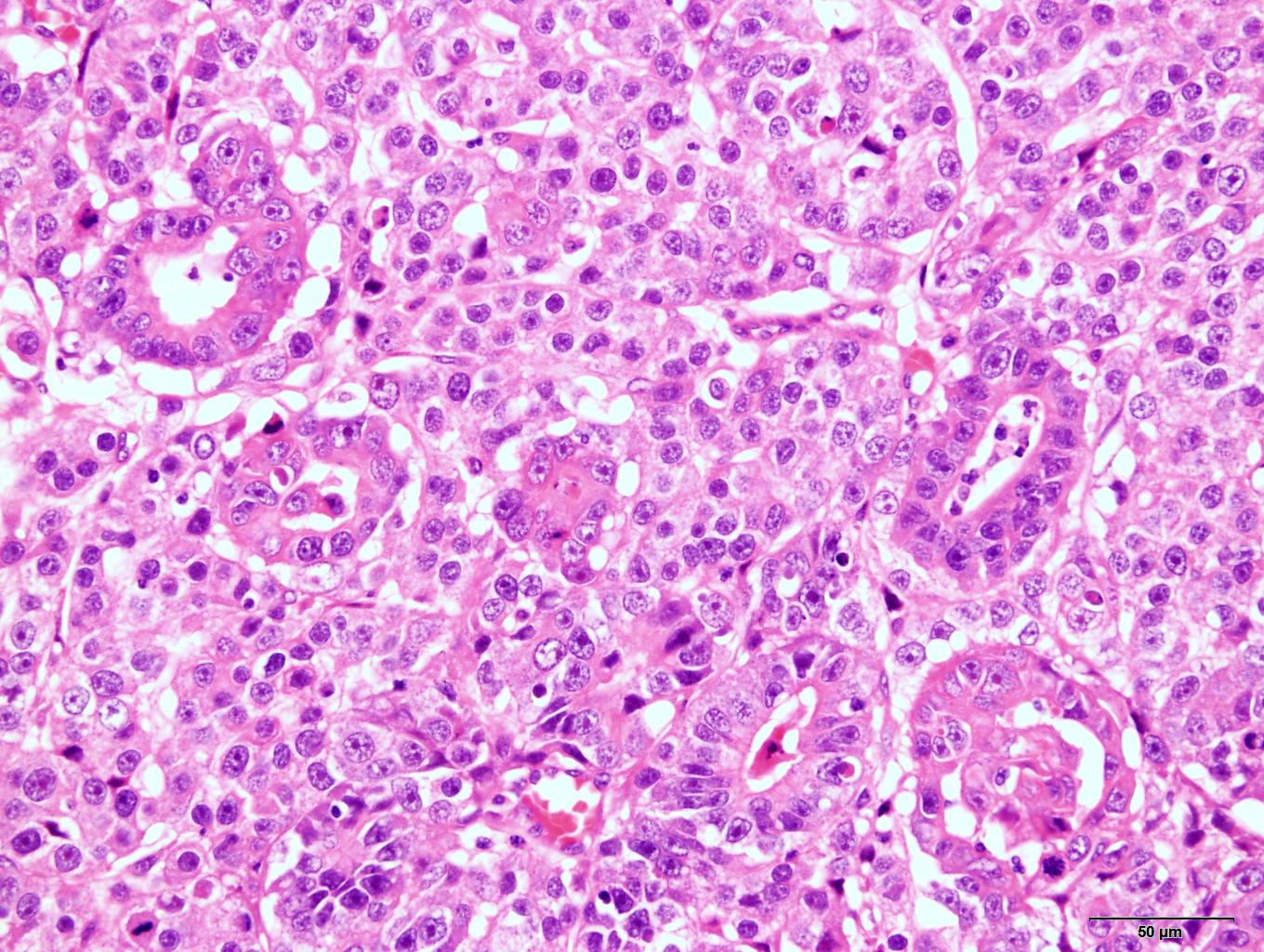
mainly composed of solid and irregular nests of round tumor cells. Ductal
structures and keratinizing epithelial cell nests were often mingled. Neoplastic
round tumor cells showed a high N/C ratio and resembled to germ cells of
seminoma/dysgerminoma. The tumor cells have large round nuclei with scattered
chromatin and one or a few large nucleoli. The cytoplasm is abundant with
weak-eosinophilic or clear, and infrequently vacuolated. Mitotic figures are
frequently seen. The nuclear figure of epithelial tumor cells are similar to
that of round tumor cells.
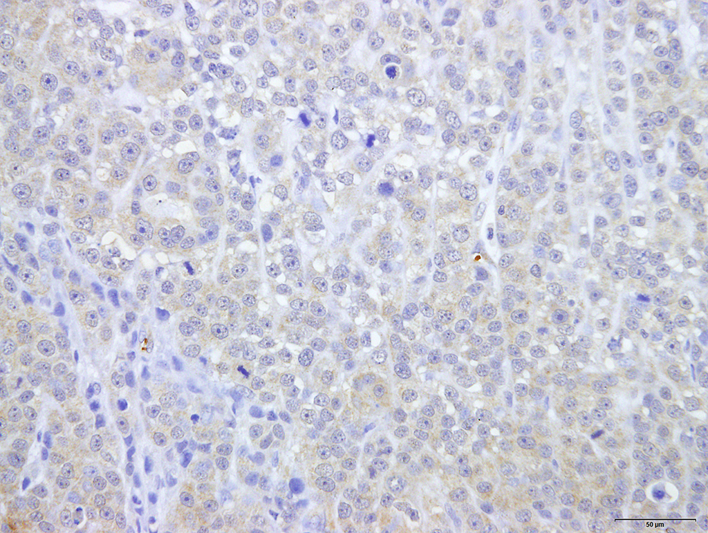
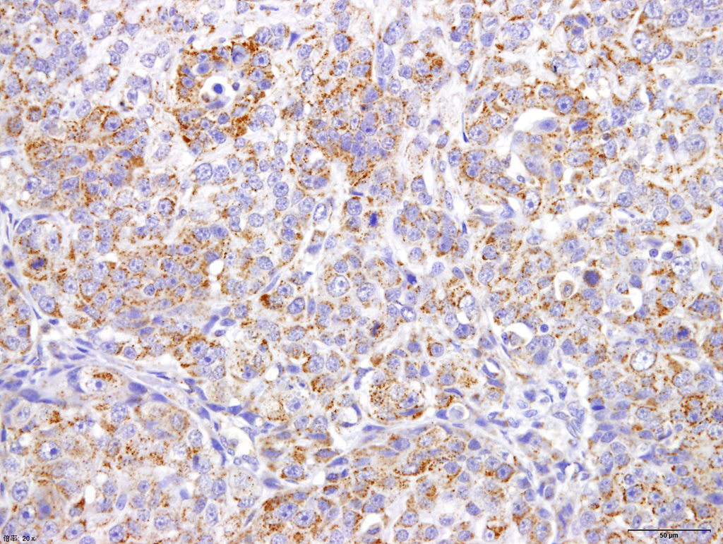
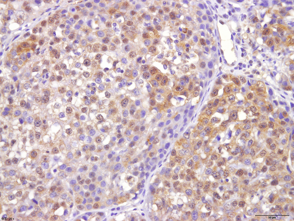
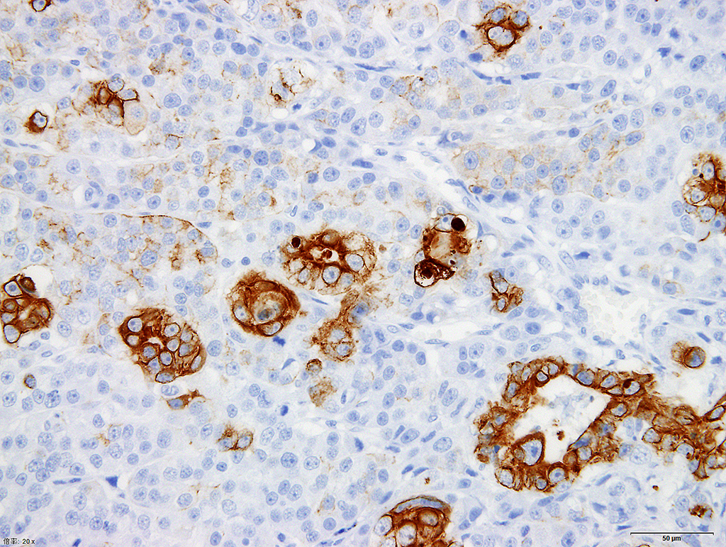
Immunohistochemically, both round and
epithelial tumor cells are cytoplasmic weakly positive for alpha-fetoprotein and cytoplasmic granular positive for CD30. Each tumor cell types also are positive
for octamer 4 (OCT4). Cytokeratin AE1/AE3 and CAM5.2 is strongly-expressed in
the cytoplasm of epithelial tumor cells and weakly positive in less than 50% of
round tumor cells. However, cytokeratin 7 and 20 are negative in both tumor
cells. Vimentin expression is seen in some part of round tumor cells, but is
not observed in epithelial tumor cells.
Morphologic Diagnosis:
Mixed germ cell tumor in canine ovary (dysgerminoma
with embryonal carcinoma).
Lab Results:
N/A
Condition:
Ovarian mixed germ cell tumor
Contributor Comment:
Canine ovarian
tumors are divided into sex cord-stromal (gonadostromal) tumors, germ cell
tumors, epithelial tumors, and mesenchymal tumors. Epithelial tumors and sex
cord-stromal (gonadostromal) tumors are the most common (80-90%). Germ cell
tumors are less common, and account for 6% to 12% of canine ovarian tumors.
Germ cell tumors are further classified to dysgerminoma, teratoma and embryonal
carcinoma according to the WHO classification.
1,2,6,9,
In addition,
yolk sac tumor and polyembryoma, chorio-carcinoma, and mixed germ cell tumor
are included among ovarian germ cell tumors of human WHO classification. In
canine ovarian germ cell tumors, dysgerminoma is most common and followed by
teratoma.
2,9
The present
tumor is mostly composed of round tumor cells, which resemble
seminoma/dysgerminoma. Positive immuno-reactivitiy for OCT-4 of round tumor
cells was also consistent with that of dysgerminoma, because OCT-4 is sensitive
and specific immunohistochemical marker for dysgerminoma, However, ductal
structures and keratinizing epithelial cell nests were often mingled, and these
tumor cells including round tumor cell showed positive for embryonal carcinoma
markers (AFP and CD30). Embryonal carcinoma composed of undifferentiated cells
of epithelial appearance with solidly cellular areas, glands, and papillary projections.
8,13,14
Areas of solid
growth in embryonal carcinomas histologically resemble dysgerminomas.
8
In humans, embryonal carcinoma is a rare germ cell tumor and occurs as a
component of mixed germ cell tumors more than pure embryonal carcinoma.
14
In animals, no pure embryonal carcinomas have been reported, but a combination
(mixed) germ cell tumor with embryonal carcinoma has been reported in only two
rats and a cynomolgus monkey.
11,15
Embryonal
carcinomas are immuno-histochemically distinguishable from dysgerminoma based
on testing for AFP, CD30, cytokeratin AE1/AE3, CAM5.2 and cytokeratin 7, which
are positive in embryonal carcinoma.
3,6,7 Thus, in the present case,
immunoreactivity of tumor cells did not perfectly satisfy the diagnostic
criterion for both dysgerminoma and embryonal carcinoma. In addition, round
tumor cells, which resemble dysgeminoma, are only partially positive for vimentin. In contrast, epithelial
tumor cells forming ducts and nests were mostly negative for vimentin.
Vimentin is immunopositive in dysgerminoma, and the reactivity is higher than
embryonal carcinoma.
4,8,13 We considered that the present tumor was partially
differentiated from dysgerminoma, and have the characteristics of both
dysgerminoma and embryonal carcinoma. Thus, the present tumor was diagnosed as
dysgerminoma with embryonic differentiation (mixed germ cell tumor composed of
dysgerminoma and embryonal carcinoma) rather than pure embryonal carcinoma.
JPC Diagnosis:
Ovary: Mixed
germ cell tumor, papillon,
Canis familiaris.
Conference Comment:
The contributor provides a challenging diagnostic case rare ovarian neoplasm in a dog. Due to near effacement of the
normal structures of the ovary by the neoplasm, some conference participants
had trouble identifying the tissue as ovary. However, at the periphery of the
neoplasm in all examined sections, there is a small amount of subsurface
epithelial structures and granulosa cell islands characteristic of canine
ovary. As mentioned by the contributor, tumors of the ovary are
uncommon and have been described in many species. They typically originate from
three distinct embryologic cell types: epithelial tumors of Mullerian origin
(adenoma or carcinoma), sex cord stromal tumors (granulosa cell tumor and
thecoma), and germ cell tumors (dysgerminoma, teratoma, yolk sac tumors). Mixed
germ cell tumors are a combination of germ cells and sex cord stromal cells. In
male dogs, mixed germ cell tumors are the fourth most common primary testicular
neoplasm and are typically characterized by a
combination of seminoma and Sertoli cell tumor, with the tubular structures of
Sertoli cell tumors containing neoplastic germ cells.5,10,12 They
are extremely rare in the ovary with reported cases in a Labrador retriever and
a cynomolgus
monkey.10,11,15
Among canine ovarian tumors, granulosa cell tumors and
epithelial tumors are by far the most common.12 In this case, the contributors posit that this is a dysgerminoma mixed
with an embryonal carcinoma, favoring the diagnosis of a mixed germ cell tumor. This interesting case stimulated discussion
among conference participants. Some favored the diagnosis of mixed germ cell
tumor and others favored a sex cord stromal tumor, dysgerminoma, or a collision
tumor. We reviewed this case in consultation with physician genitourinary
pathologists at the Joint Pathology Center, who agreed with the contributor and
the majority of conference participants, that there are foci suggesting a yolk
sac tumor (5%), embyronal carcinoma (~25%) and predominantly dysgerminoma
(70%), thus favoring the diagnosis of a mixed germ cell tumor. This case was
also studied in consultation with Dr. Robert Foster, a board certified veterinary pathologist and recognized
expert with extensive experience in the area of veterinary reproductive
pathology. He offers a dissenting view that the highly anaplastic cells may not
be germ cells and instead favors the diagnosis of a poorly differentiated
ovarian sex cord stromal tumor. He also notes that immunohistochemistry in
ovarian tumors can be problematic in domestic species.
References:
1. Akihara Y,
Shimoyama Y, Kawasako K, et al. Immuno-histochemical evaluation of canine
ovarian tumors. J Vet Med Sci. 2007; 69:703-708.
2. Bertazzolo W,
Dell'Orco M, Bonfanti U, et al. Cytological features of canine ovarian tumours:
a retrospective study of 19 cases. J Small Anim Pract. 2004; 45:539-545.
3. Liang Cheng,
Shaobo Zhang, Aleksander Talerman,et al. Nuclear
or cytoplasmic localization of Bag-1 distinctly correlates with pathologic
behavior and outcome of gastric carcinomas HumPathol. 2010:41:716-723.
4. Cossu-Rocca P,
Jones TD, Roth LM, et al. Cytokeratin and CD30 expression in dysgerminoma. Hum
Pathol. 2006; 37:1015-1021.
5. Foster RA. Male
genital system Wilcock BP, Njaa BL. Special senses. In: Maxie MG,
ed. Jubb, Kennedy and Palmer´s. Pathology of Domestic Animals. 6th
ed Vol. 3. St Louis, MO: Elsevier Saunders; 2016:496.
6. Kennedy PC CJ,
Edwards JF, et al. Histological classification of tumor of genital system of
domestic animals. In: World Health Organization International
histological classification of tumors of domestic animals. Wasington,
DC.1998.
7. Leroy X, Augusto
D, Leteurtre E, et al. CD30 and CD117 (c-kit) used in combination are useful
for distinguishing embryonal carcinoma from seminoma. J Histochem Cytochem. 2002;
50:283-285.
8. Lifschitz-Mercer
B, Walt H, Kushnir I, et.al. Differentiation potential of ovarian dysgerminoma:
an immuno-histochemical study of 15 cases. Hum Pathol. 1995; 26:62-66.
9. Patnaik AK,
Greenlee PG: Canine ovarian neoplasms: a clinicopathologic study of 71 cases,
including histology of 12 granulosa cell tumors. Vet Pathol.1987; 24:509-514.
10. Robinson NA,
Manivel JC, Olson EJ. Ovarian mixed germ cell tumor with yolk sac and teratomatous
components in a dog. J Vet Diagn Invest. 2013; 25:447-452.
11. Sawaki M,
Shinoda K, Hoshuyama S, et al. Combination of a teratoma and embryonal
carcinoma of the testis in SD IGS rats: a report of two cases. Toxicol
Pathol. 2000; 28:832-835.
12. Schlafer DH,
Foster RA. Female genital system. In: Maxie MG, ed. Jubb, Kennedy and
Palmer´s. Pathology of Domestic Animals. 6th ed Vol. 3. St Louis,
MO: Elsevier Saunders; 2016:377-378.
13. Suster S, Moran
CA, Dominguez-Malagon H, et al. Germ cell tumors of the mediastinum and testis:
a comparative immunohistochemical study of 120 cases. Hum Pathol.
1998;29:737-742.
14. Ulbright TM:
Germ cell tumors of the gonads: a selective review emphasizing problems in
differential diagnosis, newly appreciated, and controversial issues. Mod
Pathol. 2005.18(Suppl 2): 61-79.
15. Yokouchi Y,
Imaoka M, Sayama A, et al. Mixed germ cell tumor with embryonal carcinoma,
chorio-carcinoma, and epithelioid trophoblastic tumor in the ovary of a
cynomolgus monkey. Toxicol Pathol. 2011; 39:553-558.