Signalment:
Late term fetus, unknown gender, Red Angus,
Bos taurus, bovineFour abortions have occurred in this herd. Some of the aborted calves have had a short lower jaw.
Gross Description:
The mandible was between 2 and 3 cm shorter than the maxilla. Both the upper and lower molars were impacted. On cut surface, the marrow cavities of the bones at the base of the skull were filled with excessive bone. The marrow cavity of the tibia and femur was filled with bone, leaving no marrow cavity. The cortex of the tibia and femur is thin.
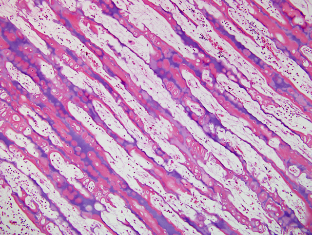
Histopathologic Description:
There is lack of resorption of the primary spongiosa in the distal metaphysis, with retention of cartilage deep into the distal metaphysic(
Fig. 3-1). Surfaces of primary spongiosa lack osteoclasts.
Morphologic Diagnosis:
Metaphysis - persistence of primary spongiosa
Lab Results:
Immunohistochemistry negative for BVD virus
Condition:
Osteopetrosis
Contributor Comment:
Osteopetrosis, also referred to as marble bone disease, occurs in multiple species of animals, and may have an inherited basis, or may have an acquired cause.(5) The osteopetroses are a heterogeneous group of bone remodeling disorders characterized by an increase in bone density due to a defect in osteoclastic bone resorption.(1) In cattle, osteopetrosis occurs in black and red Angus cattle in North America, and is assumed to be inherited as an autosomal recessive trait.(2) Affected calves are small, premature (251-276 days of gestation), and usually stillborn. Clinically, they show brachygnathia inferior, impacted molar teeth and protruding tongue. The long bones are shorter than normal, and easily fractured. Radiographically, the medullary cavities are dense, without clear differentiation between the cortex and medulla. Vertebrae are shortened, frontal and parietal bones are thick, and bones of the cranial base are thick and dense. On cut surface, the metaphyses and diaphyses of long bones are filled with dense, unresorbed cones of primary spongiosa extending from the metaphysis to the center of the diaphysis. The bones are more fragile than normal and may be fractured. As a result of the skull abnormalities, the cerebral hemispheres are rectangular with flattened dorsal surfaces, the cerebellum is partially herniated through the foramen magnum, and the optic nerves are hypoplastic.
Osteopetrosis also occurs in Hereford and Simmental cattle, resembling that of Angus calves.(4,5) BVD virus infection can cause osteopetrosis in cattle.(4) The osteopetrotic lesions are believed to be caused by transitory virus-induced osteoclast depletion. The gross lesions of BVD virus-induced osteopetrosis are different from the inherited variety in cattle.
Osteopetrosis-like lesions with metaphyseal sclerosis may occur in association with canine distemper virus infection in pups and in lead poisoning.(5)
In humans two forms of osteopetrosis are recognized: a severe, recessively inherited, lethal (malignant) form, with lesions present at birth, and a dominant (benign) form which becomes manifest later in life. In animals, most descriptions of osteopetrosis appear analogous to the malignant form and autosomal recessive inheritance is suspected in most cases.(5)
Histologically, growth plates are essentially normal but metaphyses are relatively avascular.(5) Dense chondro-osseous tissue, consisting of cartilage matrix lined by a thick layer of woven bone, occupies the medulla. Osteoclasts are rare and when present they appear to be inactive. Failure to replace the primary spongiosa and its associated woven bone with thicker trabeculae of mature lamellar bone presumably accounts for the increased fragility of osteopetrotic bones.
The lack of osteoclastic resorption of bone can occur as a deficiency in numbers of osteoclasts, or dysfunction in osteoclasts that may be present in normal numbers. Osteoclasts are capable of breaking down both the inorganic and organic matrix of bone.(1)
JPC Diagnosis:
Bone: Osteosclerosis, diffuse, severe, with retention of cartilage cores
Conference Comment:
Osteopetrosis has also been reported in four Peruvian Paso foals and one Appaloosa. The disease in horses has the same clinical and pathologic manifestations of the disease seen in the severe lethal form in Angus calves. Histologically, the only major difference is the presence of normal to increased numbers of osteoclasts. Ultrastructurally, osteoclasts in these foals do not have a ruffled border suggesting a functional defect in osteoclasts.(5)
Osteopetrosis has also been reported in white-tailed deer, Dachshund puppies, in an Australian Shepherd and a Pekingese dog, and in cats. In cats, osteopetrosis has been linked to vitamin D toxicosis and feline leukemia virus.(5)
References:
1. Balemans W, VanWesenbeeck L, VanHul W: A clinical and molecular overview of the human osteopetroses. Calcif Tissue Int
77:263-274, 2005
2. Greene HJ, Leipold HW, Hibbs CM, Kirkbride CA: Congenital osteopetrosis in Angus calves. J Am Vet Med Assoc
164:389-395, 1974
3. Ojo SA, Leipold HJ, Cho DY, Guffy MM: Osteopetrosis in two Hereford calves. J Am Vet Med Assoc
166:781-783, 1975
4. Scruggs DW, Fleming SA, Waslin WR, Grace AW: Osteopetrosis, anemia, thrombocytopenia, and marrow necrosis in beef calves naturally infected with bovine virus diarrhea virus. J Vet Diagn Invest 7:555-559, 1995
5. Thompson K: Diseases of bones and joints.Â
In: Pathology of Domestic Animals, ed. Maxie G, 5th ed., vol. 1, pp 38-40. WB Saunders, Edinburgh, Scotland, 2007