Joint Pathology Center
Veterinary Pathology Services
Wednesday Slide Conference
2019-2020
Conference 14
15 January 2020
Dr.
Neel Aziz
Supervising Veterinary Pathologist
Smithsonian Conservation Biology Institute
National Zoological Park
Washington DC, 20008
CASE III: 71618 (JPC 4136426).
Signalment: Adult male big brown bat (Eptesicus fuscus); 24 g, wet fixed.
Wild caught, Maryland, USA. (Submitted partially fixed in 10% neutral buffered Formalin)
History: This adult male big brown bat (Eptesicus fuscus) was found dead two months after being captured in Maryland for use in a research colony. It had been vaccinated for rabies and treated for mites, but no experimental manipulation had been performed.
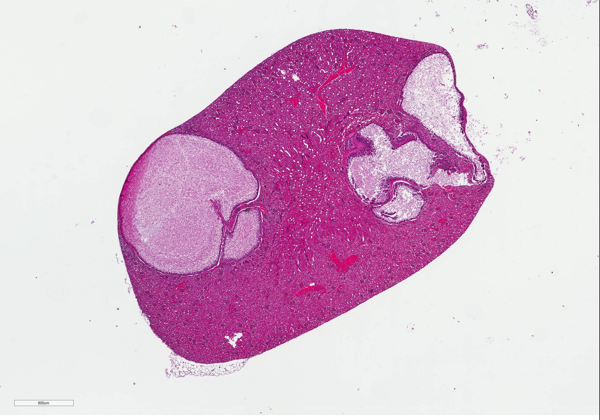
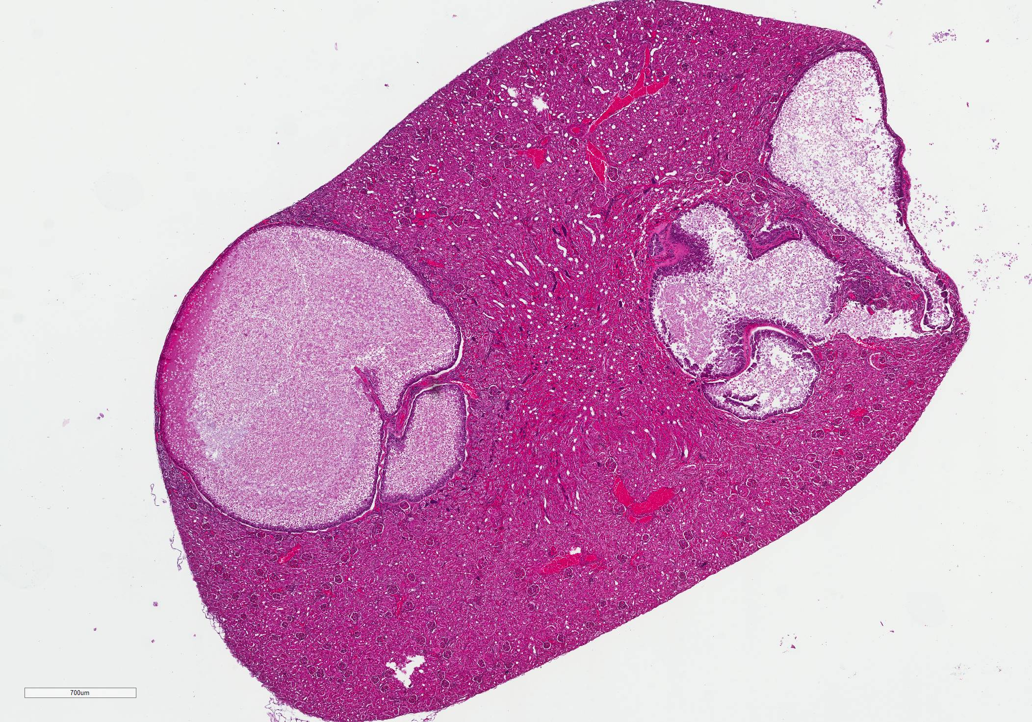
Gross Pathology: The right kidney has 4 bulging, rounded, pale tan structures, 2-5 mm diameter. On cut section these are restricted to the cortex and contain pale tan, turbid fluid.
Laboratory results:
- Blood examination: increased CK 1199U/L (99-436), increased GOT/GPT and bile acids (no value available), thrombocytopenia 23 K/?L (148-484) and increased PT 18s (11-17) and aPTT 144s (72-102).
-Toxoplasmosis and Neosporosis were excluded (IgG/M-determination.)
- Parvovirosis and Angiostrongylosis were tested with a negative result.
Microscopic Description:
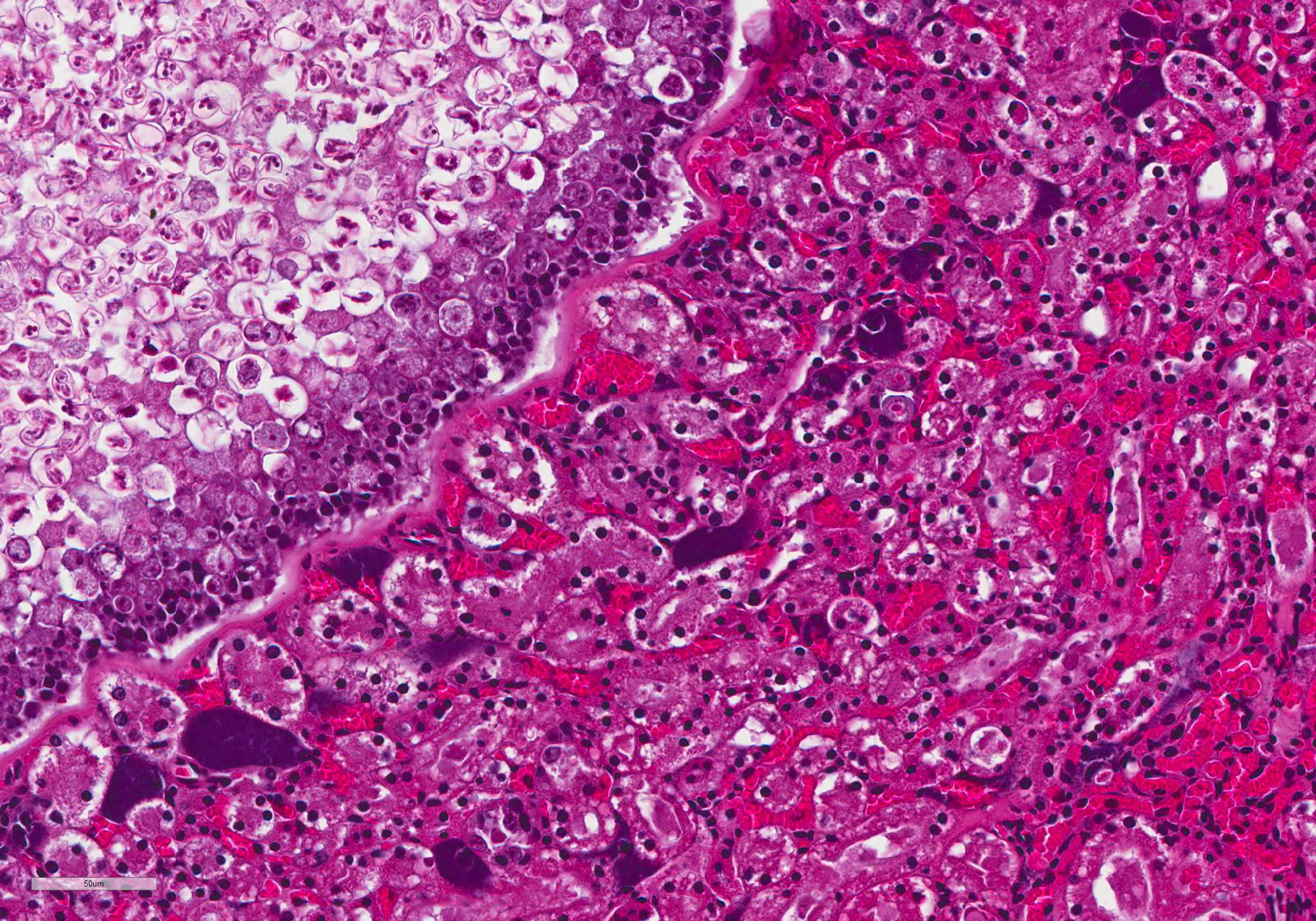
Expanding and compressing the renal cortex are multifocal cystic structures, which vary in size from 0.5 to 2 mm in diameter, and are surrounded by a 1-5 micron hyaline wall. The inner surface of the cyst is lined by 2-3 layers of hypertrophied and hyperplastic renal tubular epithelial cells. Epithelial cells contain numerous meronts and macrogametocytes, and rare microgametocytes. Within the cyst lumen are numerous round-to-oval, ~15-micron diameter oocysts with a thin, delicate eosinophilic capsule, which contain 2 sporocysts each containing 4 sickle-shaped sporozoites. Within the cyst lumen are occasional erythrocytes and abundant eosinophilic debris. Scattered throughout the cortex and medulla of both kidneys are multiple rafts of deeply basophilic bacteria, with no tissue response.
Contributor Morphologic Diagnosis:
Kidney: coccidiosis, chronic, multifocal, moderate, with cystic renal tubules
Contributor Comment: Renal coccidiosis has recently been described as an incidental postmortem lesion in wild big brown bats, caused by the coccidian parasite Nephroisospora eptesici.5 The organism was first described in 2010, after being identified in the kidneys of 29 wild big brown bats in Minnesota.5 Although renal coccidiosis had been previously reported in other species of bats, the causative organisms had not been fully characterized.1,2,3
Nephroisospora eptesici is a member of the family Sarcocystidae, most closely related to Besnoitia spp. Unlike related apicomplexans, Nephroisospora eptesici has a single-host life cycle, with the complete life cycle occurring in the kidney.5 Unlike other coccidians with a single-host life cycle, such as Eimeria spp., Nephroisospora eptesici is believed to have evolved from an ancestor which required two hosts.5 Thus, the single-host life cycle is a derived trait. The biology and host specificity of this organism remain to be elucidated. However, reports of morphologically similar coccidian parasites in other bats and the recent identification of Nephroisospora genetic material contaminating a published bat genome suggest that related organisms exist, although they have not been fully described.1,2,3,4
The associated gross lesions are white-to-beige spherical, well-demarcated cystic structures within the renal cortex.5 Histologically, these structures represent cystically dilated renal tubules enclosed by a PAS-positive hyaline membrane, which contain numerous intracellular and extracellular coccidian organisms representing all life stages.5 Oocysts within the lumen of the cystic tubules contain 2 sporocysts, which each contain 4 sporozoites.5 Periodic acid-Schiff (PAS) and Feulgen special stains can be used to better demonstrate protozoal structures.
Renal coccidiosis due to Nephroisospora eptesici is rarely associated with inflammation, and is not believed to be responsible for clinical disease.5 The primary significance of renal coccidiosis in bats is as an obfuscating factor in research. Genetic material of an undescribed Nephroisospora species has recently been identified as a contaminant in the published genome of David?s myotis (Myotis davidii).4
Abundant intravascular bacteria, without associated inflammatory response, were noted in the kidneys and other tissues. These were considered to represent postmortem overgrowth, as no wounds or other nidus of bacterial infection were identified. Another bat autopsied from this colony had similar cystic structures in the kidneys. In that case, the structures had central mineralization, indiscernible organisms, and associated lymphoplasmacytic inflammation. These were considered to represent degenerating parasitic cysts.
Contributing Institution:
Johns Hopkins University, School of Medicine
Department of Molecular and Comparative Pathobiology
Broadway Research Building, #811
733 N. Broadway
Baltimore, MD 21205
Phone: 443-287-2953
Fax: 443-287-5628
JPC Diagnosis: Kidney, tubules: Cysts, focally extensive, with numerous intraepithelial microgamonts and schizonts and sporulating intraluminal oocytes.
JPC
Comment: The contributor
has provided an excellent review of the species-specific renal coccidian Nephroisospora eptesici.
Coccidia that parasitize the kidney in
their natural hosts are uncommon; the vast majority infect the gastrointestinal
tract in natural hosts. Renal apicomplexans typically infect the renal
epithelium, with a number of species undergoing schizogony in more proximal
segments of the tubular epithelium and gametogony in more distal aspects.
Bradyzoite-laden cysts of a number of apicomplexan may be found within the
kidneys, such as Neospora or Besnotia, but these cysts are
usually found within inflammatory or mesenchymal cells and do not infect renal
epithelium.
Klossella is an apicomplexan that infects renal tubules of horses (K. equi), and guinea pigs (K cobayae), as well as mice (K. muris and mabokensis), opossum (K. tejara), Australian water rats (K. hydromyos), snakes (K. boyae), and a number of unidentified species in other rodents and marsupials. Klossiella sp. characteristically undergo the first wave of schizogony in glomerular epithelial cells, with second waves of schizogony in proximal convoluted tubles and gametogony in the loop of Henle and beyond. Like most renal coccidians, clinical disease is rare; tubular nephrosis and interstitial nephritis is generally subclinical. 6
Eimeria also has several species that infect the kidney, with E. truncata of geese the most well known, but also E. boschai and somatarie in ducks, and E. christianseni in swans and several unnamed species in owls. . These apicomplexans also rarely cause clinical disease except in young and stsress birds, which may manifest with emaciation. Severe outbreaks may result in mortality.7
Not all participants had seen the concentration of bacilli within the renal vessels as a postmortem finding, and their identity generated enthusiastic discussion. A Von Kossa Stain was negative, and the bacilli stained with a Brown-Brenn Gram stain.
References:
1. Boulard Y. Etude morphologique des coccidies (Adeleidae). Klossiella killicki n. sp. chez des microchiropterae Africans et Klossiella tejerai Scorza, 1957, chez un marsupial sud-americain. Bulletin of the Museum of Natural History, 3rd series no. 284, Zoology. 1975:194:83?89.
2. Gruber AD, Schulze CA, Br?gmann M, Pohlenz J. Renal coccidiosis with cystic tubular dilatation in four bats. Vet Pathol. 1996;33:442?445.
3. Kusewitt DF, Wagner JE, Harris PD. Klossiella sp. in the kidney of two bats (Myotis sodalist). Vet Parasitol. 1977;3:365?369.
4. Orosz F. Two recently sequenced vertebrate genomes are contaminated with apicomplexan species of the Sarcocystidae family. Int J Parasitol. 2015;45(13):871-8.
5. W?nschmann A, Wellehan JF Jr, Armien A, Bemrick WJ, Barnes D, Averbeck GA, Roback R, Schwabenlander M, D'Almeida E, Joki R, Childress AL, Cortinas R, Gardiner CH, Greiner EC. Renal infection by a new coccidian genus in big brown bats (Eptesicus fuscus). J. Parasitol. 2010;96(1):178-183
6. http://www.askjpc.org/vspo/show_page.php?id=WDJ3V1B1eVJidHQyV3AwNGVZZHdmUT09
7. http://www.askjpc.org/vspo/show_page.php?id=V0Uzd2w5eFJvb3I5UllaWG9aUTNRZz09