Signalment:
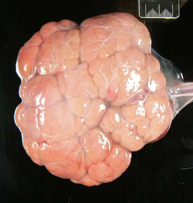
Gross Description:
Histopathologic Description:
Morphologic Diagnosis:
Condition:
Contributor Comment:
Mole is a term -�-�pirated from the human literature which refers to the gestational trophoblastic diseases that are characterised by formation of an irregular mass of chorionic villi and varying degrees of trophoblastic proliferation, often with disruption of lymphatic drainage and subsequent cystic transformation of the mass.(6) The latter form is referred to as hydatidiform. In bovid species, all forms of moles are rare, with fewer than 10 published reports. All of these reports have been associated with a co-twin and its own placenta.(4,5) The absence of any detectable fetal structures such as skin distinguishes them from the more common amorphous globosus, which is often referred to as a fetal mole.(3)
Two forms of hydatidiform mole have been identified in humans: the complete mole and the partial mole. The former results from fertilisation of an unviable oocyst by one or more sperm. Thus, the genome of a complete hydatidiform mole comes entirely from the paternal side. Greater than 90% of such moles have a 46 XX diploid karyotype.(6) Partial moles, on the other hand, are nearly always triploid and usually result from fertilisation of an haploid ovum either by two sperm, or one sperm which duplicates its genome.(1) The rarity of the bovid condition has prevented similar detailed cytogenetic investigation in cows. In complete trophoblastic moles of humans, because of the absence of a viable maternally derived genome, no viable embryo is produced and no fetal tissues develop. Instead, the paternally derived genome is able to dominate, resulting in excess development of the extra-embryonic tissues, particularly the placenta and its trophoblast. Thus, the mole becomes an hyperplastic mass of trophoblastic tissue.(1)
In humans, 20% of patients with hydatidiform moles can be expected to develop malignant sequelae but such sequelae have not been reported in cattle.Â
JPC Diagnosis:
Conference Comment:
In the human literature, a hydatiform mole is defined as a cystic swelling of chorionic villi accompanied by trophoblastic proliferation.(2) This description does not quite fit the histologic features of this lesion. The human literature further subdivides these moles into complete and partial moles. Complete moles are characterized histologically by diffusely edematous villi with diffuse trophoblastic hyperplasia, whereas partial moles have a mix of edematous and non-edematous villi with focal trophoblastic proliferation. The contributor mentioned the different karyotypes of these two moles. Because of the lack of female chromosomes, complete moles have no development of fetal parts. In contrast, partial moles can have development of some fetal parts because they have a karyotype with X and Y chromosomes allowing at least partial fetal development.(2)
In human medicine, it is extremely important to classify moles as either complete or partial because complete moles may precede choriocarcinoma.(2)
References:
2. Kumar V, Abbas AK, Fausto N: Robbins and Cotran Pathologic Basis of Disease, 7th ed., pp. 1110. Elsevier Saunders, Philadelphia, PA 2005
3. Long S: Abnormal development of the conceptus and its consequences. In: Veterinary Reproduction and Obstetrics, eds. Arthur GH, Noakes DE, Pearson H, Parkinson TJ, 8th ed., pp 12930. WB Saunders, London, UK, 2001
4. Meinecke B, Kuiper H, Dr+�-�gem+�-+ller C, Leeb T, Meinecke-Tillmann S: A mola hydatidosa coexistent with a foetus in a bovine freemartin pregnancy. Placenta 24:10712, 2002
5. Morris FJ, Kerr SM, Laven RA and Collett MG: Large hydatidiform mole: An unusual finding in a calving cow. New Zealand Veterinary Journal (in press).Â
6. Robboy SJ, Duggan MA, Kurman RJ: The female reproductive system. In: Pathology, eds. Rubin E, Farber JL, 2nd ed., pp 96770. JB Lippincott Company, Philadelphia, USA, 1994
7. Schlafer DH, Miller RB: Female genital system. In: Jubb, Kennedy, and Palmer's Pathology of Domestic Animals, ed. Maxie MG, vol 3., pp. 474-480. Elsevier Limited, Philadelphia, PA, 2007