Joint Pathology Center
Veterinary Pathology Services
Wednesday Slide Conference
2018-2019
Conference 2
September 5th, 2018
CASE IV: 1/12 (JPC 4066919-00).
Signalment: Ten-month old, mixed breed, female spayed, canine
History: Multiple diffuse papules. No pruritus.
Gross Pathology: Not available.
Laboratory results: None.
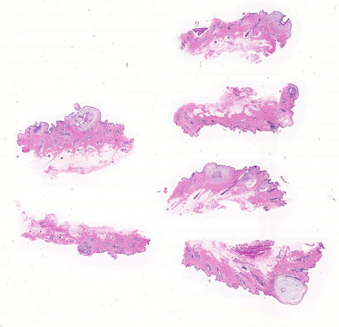
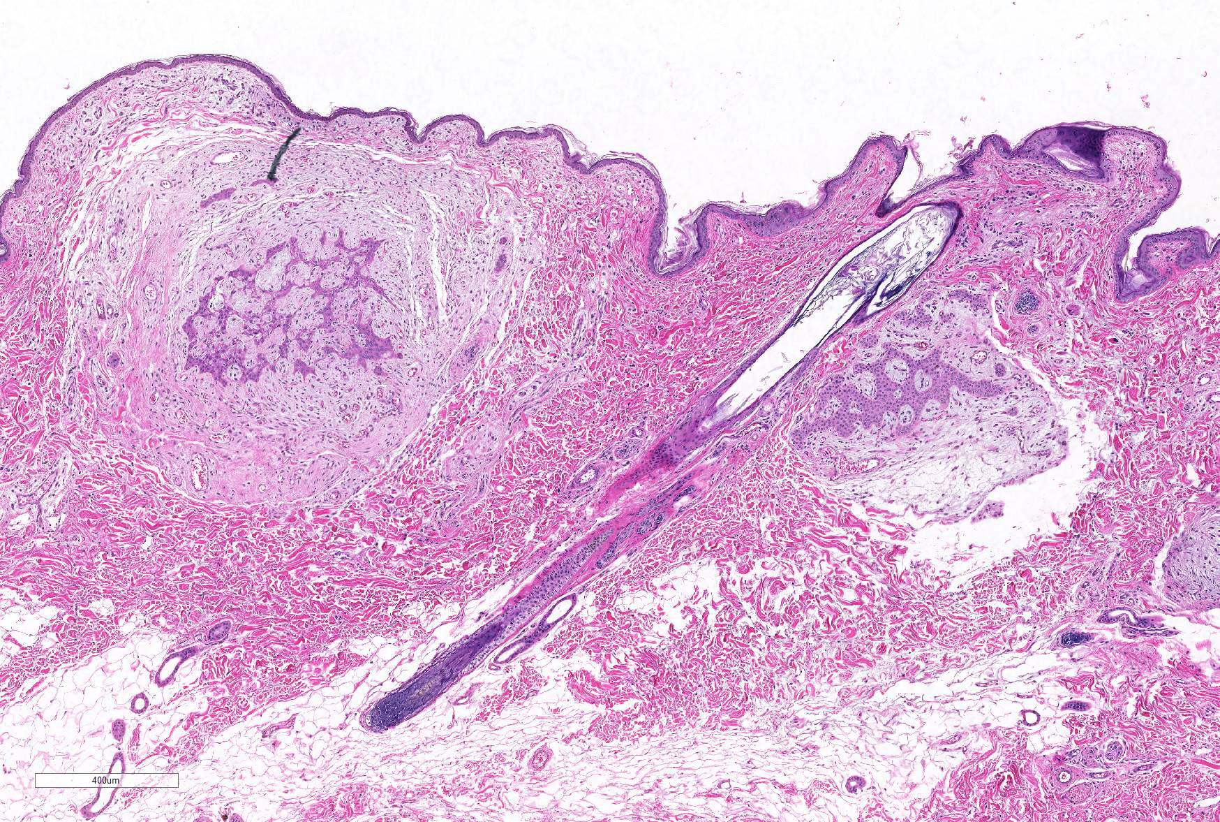
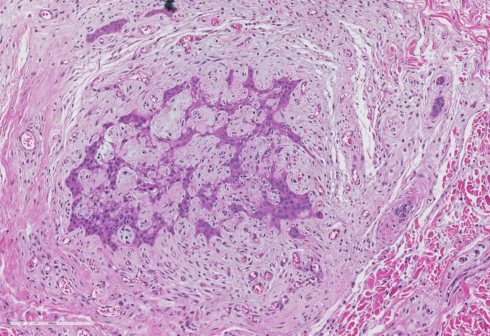
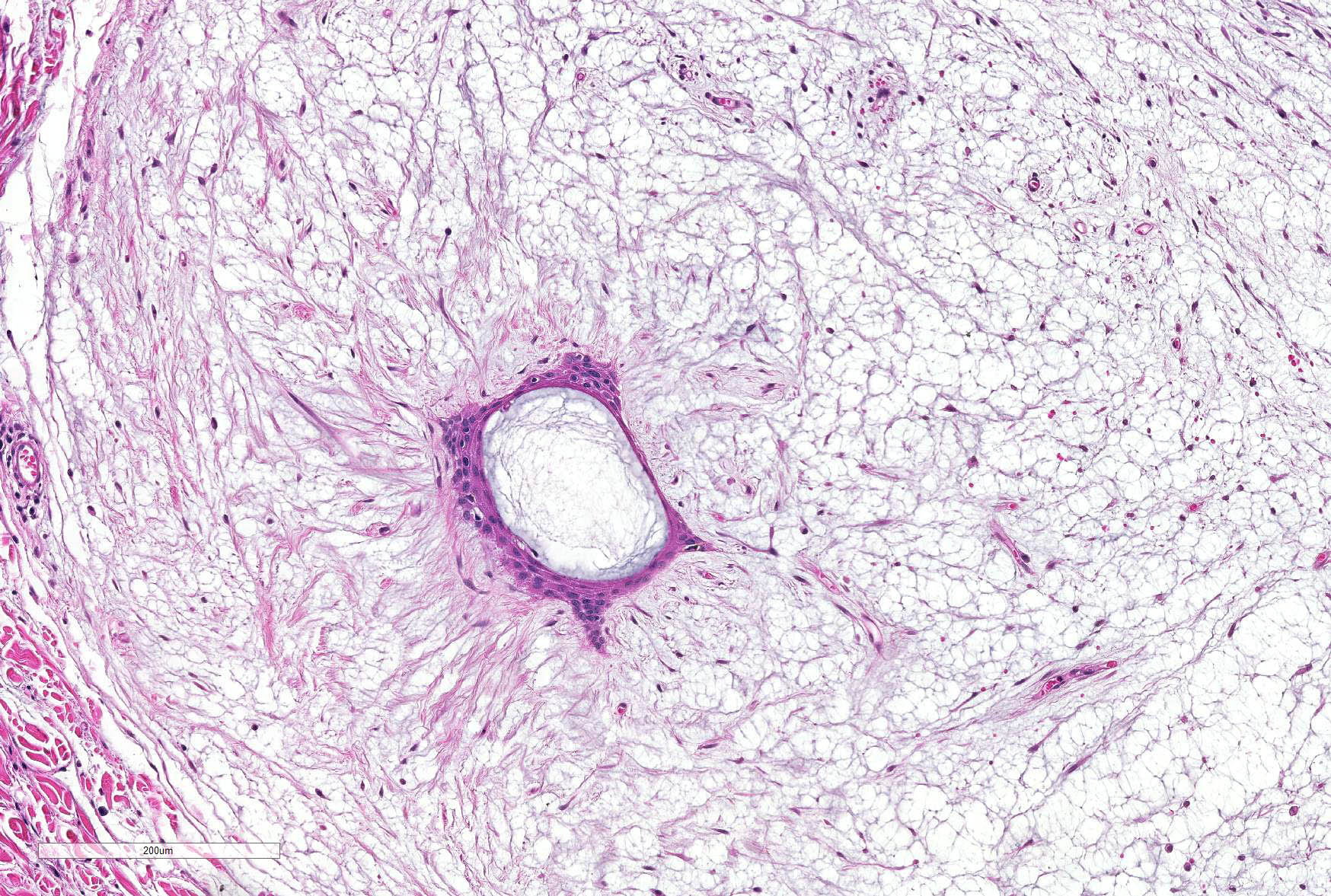
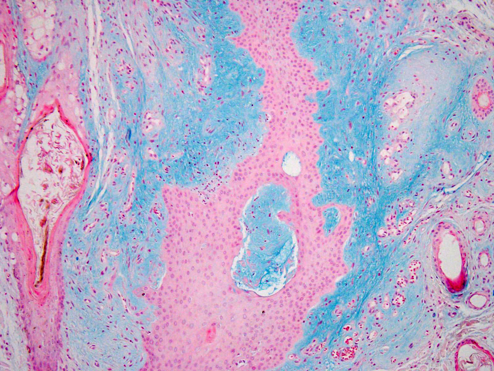
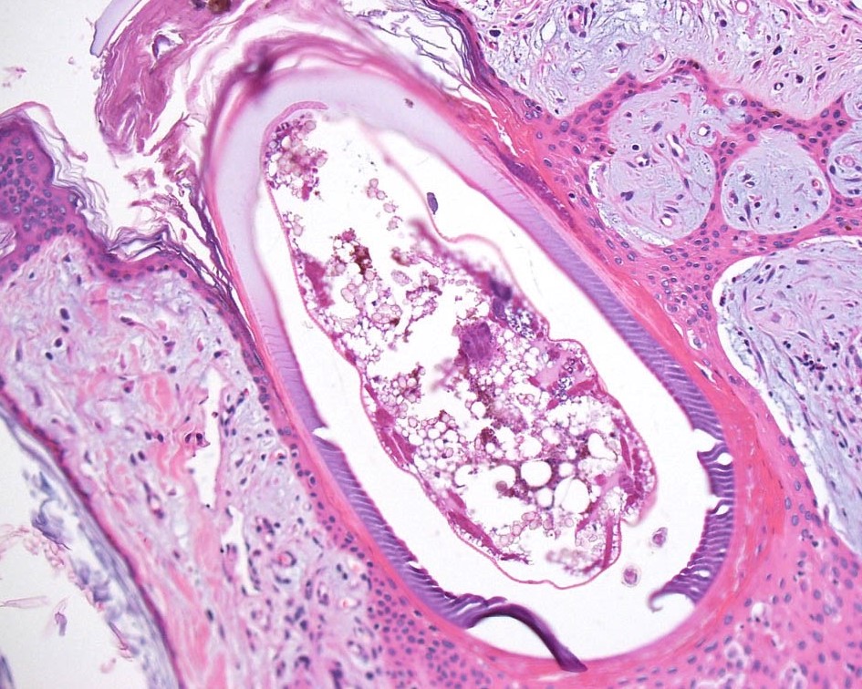
Microscopic Description: Haired skin: All sections have similar histological characteristics with variations in intensity. The epidermis is minimally hyperplastic to normal. There is formation of multiple raised dermal nodules, varying from 0.5 to 2mm in diameter. The nodules consist of a large dilated hair follicle with hyperplastic epithelium forming arborizing cords growing into a perifollicular accumulation of myxomatous material. The latter constitutes the bulk of the mass and produces its nodular shape. In several nodules there is a central 20-50µm thick band of deep eosinophilic hyaline material. This hyaline band surrounds a focus of necrotic debris, or, in rare follicles, a degenerated mite larva. There is minimal superficial perivascular dermal mononuclear cell infiltration.
Contributor’s Morphologic Diagnoses: Skin, Arborizing follicular hyperplasia with myxomatous degeneration and dilation, intrafollicular hyaline material and arthropod larvae, canine, mixed breed - findings typical of canine straelensiosis
Contributor’s Comment: Infection with the trombidioid mite Straelensia cynotis has been reported in dogs in France, Portugal and Spain.2,4,10 The findings described in these reports are virtually identical to several cases we have seen in Israel.
Straelensiosis is a trombidiosis caused by the “chigger” mite Straelensia cynotis, and is a relatively newly described etiology for nodular dermatitis in dogs in the Old World.2,4,10 Nodular trombiculiasis has also been described in white tailed deer and birds.5,7 The findings are similar to those described for the dog. The first publication of the morphologic characteristics of these mites was by Le Net and Fain who proposed the classification of a new species: S. cynotis, superfamily Trombidioid, family Leeuwenhoekiidae. Straelensiosis was first identified as a cause of nodular dermatitis in dogs in France by Le Net et al.1,4
In general, the nymphs and adults of trombiculid arthropods are free-living, or parasitize plants or other arthropods. The parasitic stage is the larvae which are known as ‘harvest mites’, ‘chiggers’ or ‘red bugs’.3,9 Infected animals may present with accumulation of orange granular material or pin-sized red spider-like foci in the canthi of the eyes.3,5,9 The larvae attach themselves to areas of the host’s skin in contact with the ground e.g. legs, feet, head, ears or ventrum and make a tunnel to the epidermis, called stylosome, through which salivary enzymes are injected and digested tissue fluids are withdrawn. The larvae engorge through a period of 3-5 days after which they drop off to become nymphs and complete their life cycle in the soil. Wild mammals are the usual hosts for trombiculid mite larvae, but pets and people may be accidentally infected.3,9
The histologic finding in skin infection with trombiculid larvae is the presence of tunnels within the stratum spinosum or stratum corneum, inducing degenerative and hyperplastic changes in the epidermis. The stylostome is described as a hyalinized tube oriented vertical to the skin surface.3
It is thought that the fox is the normal host for Straelensia and that hunting and rural dogs are predisposed to aberrant infection.4 There is a clear geographical pattern of infection in certain areas within the countries reported and an obvious tendency for infection to occur in the fall and spring.7 The nodules may last 1 to 12 months. On average, they are reported to last 3 months,4 which is much longer than the course described for chiggers in the US. The follicular reaction to the presence of the larvae is highly characteristic and allows diagnosis to be made in the absence of larvae in the section. The follicle is dilated and contains a ring of hyalinized material identified as the stylosome.4,8,10 The follicles exhibit epithelial hyperplasia with formation of arborizing epithelial cords within a perifollicular accumulation of myxomatous material. 4,8,10 Associated pyogranulomatous, suppurative and eosinophilic perifollicular infiltration is described. 4,8,10
In this particular case inflammation is negligible, but we have observed pyogranulomatous and suppurative perifollicular inflammation in most of the dogs infected in Israel.
Prognosis is variable.2, 4,8,10 Some dogs may run the course of the infection and have spontaneous regression. Some respond well to antiparasitic shampoos or Ivermectin injections, but some dogs are reported to present with persistent infection and no apparent response to therapy.2, 4,8,10 This may represent continuous exposure.
In our experience in Israel, infection is most common in the northern and in the Jerusalem areas, (both of higher altitude), in the spring and winter. Response, as described in the European reports, is variable but most dogs appear to respond well to pesticide shampoos.
JPC Diagnosis: Haired skin, follicles: Pseudoepitheliomatous hyperplasia, multifocal, severe, with marked perifollicular mucin, and occasional intrafollicular trombiculid larvae.
Conference Comment: The contributor provides an excellent description of changes associated with most common trombiculid mite infections, however the histologic findings of S. cynotis are quite different than traditional trombiculid mite infections. While other trombiculid mites generally attach to the surface epidermis, the larva of S. cynotis live and feed with in follicular ostia. They are separated from the wall of the follicle by the highly characteristic stylosome, a proteinaceous tube secreted by the mouthparts of the larval mite, through which the larva feeds by repeated cycles of extrusion of digestive salivary fluids followed by suction of digested tissue and tissue fluids.5 The extensive pseudoepitheliomatous hyperplasia of follicular epithelium and perifollicular mucinosis are additional histologic features which allow for the diagnosis of S. cynotis infection even in the absence of the larval arthropods. The larval forms of this parasites may be absent in treated cases or following the completion of the larval stage (as nymphs and adults are free living stages).8
Another interesting fact about infection by S. cynotis is the almost total lack of dermal inflammation in response to the presence of the parasite. This may be the result of the intra-follicular location of the mites as well as a total enclosure by the presence of the stylosome.8
A number of arthropod and helminth parasites inhabit hair follicles. By far the most common genus is that of Demodex. which are normal inhabitants of hair follicles and sebaceous glands in most mammals, including humans. Interestingly, their position within the hair follicle is invariably head down.6 Demodex sp. are obligate parasites which complete their entire life cycle on the host and are usually transmitted from mother to offspring within the first 3 days of life through close physical contact while nursing. Demodex mites, unlike trombiculid mites, feed harmlessly on sloughed cells, sebum, and epidermal debris. These mites may move from follicle to follicle, and transmission likely occurs while in transit. Prolonged sojourns on the epidermis may result in death of the mite through desiccation.6
Two important helminth species parasitize hair follicles in mammals. Parasitic females of the species Pelodera (Rhabditis) strongyloides, normally also free-living indicating plant matter, may indicate the skin and reside within hair follicles.6 Pelodera dermatitis occurs most often the dogs but cases are well documented in cattle, sheet, horses, and humans.6 Affected dogs are often victim supportive husband 3 and straw embedding is a common finding. Stephanofilaria species are uncommon cutaneous parasites of a wide range of ruminant species; with S. stilesi occurring in Western and Southwestern North America. While abdominal skin is characteristic for this particular species, other species each have particular sites where they affect their respective host.6 Flies are intermediate hosts for these parasites and deposit infective larva on both intact and broken skin of the host species. The adults of S. stilesi and similar species live within cystic hair follicles and microfilariae occur free in the dermis or within dermal lymphatics, where they may be ingested by flies and complete their life cycle. Maturation from microfilaria to infective larva occurs within the intermediate host. A marked inflammatory reaction occurs if the adults exit the hair follicles, resulting in profound lymphoplasmacytic and eosinophilic inflammation, epidermal hyperplasia, and often alopecia. The presence of the adults within the follicle or of microfilariae within the dermis generally results in little inflammation.6
Contributing Institution:
Department Vet Resources, Weizmann Institute, Rehovot, Israel
http://www.weizmann.ac.il/vet/
References:
1. Fain A, Le Net JL. A new larval mite of the genus Straelensia Vercammen-Grandjean and Kolebinova, 1968 (acari: Leeuwenhoekiidae) causing nodular dermatitis of dogs in France. Int Jour of Acarology . 2000;26: 339–45.
2. Font A, Straelensiosis in a dog in Spain. Vet Derm. 2007;18: 67-68.
3. Ginn PE et al, Skin and appendages. In: Jubb, Kennedy and Palmer’s Pathology of Domestic Animals, 5th ed. Maxie Ed. Saunders Elsevier 2007;Vol. 1:7272-728.
4. Le Net JL et al. Straelensiosis in dogs: a new deA hyalscribed nodular dermatitis induced by Straelensia cynotis. Vet Rec. 2002;150:205-209.
5. Little SE et al, Trombidiosis-induced Dermatitis in White-tailed Deer (Odocoileus virginianus). Vet Pathol 1997;34:350-352.
- Mauldin EA, Peters-Kennedy J.. Integumentary System, In: Maxie MG, ed. Jubb, Kennedy and Palmer’s Pathology of Domestic Animals, 6th 2016; St. Louis MO, Elsevier Press, vol 1, pp. 666-690.
- Ornelas-Almeida MA et al. Nodular trombiculinosis caused by Apolonia tigipioensis,Torres and Braga (1938), in an ostrich (Struthio camelus) and a house sparrow (Passer domesticus). Vet Parasit. 2007;150: 374–377.
- Ramirez GA, Clinical, histopathological and epidemiological study of canine Straelensiosis in the Iberian Peninsula (2003-2007). Vet Derm. 2009;20:35-41.
- Scott et al, Parasitic skin diseases. In: Muller and Kirk’s Small Animal Dermatology. 5th ed. Saunders 1995;407-408.