Signalment:
3-year-old female red kangaroo (
Macropus rufus).This kangaroo died acutely with no observed clinical signs. Four animals died at this private zoo over the past 3 months, also without any clinical signs. This zoo has a large feral cat population.
Gross Description:
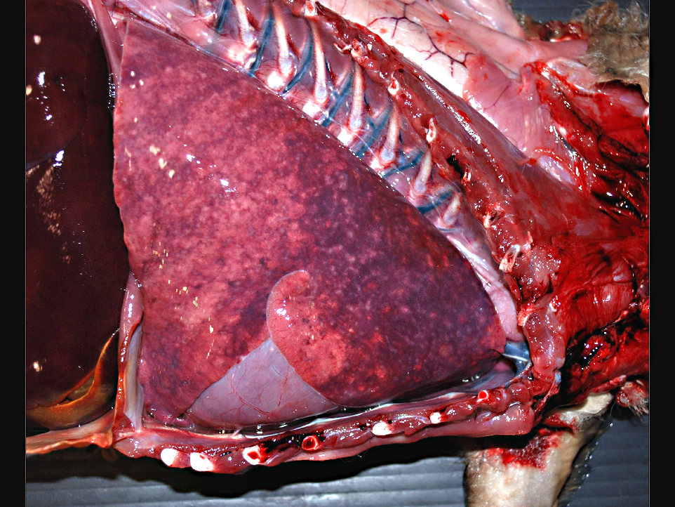
The animal was in a mild state of decomposition. Gross examination revealed the animal was in good nutritional condition. The lungs failed to collapse upon opening the thoracic cavity. There were hundreds of firm, 1-2 mm in diameter, white to tan, slightly raised foci disseminated throughout the pulmonary parenchyma. Mediastinal and tracheobronchial lymph nodes were enlarged and on section were hemorrhagic. Multiple white radiating streaks were identified on the epicardial surface extending into the myocardium.
Histopathologic Description:
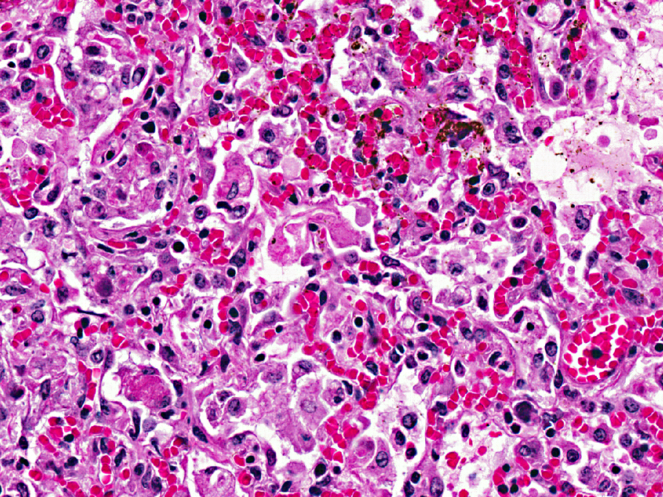
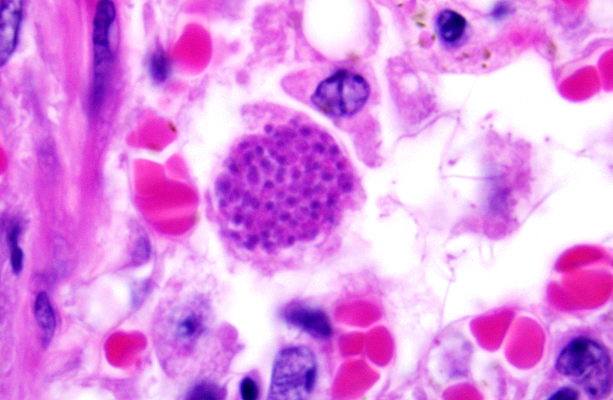
Lung: The normal architecture of the lung is disrupted by interstitial and alveolar inflammation, necrosis, fibrin deposition, edema, and intralesional protozoa. Expanding and occasionally forming nodular aggregates within the alveolar interstitium and alveoli are a large number of macrophages, lymphocytes, and plasma cells. Rare degenerate neutrophils are observed. Often the alveolar interstitium contains abundant amounts of fibrin. There is marked necrosis within the sections, characterized by eosinophilic cellular debris and basophilic nuclear debris. Admixed within the inflammation are 1-2μm, oval to round organisms arranged individually (tachyzoites) or grouped together into packets or cysts (bradyzoites) 20-25 μm in diameter. There is a mild amount of pulmonary edema. Alveolar interstitium are markedly congested. [
Organisms are present in all slides; conspicuous in some, inconspicuous in others]
Morphologic Diagnosis:
Lung: Necrotizing interstitial pneumonia, lymphohistiocytic, diffuse, acute, severe, with multifocal necrosis and intralesional
Toxoplasma gondii protozoa.
Lab Results:
Immunohistochemical stains for
Toxoplasma on lung tissue were positive. Fluorescent antibody staining for CAV-1 on lung tissue was negative.
Condition:
Toxoplasma gondii
Contributor Comment:
Toxoplasmosis is caused by
Toxoplasma gondii, an obligate intracellular coccidian parasite.Â
Toxoplasma gondii has a broad intermediate host range and the only known definitive hosts for
T. gondii are domestic cats and other felidae.(2) Infection with
T. gondii within the cat is dependent on enteroepithelial or extraintestinal life cycle. In kangaroos, similar to other intermediate hosts, infection is dependent on the extraintestinal life cycle.Â
Toxoplasma gondii has a worldwide distribution and causes high morbidity and mortality in Australian marsupials, particularly macropods.(1)
Kangaroos are herbivores and presumably acquire
T. gondii by ingestion of oocysts from their environment. In this case, it was difficult to determine whether there was contamination of the foods, grazing areas, or water, or if oocysts were ingested during grooming activities. However, it was likely that feral cats defecated in the enclosure area, resulting in contamination of the ground or feeding stations potentially exposing these animals to infectious oocysts.Â
At necropsy, kangaroos with toxoplasmosis have gross lesions similar to other domestic animal species; including pulmonary congestion and edema, pulmonary consolidation, pale myocardial streaks and hemorrhage, a miliary pattern of white pulmonary foci, lymphadenomegaly, and splenomegaly. In addition to the lung lesion, this patient exhibited inflammatory lesions with
Toxoplasma organisms in the brain, heart, liver and kidney. Similarly, disseminated toxoplasmosis was confirmed in the other four kangaroos that had died previously.
JPC Diagnosis:
Lung: Pneumonia, interstitial, necrotizing, diffuse, moderate, with rare intrahistiocytic apicomplexan cysts and free zoites.
Conference Comment:
A characteristic finding of toxoplasmosis is necrosis, which is caused both by rupture of infected cell membranes by rapidly dividing tachyzoites, and by ischemia secondary to vasculitis. The moderator emphasized that the most susceptible captive zoo species are the squirrel monkey, macropods, lemurs, and the Thomsons gazelle, that protozoal cysts are most readily found in skeletal muscle and the central nervous system, and that pulmonary infection is the most common cause of death. Characteristic gross findings in the lung include pulmonary consolidation with multifocal to coalescing green foci; lymphadenomegaly, and hemorrhage and necrosis of the lymph nodes are also commonly noted. The moderator also discussed the serious problem that toxoplasmosis causes in zoos, with the parasite reservoirs being feral cats, opossums, rats, and cockroaches.
References:
1. Canfield P, Hartley W, Dubey P. Lesions of toxoplasmosis in Australian marsupials.Â
J Comp Path. 1990;103:157-167.
2. Dubey J, Lappin M. Toxoplasmosis and Neosporosis In: Green CE, ed.Â
Infectious Diseases of the Dog and Cat Saunders Elsevier, St. Louis, MO, 2006:754-775.
3. Schlafer DH, Miller RB. Female genital system. In: Maxie MG, ed.Â
Jubb, Kennedy, and Palmers Pathology of domestic Animals. 5th ed. Edinburgh, Scotland: Saunders Elsevier; 2007:513-4.