Signalment:
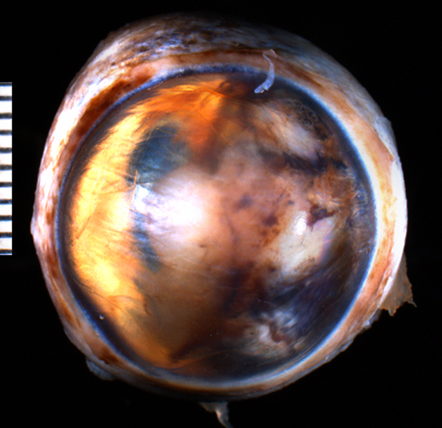
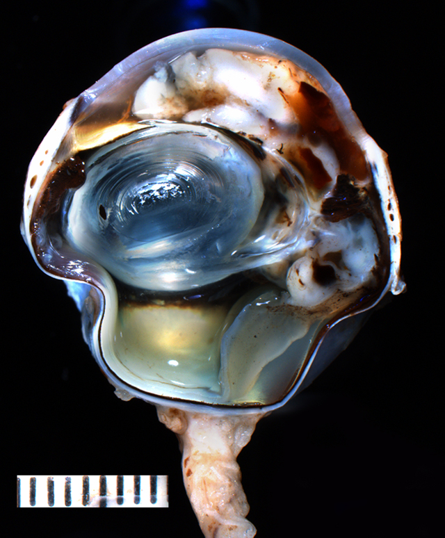
Gross Description:
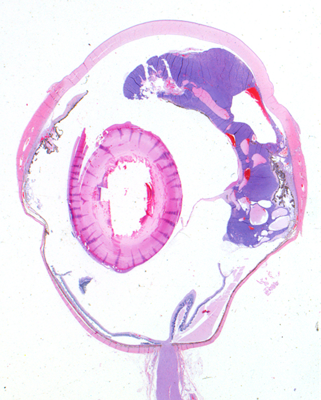
Histopathologic Description:
Morphologic Diagnosis:
1. Feline iridociliary adenoma
2. Equatorial cortical cataract
3. Segmental retinal detachment
4. Epithelial cysts of the pars plana
Condition:
Contributor Comment:
The epithelium of the iris and ciliary body is derived from the neuroectoderm by expanding forward from the margins of the optic cup.(2) Neoplasia of the neuroectoderm of animals is considered uncommon (iridociliary adenomas and carcinomas) or rare (medulloepitheliomas, retinoblastomas, astrocytomas and gliomas).(3,4) With the exception of ciliary adenocarcinomas, these neoplasia are considered benign.(3) Iridociliary epithelial tumors are the second and third most common primary intraocular tumor in the dog and cat, respectively.(1) They arise from the pigmented or non-pigmented epithelial cells, usually from the pars plicata of the ciliary body.(1)
Clinically, these tumors usually present as white to brown or black, fairly well delineated, sometimes pedunculated, slow growing masses, that usually grow into the vitreous behind the lens.(4) They are usually visible though the pupil in the posterior chamber. Invasion through the iris or protrusion through the pupil can lead to a localized mass visible in the anterior chamber.(3) Clinical signs usually include a retro-iridal mass that may displace the iris or lens by expansive growth, and if the tumor is large, secondary glaucoma, ocular pain and/or intraocular hemorrhage may be noted.(4) Hyphema and aqueous flare may be present due to the common formation of preiridal fibrovascular membranes.(3) In cases where the intraocular hemorrhage and flare impair the direct observation of the tumor, ultrasonographic imaging may prove helpful in delineating a mass lesion in the posterior chamber.(1)
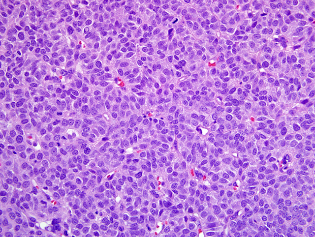
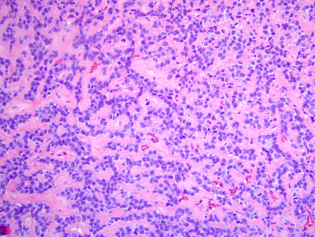
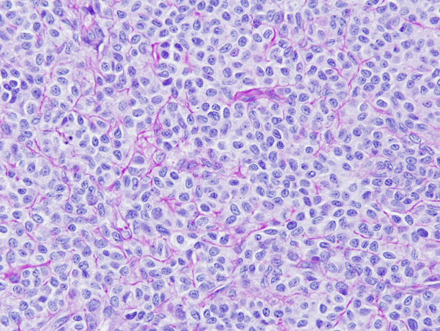
Histologic features of feline iridociliary adenomas include a solid non-pigmented epithelial tumor with packets of cells surrounded by thin PAS positive basement membrane structures.(2) The defining feature of adenocarcinoma, versus adenoma, is invasion into the sclera by the neoplastic population rather than features of cellular atypia.(2) Thus, most iridociliary adenocarcinomas in both cats and dogs have relatively benign cellular features.
A retrospective study of 101 cases of feline iridociliary tumors conduced in the Comparative Ocular Pathology Laboratory of Wisconsin (COPLOW) (unpublished data) identified other common features of these tumors. Of the 101 cases, 84 (83.1%) of the tumors were classified as adenomas. Seventeen (16.9%) presented scleral or choroidal invasion and were classified as adenocarcinomas. Adenomas presented a solid pattern in 97.6% (82/84) of the cases. They were either classified as uveoinvasive (52.3%) and non-uveoinvasive (47.7%). The most common histological features of adenomas were PAS positive basement membranes (present in 82.3% of the cases), cavitated spaces filled with blood and/or proteinaceous material (77.3%), presence of osteoid matrix (42.8%), osseous metaplasia (12%), and formation of pseudo-rosettes (26.2%). Mitotic figures were rare in both adenomas and adenocarcinomas. Two cases of iridociliary adenomas presented atypical patterns, one papillary and the other a mucoid variant that presents PAS positive basement membrane and positivity for vimentin and cytokeratin. Most tumors were positive for vimentin and only carcinomas presented variable positivity for cytokeratin. A previous study demonstrated that 50% of feline and canine iridociliary adenomas were positive for S-100, 1/3 of the feline cases were positive for glial fibrillary acidic protein (GFAP) and all canine cases were positive for neuron-specific enolase (NSE).(2)
In the recent unpublished study, the age of the animals ranged from 2 to 18 years (average 9.1). The most common secondary abnormalities detected were glaucoma, preiridal fibrovascular membrane, hyphema and retinal detachment. Presence of an intraocular mass was the main reason for presentation to the veterinarian and enucleation. Glaucoma is a common secondary condition associated with iridociliary tumors, in part because these tumors are associated with the formation of fibrovascular membranes that can lead to peripheral anterior synechia and obstruction of the iridocorneal angle.(1)
In the case presented, the evidence of glaucoma is equivocal. The most reliable microscopic evidence of glaucoma is absence or reduced numbers of retinal ganglion cells. In cats, even cases of chronic glaucoma can show an otherwise robust retina that lacks ganglion cells. In contrast, the glaucomatous canine globe frequently shows atrophy of the inner retinal layers to full-thickness atrophy in chronic cases. Gliosis of the optic nerve is also reliable evidence of glaucoma. Atrophy of the optic nerve with cupping of the optic nerve head is also common, particularly in dogs. Finally, a feature of glaucoma seen characteristically in dogs with primary glaucoma (goniodysgenesis) is tapetal sparing in which the tapetal (superior) retina is less profoundly atrophied than the non-tapetal (inferior) retina. Presence of epithelial cysts of the pars plana is a common finding in older cats and presumed to be a degenerative condition not related to the tumor in this case.
In sum, solid nonpigmented tumors arising from the ciliary body or iris epithelium with small epithelial cells packed by thin PAS-positive membranes and staining positive for vimentin are significant features defining iridociliary tumors in cats.
JPC Diagnosis:
1. Eye: Iridociliary adenoma.
2. Eye, retina: Detachment, segmental.
Conference Comment:
References:
2. Dubielzig RR, Steinberg H, Garvin H, Deehr AJ, Fisher B: Iridociliary epithelial tumors in 100 dogs and 17 cats: a morphological study. Vet. Ophth 1:223-231, 1998
3. Grahn BH, Peiffer RL: Fundamentals of veterinary ophthalmic pathology. In: Veterinary Ophthalmology, ed. Gellat KN, 4th ed., pp. 355-437. Blackwell Publishing, Ames, IA, 2007
4. Miller PE, Dubielzig RR: Ocular tumors. In: Withrow & McEwens Small Animal Clinical Oncology, ed. Withrow SJ, Vail, DM, 4th ed., pp. 686-698. Elsevier, St. Louis, MO, 2007
5. Zarfoss MK, Dubielzig RR: Metastatic iridociliary adenocarcinoma in a Labrador retriever. Vet Pathol 44:672-676, 2007