Signalment:
Gross Description:
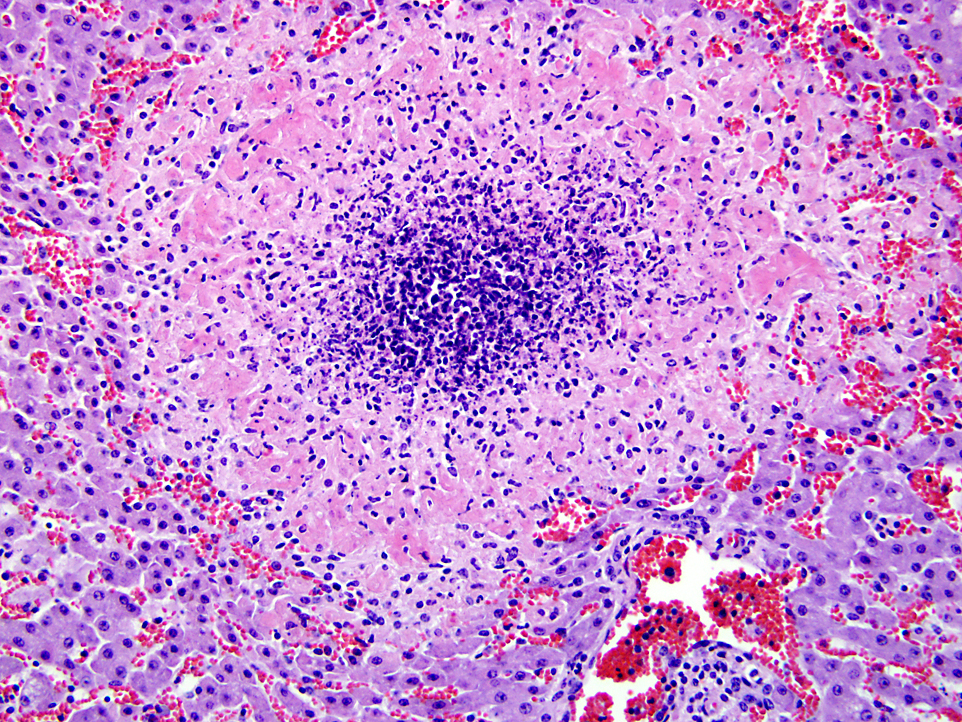
Histopathologic Description:
Morphologic Diagnosis:
Lab Results:
Condition:
Contributor Comment:
The three main types of disease caused by Listeria are meningoencephalitis, abortions and septicemia. Meningoencephalitis is the most common form of the disease and occurs mainly in adult ruminants. Abortions in the last third of the pregnancy or stillbirths are most often detected in sheep, goats and cattle. Septicemia occurs in young ruminants and neonate monogastric animals. Less common forms of the disease are conjunctivitis, keratitis or uveitis in ruminants and horses(1); mastitis, endocarditis, meningitis in young animals; spinal myelitis in sheep; and enteritis in adult sheep. Recently, a fatal mesenteric lymphadenitis was described in adult cattle.(5) Also, skin infections have been described in veterinarians. L. monocytogenes septicemia occurs also in rabbits, chinchillas, hares, and has been described in semi-domesticated reindeer calves associated with silage feeding.(3) The disease is often sporadic but small epidemics occur.
Infection occurs via ingestion, from the contaminated feed or udder, via milk or umbilical cord, or in the uterus. The bacteria penetrate the intestinal mucosa and cause a subclinical bacteremia. Encephalitis in ruminants has a different pathogenesis and is probably acquired via cranial nerves from oral mucosal abrasions or an infected tooth cavity. In these cases the bacteria reach the brain stem by using axonal transport. This is supported by the special distribution of the lesions in the brain stem. The incubation period varies from 2 days to several weeks. Only a small proportion of infected animals develop clinical disease. The predisposing factors include stress, pregnancy, parturition, poor nutritional state, large dose and failure of passive transfer. In humans the infection is usually foodborne and pregnant women, elderly and immunocompromised individuals are predisposed.
Listeria is a facultative intracellular parasite and invades cells, macrophages, monocytes, neutrophils and epithelial cells by using a special surface protein, internal in.(2) In the cell, bacteria escape from the phagosomes by producing a haemolysin called listeriolysin O and phospholipases. The bacteria are motile and have a special cell to cell movement system. Cell-mediated immunity has a role in causing tissue damage. Gross pathological findings in septicemic forms of the disease consist of multiple random pale necrotic foci or microabscesses, most commonly in the liver and spleen.Â
Histologically, in the septicemic form, multiple necrotic foci or microabscesses with variable numbers of neutrophils and macrophages are seen in the liver, and are often also in the spleen or other tissues.Â
The differential diagnoses for necrotic hepatitis include Salmonella, Bacillus piliformis (Tyzzers disease), Yersinia pseudotuberculosis, and in rodents also Toxoplasma gondii.
JPC Diagnosis:
Conference Comment:
References:
2. Maxie MG, Youssef S. Nervous system. In: Maxie MG, ed. Jubb, Kennedy and Palmers Pathology of Domestic Animals. 5th ed. Vol 1. Philadelphia, PA: Saunders Elsevier; 2007:405-8.
3. Nyyss+�-�nen T, Hirvel+�-�-Koski V, Norberg H, et al. Septicaemic listeriosis in reindeer calves- a case report. Rangifer. 2006;26:25-28.
4. Schlafer DH, Miller RB. Female genital system. In: Maxie MG, ed. Jubb, Kennedy, and Palmers Pathology of Domestic Animals. 5th ed. Vol 3. Philadelphia, PA: Saunders Elsevier; 2007:492-3.
5. Thompson H, Taylor DJ, Philbey AW. Fatal mesenteric lymphadenitis in cattle caused by Listeria monocytogenes. Vet Rec. 2009;164:17-18.Â
6. Wilkins PA, Marsh PS, Acland H, et al. Listeria monocytogenes septicaemia in a thoroughbred foal. J Vet Diagn Invest. 2000;12:173-176.
7. Zachary JF. Mechanisms of microbial infection. In: Zachary JF, McGavin MD, eds. Pathologic Basis for Veterinary Disease. 5th ed. St. Louis, MO: Elsevier Mosby; 2011:195.