Joint Pathology Center
Veterinary Pathology Services
Wednesday Slide Conference
2017-2018
Conference 19
April 4th, 2018
CASE II: 3855-16 (JPC 4101305).
Signalment: 13-year-old female spayed Australian shepherd (Canis familiaris), canine.
History: This dog had a soft tissue mass on the dorsal left chest wall. The mass was deep and attached to chest wall muscles and invaded into adjacent fat. The surgeon dissected around mass and removed muscle where it was attached.
Gross Pathology: Mass measured 5 x 4 x 3 cm
Laboratory Results (clinical pathology, microbiology, PCR, ELISA, etc.): None provided.
Microscopic Description:
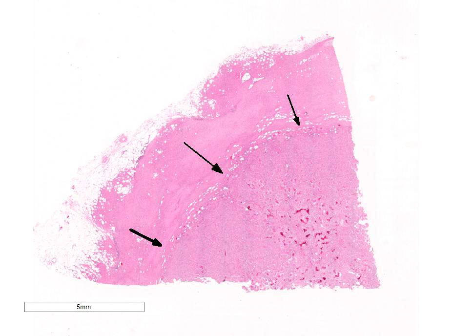
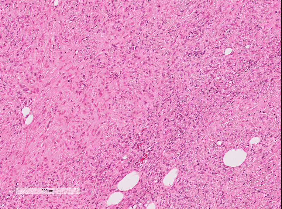
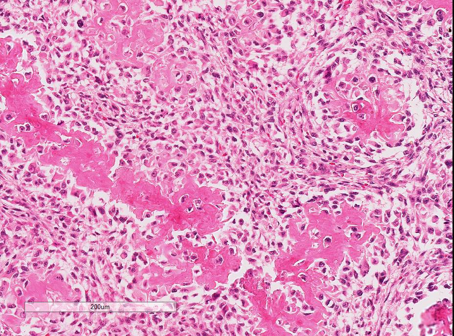
A 5 x 4 x 3 cm subcutaneous mass from behind the left shoulder blade is examined in four sections representing a cross section and lateral margins. The specimen has two distinct masses with a line of demarcation at their collision point. The peripheral mass is densely cellular, poorly circumscribed, and unencapsulated and is infiltrating into the adjacent subcutaneous adipose tissue. The mass consists of spindle-shaped cells arranged in bundles, streams, storiform patterns and whorls accompanied by collagen; the collagenous stroma varies from delicate to thick bands, in different regions of the tumor. The neoplastic cells are medium-sized with indistinct cellular margins and small amounts of fibrillar eosinophilic cytoplasm which blends with the stroma. The nuclei are medium-sized and oval with stippled chromatin and 1-5 nucleoli. Eleven mitotic figures are seen in ten high powered fields including a few atypical mitotic figures. The central mass is densely cellular, circumscribed, unencapsulated, and slightly compresses the surrounding malignant peripheral nerve sheath tumor. The mass consists of plump spindle-shaped cells arranged in short streams and sheets accompanied by osteoid matrix. The neoplastic cells are medium to large with distinct cellular margins and moderate to large amounts of finely granular to fibrillar eosinophilic cytoplasm. The nuclei are large and oval with vesicular chromatin and 1-5 nucleoli. Thirty-two mitotic figures are seen in ten high powered fields, including a few atypical mitotic figures. The mass has moderate anisocytosis and anisokaryosis with rounded multinucleated giant cells occasionally scattered throughout it. The mass touches the deep and lateral margins.
Contributor’s Morphologic Diagnosis:
Malignant peripheral nerve sheath tumor and osteosarcoma – collision tumor
Contributor’s Comment: This neoplastic mass has a very unique histological appearance where the outer part has differentiated into a malignant peripheral nerve sheath tumor and the inner part has differentiated into an osteosarcoma. As there is a distinct junction between the two masses, this neoplasm was classified as a collision tumor.
Collision tumors are the result of two tumors in the same anatomic site which abut one another but have a distinct demarcation between the two. Collision tumors are rare in domestic animals. Compared to collision tumors, biphasic or mixed tumors exist in which there are two intermixing phenotypically distinct populations of neoplastic cells. Reports of collision tumors involving melanomas are common.3,6,8 Additionally, mixed Sertoli-seminoma tumors in the testes of dogs comprised nearly seven percent of all tumors in one study.7 These collision and mixed tumors present a diagnostic dilemma as to how they arise, particularly if the tissue of origin is embryologically different.
It is uncertain if this case represents a mass with growth of each neoplasm de novo, resulting in a collision of the two tumors or if there was metaplasia leading to development of a second tumor with characteristics of malignancy. Peripheral nerve sheath tumors are a heterogeneous group of tumors and their classification is confusing.5 As the various classifications imply, there may be mesenchymal origin of these tumors that may undergo metaplasia. An example of cartilaginous differentiation exists.5 It is feasible osseous metaplasia may, too, occur and develop into what appears to be a collision tumor.
JPC Diagnosis: 1. Fibrovascular tissue: Sarcoma (favor peripheral nerve sheath tumor), Australian shepherd (Canis familiaris), canine.
- Fibrovascular tissue: Osteosarcoma.
Conference Comment: There have been several collision tumors reported in the veterinary literature within the last five years, including: malignant trichoblastoma/melanoma in a rabbit,2 oral squamous cell carcinoma/malignant melanoma in a dog,8 uterine adenocarcinoma/leiomyosarcoma in a goat,1 perianal gland carcinoma/hemangiosarcoma in a dog,9 and fibrosarcoma/mast cell tumor in a dog.9 In most cases of collision tumors, the authors postulate that one tumor is primary and a second separate tumor occurs due to chronic inflammation, repetitive mitotic stimulation, and eventual neoplastic transformation of various local cell types. This differentiates collision tumors from mixed tumors, which are single tumors that include a mixture of different neoplastic cell types.
Collision tumors are also recognized in humans where much research is being done regarding their genetic profiles. For instance, it has been shown that intracranial collision tumors are composed of two distinct components. In the case of a combined meningioma and oligodendroglioma, there is deletion of chromosome 22q and 19q in both tumors initiating neoplastic transformation of both cell types.4
Attendees discussed the use of cytology with ALP staining for rapid diagnosis of osteosarcoma during surgery, remarking that sometimes biopsies can resemble reactive bone, especially if the sample is obtained from the periphery of the neoplasm. Additionally, participants discussed the special stains utilized in this case, including S-100, which exhibited strong, intracytoplasmic immunoreactivity for the spindle cell population of the sarcoma.
Contributing Institution:
Veterinary Diagnostic Center
School of Veterinary Medicine and Biomedical Sciences
University of Nebraska-Lincoln
http://vbms.unl.edu/nvdls
References:
- Dockweiler JC, Cossic B, McDonough ST, Fubini SL, et al. Tumor collision of uterine adenocarcinomoa and leiomyosarcoma in a goat. J Vet Diagn Invest. 2017; 29(5):696-699.
- Golbar HM, Izawa T, Kuwamura M, Fujita D, et al. A collision tumor consisting of malignant trichoblastoma and melanosarcoma in a rabbit. J Comp Path. 2014; 151(1): 63-66.
- Jakab C, Balka G. First report of malignant collision skin tumor with malignant melanoma and anaplastic sarcoma components in a dog. Acta Vet Hung. 2012; 60:245-255.
- Kearney H, Cryan JB, Looby S, Brett FM, et al. The DNA copy number landscape of a collision tumor. Clin Neuropathol. 2018;37(2):68-73.
- Koestner A, Higgins RJ. Tumors of the nervous system. In: Meuten DJ, ed. Tumors in Domestic Animals. 4th ed. Ames, IA: Iowa State Press; 2002:697–738.
- Muscatello LV, Avallone G, Benazzi C, Sarli G, Porcellato I, Brachelente C, Brunetti B. Oral squamomelanocytic tumor in a dog: a unique biphasic cancer. J Comp Path. 2016; 154:211-214.
- Patnaik AK, Mostofi FK. A clinicopathologic, histologic, and immunohistochemical study of mixed germ cell-stromal tumors of the testis in 1 dogs. Vet Path. 1993; 30:287-295.
- Rodríguez F, Castro P, Ramírez GA. Collision Tumor of squamous cell carcinoma and malignant melanoma in the oral cavity of a dog. J Comp Path. 2016; 154:314-318.
- Scott JE, Liptak JM, Powers BE. Malignant collision tumors in two dogs. J Am Vet Med Assoc. 2017; 251(8):941-945.