CASE II:
Signalment:
8 years 3.5 months, Female, Slender-tailed Meerkat, Suricata suricatta
History:
An adult female meerkat housed in a zoological collection was found collapsed. Nine months previously, the animal had a ?fit? witnessed by the keepers, followed by a ?vacant? episode the following month. Skull radiographs during a routine health check two months later revealed a mild opacity in the region of the left caudal cerebrum. On the day of presentation, additional radiographs showed similar irregular soft-tissue densities in the same region. Due to the clinical suspicion of the cause of the neurological signs, anesthesia was not reversed, and an intracardiac barbiturate injection euthanized the animal. Additional findings included evidence of previous conspecific aggression, with crusting present over the ear pinna and distal tail.
Gross Pathology:
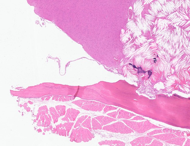
Gross examination revealed a moderately thickened calvarium, with strong adhesions to the underlying, moderately thinned and friable cerebrum, with a moderate hydrocephalus.
Laboratory Results:
Serum cholesterol measured approximately 24 months and six months before euthanasia was 16.96 mmol/L and 13.01 mmol/L, respectively (ZIMS global species RI: 4.95-18.52 mmol/L).
Microscopic Description:
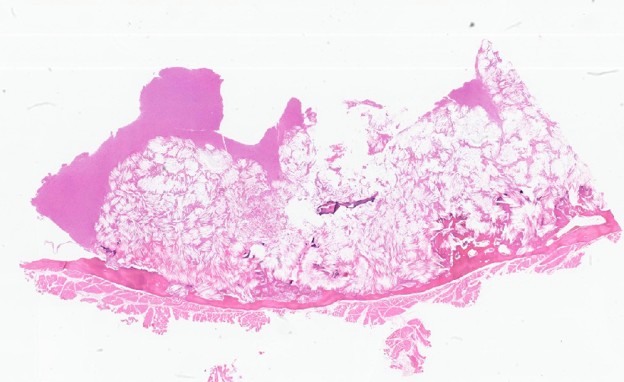
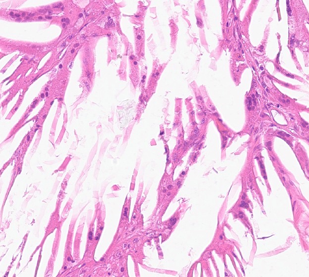
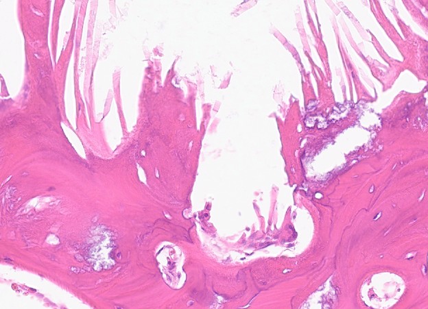
Parietal skull with brain. Two sections of the cerebrum and overlying calvarium are examined; both are histologically similar. Occupying at least 75% of the sections, this focally extensive, poorly demarcated, irregularly shaped area expands the meninges and invades and replaces the cerebrum and calvarium. This area is composed of irregularly arranged clear elongate acicular cholesterol clefts. Between aggregates of these cholesterol clefts are variable amounts of cerebral parenchyma multifocally infiltrated by a moderate number of foamy haemosiderin-laden macrophages (haemosiderophages) and occasional lymphocytes and plasma cells. Within the cholesterol clefts and associated cerebral parenchyma are variably sized areas of hemorrhage. The cerebral parenchyma contains multifocal foci of mineralization, measuring up to 0.07mm in diameter. Adjacent to cholesterol clefts replacing the calvarium are low to moderate numbers of osteoclasts associated with areas of immature woven bone (bone remodeling).
Contributor's Morphologic Diagnoses:
- Cerebrum, meninges, calvarium; focally extensive severe cholesterol granuloma with haemosiderophages and foci of mineralization
- Calvarium; multifocal moderate bone remodeling
Contributor's Comment:
Cholesterol granulomas (cholesteatomas) are non-neoplastic, often asymptomatic, slow-growing aggregates of cholesterol crystals and macrophages within tissues.8 They are most often diagnosed in older horses and are rarely found in other veterinary species, including dogs, cats, and, as in this case, meerkats.1,3,5,6,8,9 In horses, cholesterol granulomas are benign masses associated with inflammation of the choroid plexus in the base of the lateral ventricles. They are often incidental findings on computed tomography (CT) scans or post-mortem examination. In horses, the pathogenesis is thought to result from recurrent choroid plexus congestion, edema, and hemorrhage with degenerate erythrocytes, leading to cholesterol crystal formation and a foreign body-type response.2 In humans, intracranial cholesterol granulomas have been associated with chronic middle ear disease and rarely disruption of nasal ventilation and drainage7. The exact pathogenesis is unclear but is believed to be like that in horses. In previous reports of cholesterol granulomas in meerkats, an association between chronic inflammation, hemorrhage and cholesterol crystal deposition has not been proven.8 In this case, the small areas of hemorrhage and aggregates of haemosiderophages within the cholesterol granuloma may support a similar pathogenesis to that described in other species and question the role of trauma as the inciting cause. Conversely, the hemorrhage and inflammation could be secondary to the severe destruction of brain tissue and parietal bone. Previous reports of cholesterol granulomas in meerkats describe them as originating from the meninges, with subsequent invasion into the cerebrum and overlying calvarium.1,8 In this case, the severity of the destruction of brain tissue and bone hinders the diagnosis of the tissue of origin, but the invasion into both the brain and skull makes meningeal origin highly likely. Cholesterol granulomas in meerkats have also been found within the pericardium, renal cortex, pancreas, mesenteric lymph node, and spleen.1,8
In meerkats, neurological signs often preceded the diagnosis of a meningeal cholesterol granuloma and are believed to be associated with neuroparenchymal compression or destruction and obstruction of cerebrospinal fluid outflow.1,8 In this case, there was no evidence of compression of the remaining cerebrum; however, the grossly observed hydrocephalus was likely due to the obstructed flow of cerebrospinal fluid secondary to the primary pathology. In reported cases of cholesterol granulomas in meerkats, serum cholesterol levels were elevated compared with captive and wild reference ranges.1,8 Further supporting the hypothesis that elevated serum cholesterol may be associated with cholesterol cleft deposition, the captivity reference range for serum cholesterol in meerkats is suspected to be falsely elevated due to widespread hypercholesterolemia in zoological collections.4 This highlights the importance of measuring serum cholesterol in meerkats exhibiting neurological signs and the need for reconsideration of the reference ranges. The underlying cause of hypercholesterolemia in captive meerkats is unknown and may represent a combination of factors, including diet, inborn errors in lipid metabolism and species-specific physiology.8
In this zoological collection, the meerkat diet consists of ?Hills Science Plan Feline Senior 7+ biscuits?, a vegetable mix, and black crickets, with a no-mealworm policy due to their high saturated fat content.4. The decrease in this individual?s serum cholesterol level from 16.96 mmol/L (655.8 mg/dL) to 13.01 mmol/L (503.1 mg/dL) 18 months later is believed to be due to a complete reduction in improper feeding by the general public. At the same time, the zoo was closed during the first few months of the Covid-19 lockdowns.
Preventing, diagnosing, and treating cholesterol granulomas in meerkats requires multi-departmental collaboration. Observing conspecific aggression by animal care staff offers the first opportunity to reduce trauma and address the suspected pathogenesis of cholesterol granulomas in meerkats. Next, veterinary investigation of neurological signs, including advanced diagnostic imaging and serum cholesterol monitoring, is the next step toward a clinical diagnosis. If done promptly, an on-site nutritionist can help achieve successful treatment. Lastly, post-mortem diagnosis of cholesterol granulomas supports clinical suspicion and helps gauge the effectiveness of current treatment protocols and dietary management. Cholesterol granulomas are a relatively common fatal disease in captive-bred meerkats, warranting further research into their pathogenesis to support the welfare of this common, zoologically housed exotic species.
Contributing Institution:
Royal Veterinary College
"https://www.rvc.ac.uk/pathology-and-diagnostic-laboratories"
JPC Diagnoses:
Calvarium, meninges, cerebral gray matter: Cholesterol granuloma with calvarial osteolysis.
JPC Comment:
The contributor provided an excellent and thorough summary of cholesterol granulomas in both meerkats and domestic veterinary species. A notable feature of this case, consistent with previous meerkat reports, is the lesion's anatomic location and the substantial destruction of surrounding tissue relative to equine counterparts1,8. To date, five CNS cholesterol granulomas have been described in meerkats; all CNS-involved cases are located within the meninges. Destruction of the underlying gray matter and adjacent calvarium is gradual, with only minimal inflammatory reaction in the gray matter adjacent to the slowly expanding lesion1,8.
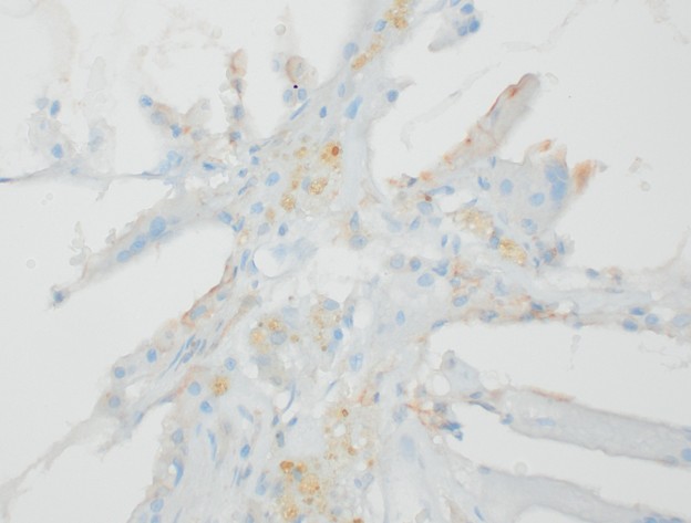
As expected, the inflammatory cells exhibited diffuse cytoplasmic IBA1 immunoreactivity, confirming their identity as macrophages and macrophage-derived multinucleated giant cells. However, the macrophage cytoplasm was also brightly PAS-positive. This finding is interesting because pure lipids do not stain with PAS and are more reliably demonstrated using Oil Red O or Sudan Black B on frozen sections. Because PAS primarily stains carbohydrates and mucosubstances (glycoproteins), there are two potential mechanisms explaining this brilliant cytoplasmic PAS reactivity11. This intense positivity most likely reflects accumulated glycolipids, lipofuscin deposition, or a combination of both. Lipofuscin is a membrane-bound, insoluble wear-and-tear pigment containing carbohydrate and protein moieties that impart variable PAS positivity. Finally, while cholesterol processing is complex and cholesterol is predominantly stored in macrophages as cholesterol esters or lipoproteins, in environments of cholesterol saturation and poor drainage, lipid is likely also stored as glycolipids, which would be intensely PAS-positive10.
References:
- Allan KJ, Waters M, Ashton DG, Patterson-Kane JC. Meningeal cholesterol granulomas in two meerkats (Suricata suricatta). Vet Rec. 2006;158:636-637.
- Cantile C, Youssef S. Nervous System. In: Maxie M. G. Jubb, Kennedy and Palmer?s Pathology of Domestic Animals, 6th ed; 1: 304.
- Davis AM, Lahmers K, Trusiano B, Zimmerman K, Romeiser J, Tuohy J. Cholesterol Granuloma of the Cranial Mediastinum in a Dog. J Am Anim Hosp Assoc. 2022;58:129-136.
- Dobbs P, Liptovszky M, Moittie S. Dietary management of hypercholesterolemia in a bachelor group of zoo-housed slender-tailed meerkats Suricata suricatta. J Zoo Aquar Res. 2020;8:294-296.
- Fliegner RA, Jubb KVF, Lording PM. Cholesterol Granuloma Associated with Otitis Media and Destruction of the Tympanic Bulla in a Dog. Vet Pathol. 2007;44:547-549.
- Greci V, Travetti O, Di Giancamillo M, et al. Middle ear cholesteatoma in 11 dogs. Can Vet J. 2011;52:631-636.
- Leon ME, Chavez C, Fyfe B, Nagorsky MJ, Garcia FU. Cholesterol granuloma of the maxillary sinus. Arch Pathol Lab Med. 2002;126:217-219.
- Sladky KK, Dalldorf FG, Steinberg H, Wright JF, Loomis MR. Cholesterol granulomas in three meerkats (Suricata suricatta). Vet Pathol. 2000;37:684-686.
- Van der Heyden S, Butaye P, Roels S. Cholesterol granuloma associated with otitis media and leptomeningitis in a cat due to a Streptococcus canis infection. Can Vet J. 2013;54:72-73.
- Chistiakov DA, Bobryshev YV, Orekhov AN. Macrophage-mediated cholesterol handling in atherosclerosis. J Cell Mol Med. 2016;20(1):17-28.
- Pernick N. Stains (IHC & specials) Mucins. PathologyOutlines.com, Inc. June 1, 2025. Accessed September 2, 2026. https://www.pathologyoutlines.com/topic/stainsmucins.html