Signalment:
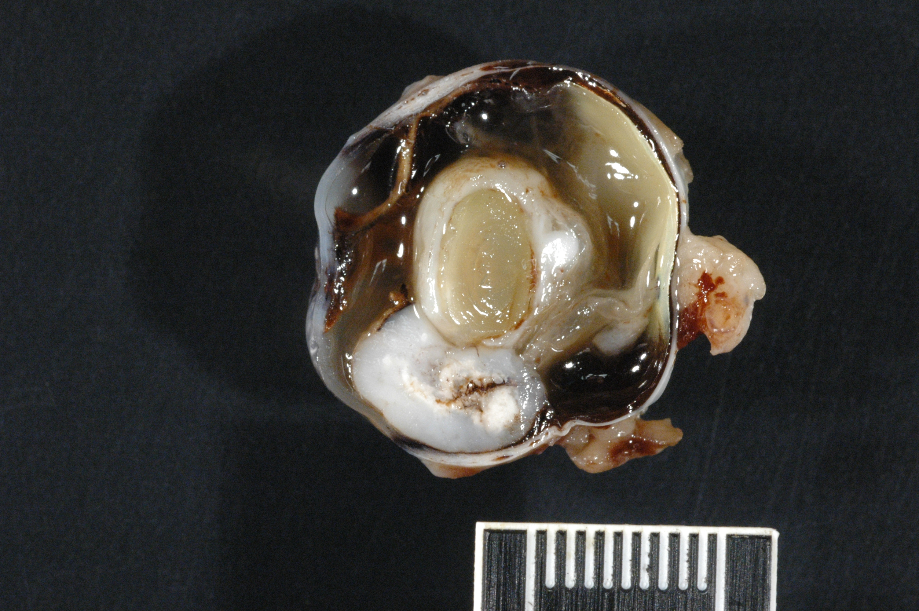
Gross Description:
Histopathologic Description:
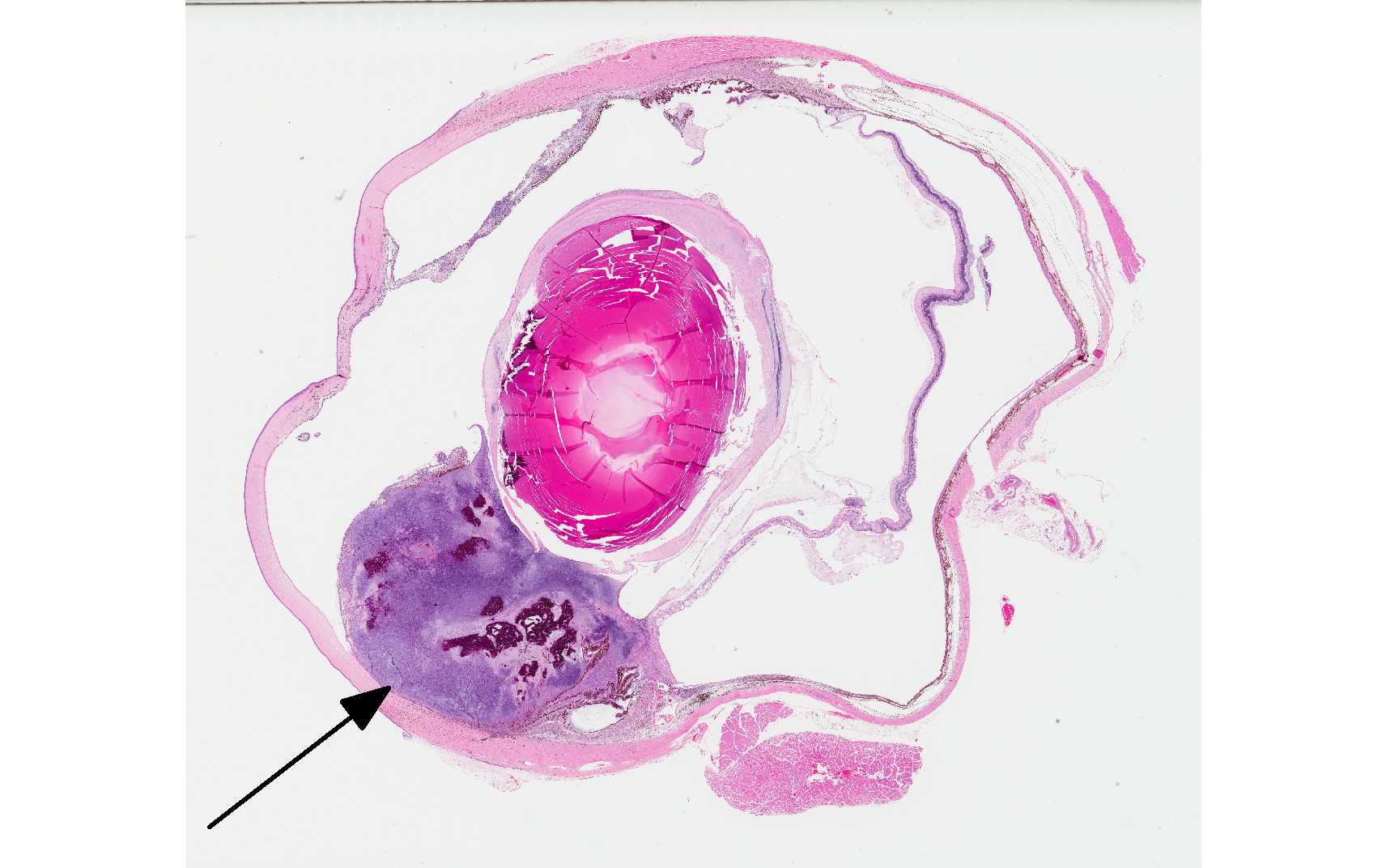
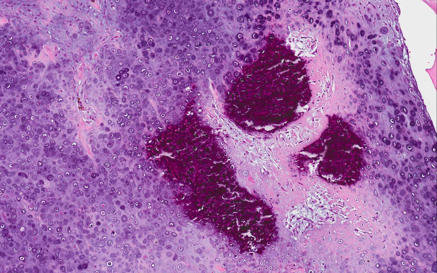
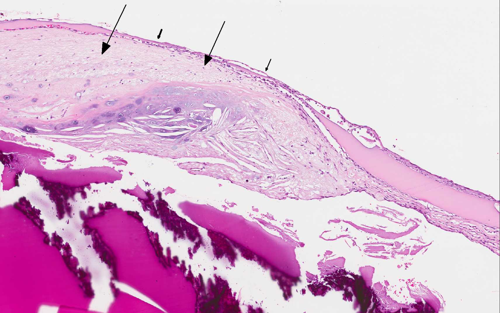
The cornea is distorted axially by implanted uveal pigment, fibrosis, and segmental rupture of Descemet's membrane. Portions of smooth muscle, iris pigment epithelium, and nodules of lymphocytes and plasma cells are also adherent to the exposed posterior stroma. The surrounding stroma is densely eosinophilic and slightly hyp-ercellular (fibrosis), and the epithelium is hyperplastic. Peripherally, the stroma lacks clefting (edema) and the epithelium is thinned. The posterior chamber/uveal mass is composed of irregular clusters of chondrocytes, sheets of spindle to stellate cells, and abundant granular to hyaline basophilic chondroid matrix, which abuts the posterior cornea and is intermittently covered by extensions of an inflamed fibrovascular membrane; there is a segment of Descemet's membrane surrounded by this membrane. The cells have scant amounts of amphophilic cytoplasm, and distinct borders. Nuclei are plump and oval, with coarse chromatin and prominent nucleoli. There is moderate anisocytosis and anisokaryosis, and about 35 mitotic figures per 10 400x fields. There are many scattered cells that are shrunken and hypereosinophilic, and there are several regions of mineralization. The mass and associated matrix is contiguous with the break in the lens capsule and the variably cellular layer of fibrous tissue that lines the inner capsule that replaces the cortex; there are foci of cartilage within the lens. The lens nucleus lacks fiber definition, and has a few swollen fibers and irregular clefts. Fibrous tissue with moderate nuclear atypia and scattered mitotic figures surround the outer capsule, as well.
-
The unaffected iris leaflet is expanded by edema and mononulcear inflammation, and the iridocorneal angle is collapsed. Reactive fibrovascular tissue lines the inner ciliary body. The entire retina is detached, with variable outer retinal atrophy, minimal subretinal exudate, and diffuse hypertrophy of the retinal pigmented epithelium. Peripherally, the full thickness of the retina is disorganized with fluid filled clefts/cavities. The inner retina is largely devoid of ganglion cells, particularly the non tapetal aspect. There is perivascular retinal infiltration by lymphocytes and plasma cells. The optic nerve head is vacuolated and anteriorly displaced. The optic nerve parenchyma is essentially normal. There are a few foci of lymphocytes and plasma cells in the sclera.
Morphologic Diagnosis:
1. Right eye: Lens capsule rupture with uveal chondrosarcoma (post traumatic ocular sarcoma)
2. Right eye: Lymphonodular panuveitis
3. Right eye: Iridocorneal angle collapse with inner retinal atrophy (chronic glaucoma)
4. Right eye: Complete chronic retinal detachment and chronic retinitis
5. Right eye: Mild to moderate chronic keratitis with focal
Descemets membrane rupture and uveal implantation
Lab Results:
Condition:
Contributor Comment:
Feline ocular sarcomas are malignant, intraocular neoplasms that are usually associated with evidence of trauma (i.e. FPTOS) from the clinical history and/or histologic findings (e.g. lens capsule rupture, retinal detachment, and corneal perforation). The time from trauma to the development of FPTOS varies widely, uncommonly in as little as 2 months, and more commonly several years; up to 10 years of latency has been reported.3 The average time between trauma and enucleation is 7 years.3 Extra-ocular invasion can occur and is typically at the limbus through the sclera, or at the optic nerve.2,5 For this reason, prophylactic enucleation of a feline eye after trauma should be considered if the eye is nonvisual.3 Metastasis is uncommonly reported in this disease, but current studies are small and/or have limited follow up.
FPTOS have a typical distribution. Tumors commonly occupy the posterior iris with expansion to the posterior chamber, posterior lenticular capsule, retina, and choroid.2 Invariably the lens is destroyed, either from initiating trauma or from tumor invasion, and significant inflammation accompanies these tumors (i.e. lens-induced uveitis).2,7 There are three variants of FPTOS, the most common of which is the spindle cell variant, followed by round cell variant, and lastly os-teosarcoma/chondrosarcoma.3 The cell of origin in spindle cell variants is likely lenticular epithelium.3,7
The phenomenon of FPTOS is similar to vaccine-associated feline sarcomas. The latter is associated with vaccination of cats for infectious diseases, most commonly rabies and feline leukemia virus.5 Vaccine-associated feline sarcomas are most commonly fibrosarcomas, but many different sarcoma variants have been reported, similar to FPTOS.5,7 Although the pathogenesis of both types of tumors is unknown, inflammation is an antecedent feature, suggesting a causal relationship is possible.
JPC Diagnosis:
1.Eye, globe: Chondrosarcoma (post-traumatic ocular sarcoma).
2.Eye, lens: Lenticular rupture with subcapsular chondroid metaplasia.
3.Eye: Anterior uveitis, lymp-hoplasmacytic, multifocal, mild.
Conference Comment:
Aside from cats, intraocular sarcoma has also been documented in rabbits with a history of chronic intraocular inflammatory disease.The two documented cases in rabbits were negative for E. cuniculi and bacterial infection via histochemical staining and polymerase chain reaction.The neoplasms expressed vimentin but were negative for smooth muscle actin, desmin, cytokeratin and S100. The neoplasms consisted of a population of anaplastic mesenchymal cells with the presence of lens fragments in the tumor. Trauma was not documented in the rabbits but the neoplasms were particularly aggressive and they were associated with the lens, similar to what is seen in many cases of FPTOS.Neoplastic cells extended through the sclera into adjacent extraocular tissues as well.4 Non-trauma related feline intraocular chondrosarcoma has been documented in cats, although it is very uncommon. Given that mammals do not have cartilage within the globe, the tissue origin in non-trauma related intraocular sarcoma is unclear.It is proposed to arise from multipotent mesenchymal stem cells which may be present within the trabecular meshwork, cancer stem cells, or potentially from vascular pericytes.In all four documented cases in cats the neoplasm was contained within the globe.1
Conference participants discussed the spatial association of the neoplastic mass with the lens and how it may relate to the pathogenesis of this neoplasm, as well as the origin of the mass of cartilage distant from the neoplasm at the posterior aspect of the lens.A possible origin for this area is spindle cell and chondroid metaplasia of subcapsular lens fibers separate from the neoplastic transformation on-going elsewhere.-
The trabecular meshwork of the iridocorneal angle was described as being collapsed and/or obscured by anterior uveitis, and glaucomatous changes were described in the retina including loss of ganglion cells as described above, as well as retinal atrophy.Anterior uvietisi is a common finding in the cat with quite a number of potential etiologies, and its connection to the neoplasm in this case could not be definitively proven.There are also areas with normal retina in this case, and the most severe thinning and loss of nuclei are present in the non-tapetal region.
References:
1.Beckwith-Cohen B, Teixeira LBC, Dubielzig RR.Presumed primary intraocular chondrosarcoma in cats. J Vet Diag Invest. 2014; 26(5):664-668.
2.Dubielzig RR, Everitt J, Shadduck JA, Albert DM. Clinical and morphologic features of post-traumatic ocular sarcomas in cats. Vet Pathol. 1990; 27:62-65.
3.Dubielzig RR. Non-surgical trauma. In: Dubielzig RR, Ketring KL, McLellan GJ, Albert DM, ed. Veterinary Ocular Pathology: A Comparative Review. Philadelphia, PA: Saunders Elsevier; 2010:81-114.
4.Morrison WB, Starr RM. Vaccine-associated feline sarcomas. J Am Vet Med Assoc. 2001; 218:697-702.
5.McPherson L, Newman SJ, McLean N, McCain S, et al.Intraocular sarcomas in two rabbits. J Vet Diagn Invest. 2009; 21:547-551.
6.Lin H, Ouyang H, Zhu J, Huang S, et al. Lens regeneration using endogenous stem cells with gain of visual function. Nature. 2016; 531:323-328.
7.Zeiss CJ, Johnson EM, Dubielzig RR. Feline intraocular tumors may arise from transformation of lens epithelium. Vet Pathol. 2003; 40:355-362.