Signalment:
Gross Description:
Histopathologic Description:
Morphologic Diagnosis:
Lab Results:
Urinary Analysis
| Parameter | Value |
| Density | 1.026 |
| Blood | +++ |
| pH | 6 |
| Bilirubin | + |
| Leukocytes | + |
| Macrophages | +++ |
Blood Analysis
| Parameter | Value | Normal Value |
| Urea | 0.64 g/L | 0.2-0.6 g/L |
| Creat | 13 mg/L | < 12 mg/L |
| PAL | 76 UI/L | < 200 UI/L |
| ALAT | 24 UI/L | < 80 UI/L |
| TP | 47 g/L | 55-80 g/L |
| Glucose | 0.87 g/L | 0.6-1.1 g/L |
| Na+ | 138 mmo1/L | 140-150 mmo1/L |
| K+ | 4.4 mmo1/L | 3.8-5.2 mmo1/L |
| HCO3- | 22 mmo1/L | 25 mmo1/L |
Condition:
Contributor Comment:
Table1: Immunophenotypes of the different antigen-presenting cells2
| Multipotent bone marrow stem cells | |||
| CD34+ CLA+ | CD34+ CLA- | CD34+ IL-3R | |
| Langerhans cells (epithelial dendritic cells) | Intersitial dendritic cells | Macrophages/ Histiocytes | Dendritic cells of lymphoid organs |
| CD1+ CD14+ CD11c+ MHCII Birbeck granules+ | CD1+ CD14- CD11c+ MHCII+ Thy-1+ |
CD1- CD14+ MHCII+ CD11c- CD11b+ | IL-3R MHCII+ CD45RA+ CD4+ |
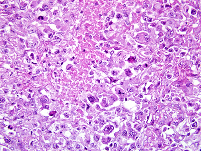
Origin lineage of histiocytic disorders in dogs is the subject of many debates. In humans, they tend to arise from both macrophages/histiocytes and dendritic cells whereas in rats, they are of macrophagic origin. In dogs and cats, they are believed to be mostly of dendritic origin.2 In dogs, the main histiocytic disorders are: cutaneous histiocytoma, cutaneous histiocytosis, systemic histiocytosis, histiocytic sarcoma (HS) and malignant histiocytosis (MH).4 Whereas cutaneous and systemic histiocytosis are non-neoplastic proliferations of activated dendritic cells, the others are classified as true neoplasms. There is a lot of confusion concerning the terms HS and MH. HS should be used when the lesion is solitary or has metastasized. MH defines a multicentric proliferation. Thus, a disseminated histiocytic sarcoma could be undistinguishable from a malignant histiocytosis. Bernese mountain dogs (BMD) show particular susceptibility to histiocytic disorders and neoplasms.6 Properties of these histiocytic disorders are summarized in Table 2. Concerning HS/MH, respiratory symptoms are the most frequent cause of consultations. Anemia can be observed as part of a regenerative hemolytic process or a nonregenerative anemia caused by bone marrow invasion and erythrophagocytosis. Hypercalcemia can be observed as a paraneoplastic syndrome. On histopathologic examination, there is proliferation of round-to-oval cells with abundant eosinophilic cytoplasm, nuclear atypia, numerous mitoses, multinucleation, phagocytosis and some infiltration by lymphocytes, plasma cells and neutrophils.4 On immunohistochemistry, along with markers of Table 1, cells are positive for lysozyme, vimentin, and negative for cytokeratin A.3 Differential diagnosis includes: anaplastic carcinoma or lymphoma, rhabdomyosarcoma, and lymphomatoid granulomatosis. This case showed original features. Indeed, MH has only been reported in two Shar Peis and prostate involvement was only reported once.2 Furthermore, no infiltration of the liver was observed, both at gross and microscopic examination. This feature is uncommon as the liver is one of the three most frequently involved organs.2
Table2: Properties of histiocytic disorders in dogs
| Diseases | Concerned breeds | Mean age | Cytonuclear atypias | Involved organs | Metastatic potential | Prognosis |
| Cutaneous histiocytoma | All, predisposition of Boxer, dachshund, cocker spaniel, Great Dane, Shetland | Mostly young dogs | Rare | Skin (mainly solitary lesion). Draining lymph nodes involvement is exceptional | None | Good |
| Cutaneous histiocytosis | All, predisposition of golden retriever and German shepherd | Mostly young dogs | Rare | Skin (multiple lesions), sometimes lymph nodes | Rare | Good |
| Systemic histiocytosis | BMD, golden retriever, Doberman pinscher, rottweiler | Adults Mean age is 7 for BMD | Moderate | Skin (multiple lesions), lymph nodes sometimes ocular tissues | Moderate | Guarded |
| Histiocytic sarcoma | Adults Mean age is 6 for BMD | Severe | Subcutaneous tissues or internal organs | Elevated to draining lymph nodes | Guarded | |
| Malignant histiocytosis | Severe | Rapid multicentric dissemination (lungs, liver, spleen) | Elevated | Poor |
JPC Diagnosis:
Conference Comment:
Canine malignant histiocytosis/histiocytic sarcoma was first reported in Bernese mountain dogs and has since been reported in various dog breeds, cats, and other species.4 Malignant histiocytosis implies multicentric origin of the neoplasm. In cases of widespread disease, it is unclear whether the neoplasm arose multicentrically or metastasized widely from a single primary site. Since conference participants were only aware of the neoplasm in the prostate, histiocytic sarcoma was considered the most appropriate diagnosis. Given the presence of widespread disease, disseminated histiocytic sarcoma is preferred. These tumors are composed of pleomorphic histiocyte-like cells that are often multinucleated or may contain phagocytized material. Two major patterns have been described: round cell predominant and spindle cell predominant.4 In the round cell variant, neoplastic cells often have abundant eosinophilic cytoplasm and round to reniform nuclei.4,5 The spindle cell variant is often composed of plump spindle cells and these tumors often resemble other sarcomas.4 Organs most commonly affected include the liver, lung, kidney, spleen, lymph node, and bones. Almost any organ may be affected by this neoplasm.5 Canine histiocytic sarcomas are of dendritic antigen presenting cell origin and express CD18, CD1, CD11c, ICAM-1 and MHC II; CD45 expression is variable. For immunohistochemistry on formalin-fixed, paraffin-embedded tissues, CD18 positivity and negative findings for CD3 and CD79a combined with characteristic histomorphology is considered diagnostic.
A variety of other histiocytic proliferative diseases have been described in dogs. Histiocytomas generally occur in dogs four years of age or younger, are extremely common and have a predilection for the head and ears. These tumors are of Langerhans cell origin and express CD1, CD11c, MHC II, and E-cadhedrin.7 At the subgross level, these tumors often appear dome shaped with aggregates of lymphocytes and plasma cells at the periphery. There is often superficial dermal edema, and tumor cells infiltrate the epidermis in a fashion similar to Pautriers microabscesses of epitheliotropic lymphoma. Neoplastic cells form sheets and generally lack a discernable stroma. Neoplastic cells have oval to reniform nuclei with a moderate to abundant amount of eosinophilic cytoplasm and mitotic figures are frequent. There is little cellular atypia.5 Multiple, persistent and recurring histiocytomas have also been described. Such tumors may progress to a malignancy characterized by dissemination to various organs. This condition has been designated Langerhans cell histiocytosis. Cutaneous histiocytosis is a nonneoplastic, reactive condition characterized by nodules of histiocytic cells that express CD45, CD18, CD1, CD11c, MHC II and E-cadherin. Mature lymphocytes and neutrophils are often scattered amongst the histiocytic cells. The mitotic rate is variable. Systemic histiocytosis is generally similar to cutaneous histiocytosis but has a more extensive distribution. The immunohistochemical profile is the same. Sites most commonly affected include lymph nodes, eyelids, sclera, nasal cavities, lungs, spleen and bone marrow.
References:
2. Affolter VK, Moore PF: Localized and disseminated histiocytic sarcoma of dendritic cell origin in the dog. Vet. Pathol 39,74-83, 2002
3. Azakami D, Bonkobara M, Washizu T, et al: Establishment and biological characterization of canine histiocytic sarcoma cell lines. J Vet Med Sci 68(12):1343- 1346, 2006
4. Gross TL, Ihrke P, Walder EJ, Affolter VK: Histiocytic tumors. In: Skin diseases of the dog and cat, 2nd edition, pp. 848-851, 2005
5. Hendrick MJ, Mahaffey EA, Moore FM, Vos JH, Walder EJ: Histological classification of mesenchymal tumors of skin and soft tissues of domestic animals. In: World Health Organization Histological Classification of Tumors of Domestic Animals, ed. Schulman FY, Second Series, vol. 2, pp. 29-31, 58-59. Armed Forces Institute of Pathology,Washington, DC, 1999
6. Moore PF., Rosin A.: Malignant histiocytosis of Bernese Mountain dogs. Vet Pathol 23:1-10, 1986
7. Moore PF: Canine histiocytosis. At: http://www. histiocytosis.ucdavis.edu/