WSC 2023-2024, Conference 14, Case 4
Signalment:
3-year-old, Thoroughbred filly (Equus caballus)
History:
Found dead in stall.
Gross Pathology:
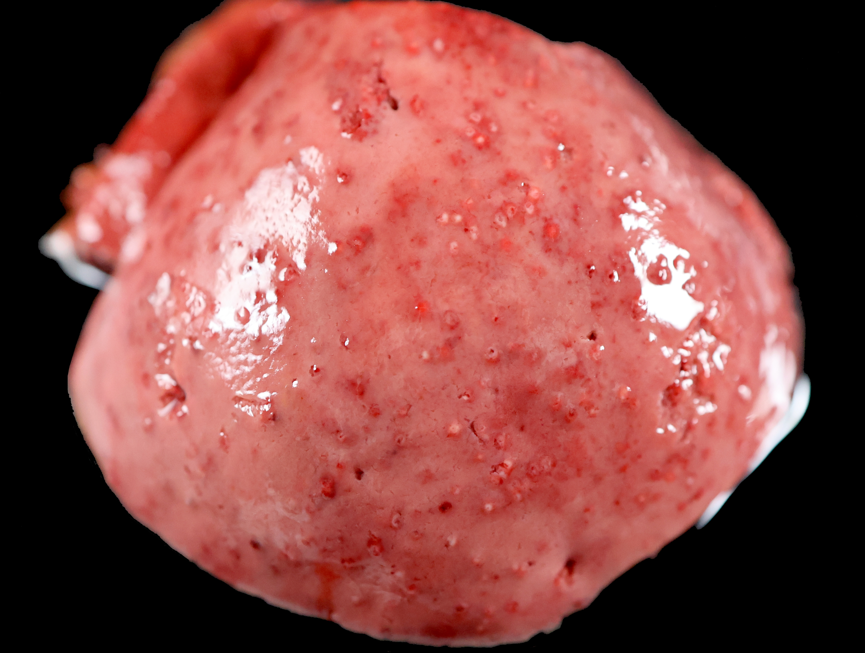
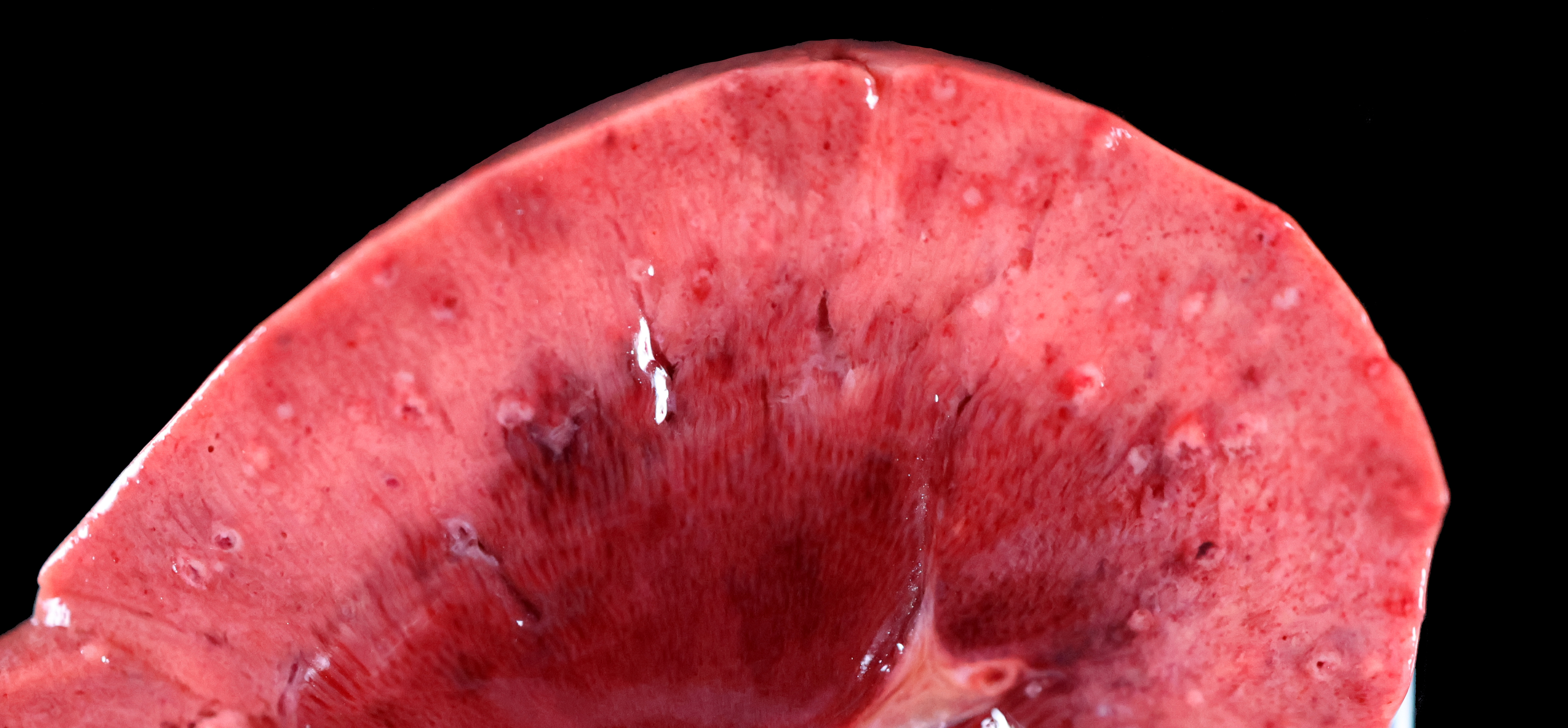
Both kidneys have numerous pinpoint raised, tan foci with a red halo scattered throughout the renal cortices.
Laboratory Results:
Aerobic culture, kidney: Actinobacillus sp., mixed flora (Actinobacillus sp. were also isolated from the liver and lung).
DNA sequence analysis: DNA sequencing of a ~600 bp region of the 16S rRNA gene identified the isolate as most closely related to Actinobacillus equuli ssp. equuli.
Microscopic Description:
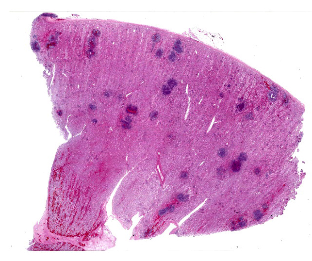
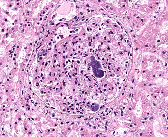
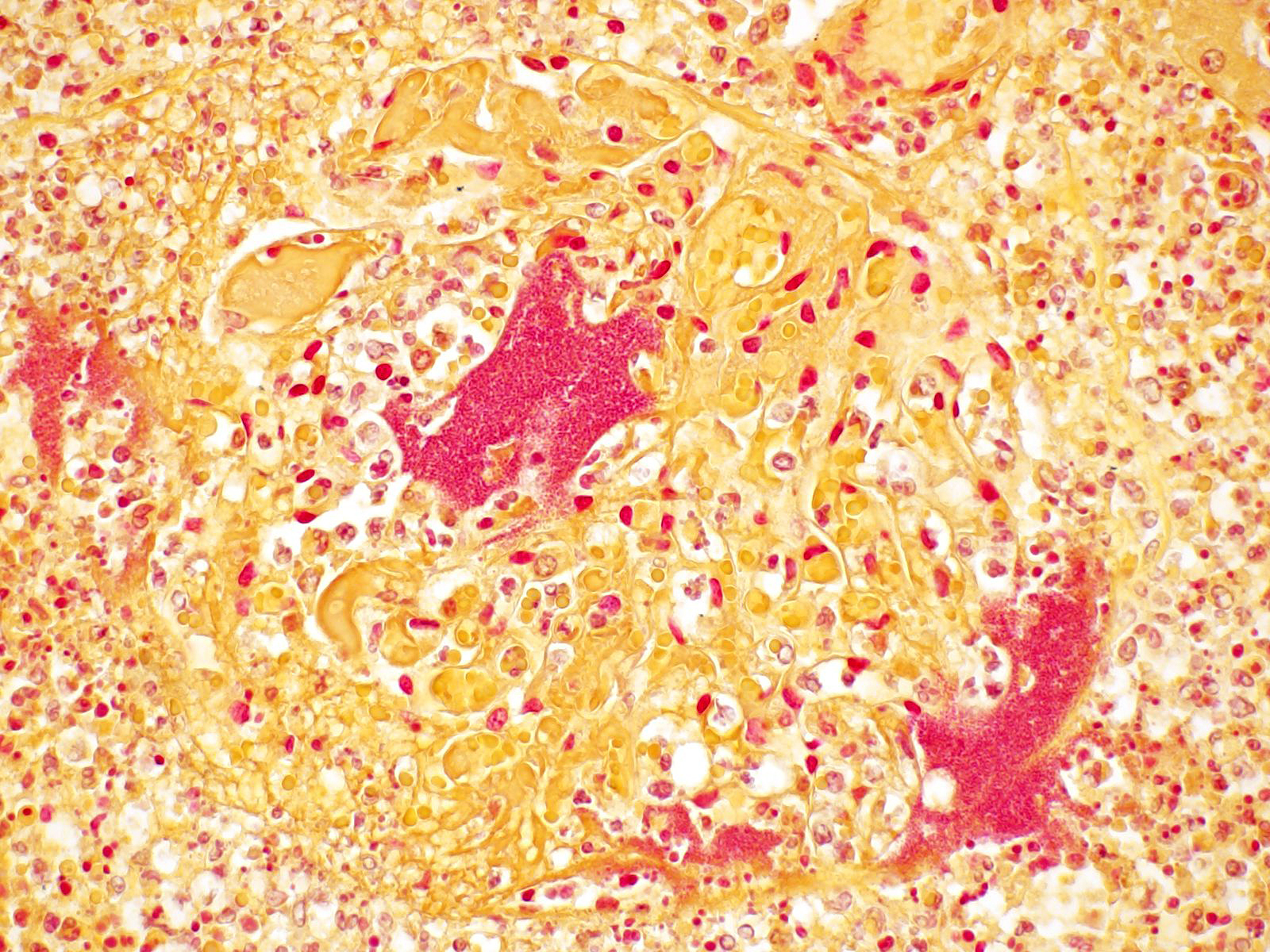
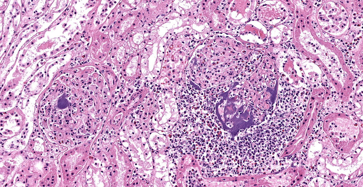
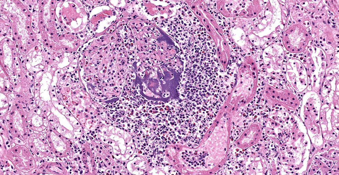
Within the renal cortical interstitium, there are randomly scattered, unencapsulated aggregates of neutrophils (measuring up to 3 mm in diameter), often with a central region of necrosis or hemorrhage, that efface the tubular and glomerular architecture. In some areas, these aggregates of neutrophils appear to be centered on glomeruli or blood vessels. There are aggregates of intra- and extravascular coccobacilli associated with these neutrophilic infiltrates. Glomeruli have similar bacteria and fibrin in the capillaries. There is also mild tubular degeneration. Coccobacilli are gram-negative.
Contributor’s Morphologic Diagnosis:
Kidney: Nephritis, embolic-suppurative, multifocal, with intra- and extravascular Gram-negative coccobacilli.
Contributor’s Comment:
Actinobacillus spp. (Family: Pasteurellaceae) are pleomorphic, non-motile, non-spore forming, gram-negative, rod-shaped bacteria characterized by their ability to grow on MacConkey agar, produce β-galactosidase, and ferment carbohydrates without the production of gas.8 There are a number of Actinobacillus species of significance within veterinary medicine, including A. lignieresii (causative agent of wooden tongue in cattle), A. pleuropneumoniae (causative agent of porcine contagious pleuropneumonia in pigs), A. suis (cause of septicemia in pigs), A. capsulatus (cause of joint disease in rabbits), and A. seminis (cause of epididymitis in rams).8 As with many pathogenic bacteria within the family Pasteurellaceae, Actinobacillus often produce repeats-in-toxin (RTX) toxins, which are typically hemolytic and cytotoxic.3
In horses, Actinobacillus are common commensals and can be isolated from the gastrointestinal, respiratory, and urogenital tracts of healthy animals.4 Actinobacillus spp. isolated from horses include: A. equuli ssp. equuli, A. equuli ssp. haemolyticus, A. arthritidis, A. lignieresii, A. suis, and A. pleuropneumoniae. Of these, A. equuli ssp. equuli and A. equuli ssp. haemolyticus are isolated most frequently from horses. These subspecies are differentiated based on the presence or absence of an RTX toxin, A. equuli toxin (or Aqx), encoded on the aqx gene.3,8
Actinobacillus equuli ssp. equuli is typically carried in the oral cavity and alimentary tract of juvenile and adult horses and is primarily associated with septicemia in neonatal foals (often referred to as “sleepy foal disease”).4,8 There are also reports of A. equuli-associated peritonitis and septicemia in adult horses, with gross and microscopic lesions similar to those described in foals.4-7,9 Predisposing factors for “sleepy foal disease” include failure of passive transfer and concurrent bacterial/viral infections.4
Similarly, actinobacillosis in adult horses can be associated with stress (such as shipping) and concurrent bacterial/viral infections; there is, however, evidence that A. equuli can act as a primary pathogen in horses in the absence of stress/concurrent disease.4
Typical light microscopic findings of A. equuli infection include: suppurative embolic nephritis, suppurative embolic pneumonia, multifocal suppurative hepatitis, lymphoid necrosis, suppurative oomphalitis, and suppurative meningoencephalitis. A. equuli is zoonotic with sporadic reports of disease in people.1
Contributing Institution:
California Animal Health and Food Safety Laboratory (San Bernardino Branch) https://cahfs.vetmed.ucdavis.edu/
JPC Diagnoses:
Kidney: Nephritis, suppurative, embolic, with numerous large colonies of bacilli.
JPC Comment:
This case is an excellent example of suppurative embolic nephritis, in which septic emboli lodge in glomerular and peritubular capillaries and produce variably sized abscesses, primarily in the renal cortex.2 In horses, Actinbacillus equuli is the most common cause of embolic suppurative nephritis, generally seen in neonatal foals after acquiring the infection in uetero, during parturition, or shortly after birth via the umbilicus.2
As the contributor notes, there are two subspecies of A. equuli that differ based on the presence of absence of an RTX toxin. A. equuli subspecies haemolyticus produces Aqx, an RTX toxin similar to the leukotoxins and hemolysins produced by related Pasteurellaceae such as Mannheimia haemolytica, A. pleuropneumonia, A. suis, and hemolytic E. coli.4 The Aqx toxin is highly cytolytic to equine erythrocytes and lymphocytes, and has less virulent lytic activity against the same cells in pigs.
The vast majority of A. equuli septicemia cases are cause by A. equuli subspecies equuli, which does not produce the Aqx toxin, indicating that other virulence factors likely play the key roles in neonatal septicemic actinobacillosis.4
Like any self-respecting gram-negative bacterium, A. equuli subspecies equuli is armed with endotoxin which, upon the destruction of the bacterium, damages endothelium and causes the vasculitis and bacterial emboli that are characteristic of septicemic actinobacillosis.4 As illustrated in this case, a particular quirk of equine actinbacillosis is the presence of large bacterial colonies present in affected vessels, unlike other gram-negative bacteria which do not form such obvious intravascular aggregates.4 This has led to speculation that A. equuli subspecies equuli may express an adhesion for endothelium. Unfortunately, other than the well-characterized Aqx toxin present in A. equuli subspecies haemolyticus, little is definitively known about the virulence factors that enable the spectacular lesions of equine actinobacillosis.4
Conference discussion focused initially on the age of the animal since, as noted above, A. equuli embolic nephritis is typically a disease of neonatal foals. The moderator noted reports of this presentation in non-stressed, non-immunocompromised adult animals and reminded residents to think broadly about differential diagnoses, even when the signalment isn’t a perfect fit. The moderator led conference participants in a discussion other famous Actinobacillus species of veterinary importance, including the species noted above by the contributor. Finally, participants discussed the presence of bacterial colonies within glomeruli and whether this would constitute glomerulonephritis. Participants felt that, since the pathogeneses behind the established categories of glomerulonephritis (membranoproliferative, membranous, etc.) did not pertain to a suppurative embolic nephritis, the term glomerulonephritis was inappropriate. Glomerulitis would be a more appropriate term; however, since suppurative inflammation was present within the glomeruli, tubules, and interstitium, the preferred morphologic diagnosis is the all-inclusive term “nephritis.”
References:
- Ashhurst-Smith C, Norton R, Thoreau W, Peel MM. Actinobacillus equuli septicemia: an unusual zoonotic infection. J Clin Microbiol. 1998;36(9):2789-90.
- Cianciolo RE, Mohr FC. Urinary System. In: Maxie MG, ed. Jubb, Kennedy, and Palmer’s Pathology of Domestic Animals. 6th ed. Vol 2. Elsevier;206:433.
- Frey J, Kuhnert P. RTX toxins in Pasteurellaceae. Int J Med Microbiol. 2002;292(3-4):149-58.
- Layman QD, Rezabek GB, Ramachandran A, Love BC, Confer AW. A retrospective study of equine actinobacillosis cases: 1999-2011. J Vet Diagn Invest. 2014; 26(3):365-75.
- Matthews S, Dart AJ, Dowling BA, Hodgson JL, Hodgson DR. Peritonitis associated with Actinobacillus equuli in horses: 51 cases. Aust Vet J. 2001;79(8):536-9.
- Means K, Townsend K, Johnson P. Multicavitary septic effusions associated with actinobacillosis in an adult Tennessee Walking Horse with weight loss. Equine Vet Educ. 2022;34(1):e60-6.
- Patterson-Kane JC, Donahue JM, Harrison LR. Septicemia and peritonitis due to Actinobacillus equuli infection in an adult horse. Vet Pathol. 2001;38(2):230-2.
- Rycroft AN, Garside H. Actinobacillus species and their role in animal disease. Vet J. 2000;159(1):18-36.
- Uchida-Fujii E, Niwa H, Kinoshita Y, Nukada T. Actinobacillus species isolated from Japanese Thoroughbred racehorses in the last two decades. J Vet Med Sci. 2019;81(9):1234-7.