Signalment:
Gross Description:
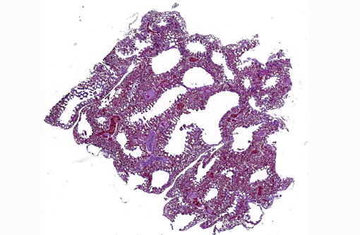
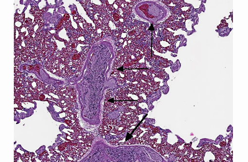
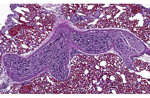
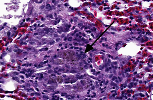
Histopathologic Description:
Morphologic Diagnosis:
Lungs:
1) Brain tissue emboli, pulmonary arterioles, multifocal, peracute, moderate to severe with arteriolar congestion.
2) Pneumonia, granulomatous, multifocal, chronic, mild (not present in all sections).
3) Anthracosis, parabronchial, chronic, multifocal, mild.
Condition:
Contributor Comment:
Cerebral tissue pulmonary embolization (CTPE) is a possible sequel to severe penetrating or closed head trauma. CTPE is most commonly associated with high impact blunt force trauma (i.e. automobile collision) in adults and instrument-assisted delivery in neonates.(4,5) Though a rare occurrence, post-traumatic pulmonary emboli can cause significant mortality (up to 43%) in the absence of prophylactic treatment.(6) Massive CTPE is detectable at autopsy and is associated with disruption of the large dorsal cerebral venous sinus in addition to brain injury. Microscopic brain emboli, however, have been identified in pulmonary arterioles and systemic veins in cases with intact dura, suggesting embolic entry through smaller cerebral and meningeal veins.(8)
The behavior, physiology and anatomy of flighted birds may increase the likelihood of CTPE in avian species compared to terrestrial animals. Behaviorally, flighted birds are prone to severe brain trauma due to in-flight speed and prevalent collision injuries. Physiologically, avian veins, unlike mammalian veins, are compliance vessels, and they actively dilate during flight to increase cardiac output.(9) A larger venous diameter permits embolization of larger tissue fragments to the lungs. Anatomically, the avian brain and spinal cord are surrounded by a series of contiguous venous sinuses including the dorsal cerebral, occipital and vertebral sinuses and the ventral sinus cavernosus. These sinuses drain blood to the heart via the jugular or vertebral veins.(10) These extensive, superficial structures are prone to rupture with severe, closed or penetrating head trauma, presenting direct venous access to injured neural tissue.
This case illustrates dissemination of central nervous system (CNS) tissue to the venous system after head trauma. Consumption of meat products contaminated with CNS tissue from cattle with bovine spongiform encephalopathy is considered to be a significant route of transmission for mutant proteinase-resistant protein (PrPsc), the proposed etiologic agent of variant CreutzfeldtJakob disease (vCJD) in humans.(2,7) Air-injection penetrating captive bolt stunning prior to terminal exsanguination has been identified as a major risk factor in CNS contamination of meat products. This method allows dislodged CNS tissue to disseminate through the bloodstream during the brief period of sustained cardiac function, followed by contamination of skeletal muscle with jugular exsanguination. For this reason, this method of cattle slaughter is currently banned in the United States and the European Union.(1)
JPC Diagnosis:
1. Lung, pulmonary arteries: Neural emboli, multiple.
2. Lung: Granulomas, parabronchiolar, multiple, with anthracosis.
Conference Comment:
Conference participants conducted an abbreviated discussion of the anatomy and physiology of the normal avian lung. The avian mesobronchus (similar to the mammalian bronchus) is an airway lined with ciliated respiratory epithelium that has hyaline cartilage and smooth muscle within its walls; it has no direct function in gas exchange. The mesobronchus gives rise to the recurrent secondary bronchi, which are analagous to mammalian bronchioles and contain smooth muscle, but no cartilage within their walls. These further divide into tertiary bronchi (parabronchi) with walls that are "scalloped" by bay-like air vesicles, where gas exchange takes place. Air vesicles are composed of simple squamous epithelium with an underlying supporting connective tissue. Air passes through numerous air capillaries in the wall of each air vesicle; these are adjacent to the blood capillaries, an arrangement that results in the establishment of a countercurrent flow. Unlike mammalian ventilation, in which a part of the ventilator volume is "stale" air, and mammalian structure with its numerous blind alleys and abundant dead space, the avian lung is a continuous flow system. Thus, avian lungs are much more efficient than mammalian, which is not surprising, considering the high demand of flight muscles for oxygenation.(3)
Participants closed with a brief summary of other reported causes of pulmonary emboli, including trophoblastic emboli (especially in guinea pigs), fibrocartilaginous emboli, neoplastic cells (especially lymphocytes in mice with tumor lysis syndrome), bone marrow elements (subsequent to injury/fracture), and allantoic fluid (in humans).
References:
2. Brown P, Will RG, Bradley R, Asher DM, Detwiler L. Bovine spongiform encephalopathy and variant Cruetzfeldt-Jakob disease: background, evolution and current concerns. Emerg Infect Dis. 2001;7:6-16.
3. Caceci T. Virginia-Maryland Regional College of Veterinary Medicine, Blacksburg, VA. VM8054 Veterinary Histology website. Respiratory System II: Avians. http://www.vetmed.vt.edu/education/Curriculum/VM8054/Labs/Lab26/lab26.htm. Accessed February 22, 2014.
4. Cox P, Silvestri E, Lazda E, Nash R, Jeffrey I, Ostojic N et al. Embolism of brain tissue in intrapartum and early neonatal deaths: report of 9 cases. Pediatr Dev Pathol. 2009;12:464-468.
5. Echeverria RF, Baitello AL, Pereira de Godoy JM, Espada PC, Morioka RY. Prevalence of death due to pulmonary embolism after trauma. Lung India. 2010;27:72-74.
6. Geerts WH, Code KI, Jay RM, Chen E, Szalai JP. A prospective study of venous thromboembolism after major trauma. N Engl J Med. 1994;331:16011606.Â
7. Jones M, Peden AH, Prowse CV, Gr+�-�ner A, Manson JC, Turner ML, et al. In vitro amplification and detection of variant CreutzfeldtJakob disease PrPSc. J Pathol. 2007;213:21-26.
8. Morentin B, Biritxinaga B. Massive pulmonary embolization by cerebral tissue after head trauma in an adult male. Am J Forensic Med Pathol. 2006;27:268-270.
9. Smith FM, West NH, Jones DR. The Cardiovascular system. In: Whittow GC, ed. Sturkies Avian Physiology. 5th ed. San Diego, CA: Academic Press; 2000:174.
10. West NH, Langille BL, Jones DR. Cardiovascular system. In: King AS, McLelland J, eds. Form and Function in Birds. Vol. 2. San Francisco, CA: Academic Press; 1981:278-283.